
Breast cancer is a cancer that develops from breast tissue. Signs of breast cancer may include a lump in the breast, a change in breast shape, dimpling of the skin, milk rejection, fluid coming from the nipple, a newly inverted nipple, or a red or scaly patch of skin. In those with distant spread of the disease, there may be bone pain, swollen lymph nodes, shortness of breath, or yellow skin.

Christiane (Janni) Nüsslein-Volhard is a German developmental biologist and a 1995 Nobel Prize in Physiology or Medicine laureate. She is the only woman from Germany to have received a Nobel Prize in the sciences.

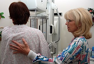
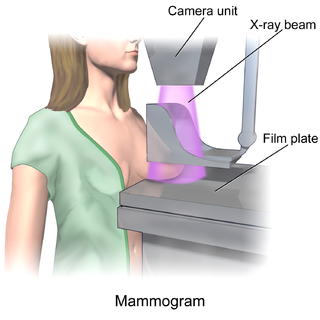
Mammography is the process of using low-energy X-rays to examine the human breast for diagnosis and screening. The goal of mammography is the early detection of breast cancer, typically through detection of characteristic masses or microcalcifications.
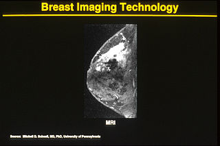
One alternative to mammography, breast MRI or contrast-enhanced magnetic resonance imaging (MRI), has shown substantial progress in the detection of breast cancer.
Breast cancer screening is the medical screening of asymptomatic, apparently healthy women for breast cancer in an attempt to achieve an earlier diagnosis. The assumption is that early detection will improve outcomes. A number of screening tests have been employed, including clinical and self breast exams, mammography, genetic screening, ultrasound, and magnetic resonance imaging.

Molecular breast imaging (MBI), also known as scintimammography, is a type of breast imaging test that is used to detect cancer cells in breast tissue of individuals who have had abnormal mammograms, especially for those who have dense breast tissue, post-operative scar tissue or breast implants.
The objective of cancer screening is to detect cancer before symptoms appear, involving various methods such as blood tests, urine tests, DNA tests, and medical imaging. The purpose of screening is early cancer detection, to make the cancer easier to treat and extending life expectancy. In 2019, cancer was the second leading cause of death globally; more recent data is pending due to the COVID-19 pandemic.
Professor Dame Janet Elizabeth Husband is Emeritus Professor of Radiology at the Institute of Cancer Research. She had a career in diagnostic radiology that spanned nearly 40 years, using scanning technology to diagnose, stage, and follow-up cancer. She continues to support medicine and research as a board member and advisor for various organisations.
Thomas M. Kolb is an American radiologist specializing in the detection and diagnosis of breast cancer in young, predominantly high-risk premenopausal women. He has served as an assistant clinical professor of Radiology at Columbia University College of Physicians and Surgeons from 1994–2010. Kolb is double board certified, having received his training in pediatrics at the Albert Einstein College of Medicine in Bronx, New York, and in diagnostic radiology at the Columbia-Presbyterian Medical Center in New York.
Automated whole-breast ultrasound (AWBU) is a medical imaging technique used in radiology to obtain volumetric ultrasound data of the entire breast.
In medicine, breast imaging is a sub-speciality of diagnostic radiology that involves imaging of the breasts for screening or diagnostic purposes. There are various methods of breast imaging using a variety of technologies as described in detail below. Traditional screening and diagnostic mammography uses x-ray technology and has been the mainstay of breast imaging for many decades. Breast tomosynthesis is a relatively new digital x-ray mammography technique that produces multiple image slices of the breast similar to, but distinct from, computed tomography (CT). Xeromammography and galactography are somewhat outdated technologies that also use x-ray technology and are now used infrequently in the detection of breast cancer. Breast ultrasound is another technology employed in diagnosis and screening that can help differentiate between fluid filled and solid lesions, an important factor to determine if a lesion may be cancerous. Breast MRI is a technology typically reserved for high-risk patients and patients recently diagnosed with breast cancer. Lastly, scintimammography is used in a subgroup of patients who have abnormal mammograms or whose screening is not reliable on the basis of using traditional mammography or ultrasound.
Fiona Jane Gilbert is a Scottish radiologist and academic.
Maryellen L. Giger, is an American physicist and radiologist who has made significant contributions to the field of medical imaging.
Nola M. Hylton is an American oncologist who is Professor of Radiology and Director of the Breast Imaging Research Group at the University of California, San Francisco. She pioneered the usage of magnetic resonance imaging for the detection, diagnosis, and staging of breast cancer by using MRIs to locate tumors and characterize the surrounding tissue.
HB 2102, also known as "Henda's Law", is a breast density (BD) notification law approved in 2011 by the FDA that mammography patients be provided educational materials on dense breast tissue can hide abnormalities, including breast cancer, from traditional screening. Henda's Law aims to promote patient–doctor discussion as well as reduce the rate of false negatives, as mammography may not detect abnormalities in dense breasts.
Etta Driscoll Pisano is an American breast imaging researcher. She is a professor in residence of radiology at the Beth Israel Deaconess Medical Center and chief research dean at the American College of Radiology. In 2008, she was elected a member of the National Academy of Medicine.
Sylvia Katina Plevritis is Professor and Chair of the Department of Biomedical Data Science at Stanford University.
Anca-Ligia Grosu is a Romanian-German radiation oncologist and professor with a research focus on the development of personalized therapy in radiation oncology. She is chair of the Radiation Oncology Department at the University Medical Center Freiburg, Germany, and member of the German National Academy of Sciences Leopoldina.
Dense breast tissue, also known as dense breasts, is a condition of the breasts where a higher proportion of the breasts are made up of glandular tissue and fibrous tissue than fatty tissue. Around 40–50% of women have dense breast tissue and one of the main medical components of the condition is that mammograms are unable to differentiate tumorous tissue from the surrounding dense tissue. This increases the risk of late diagnosis of breast cancer in women with dense breast tissue. Additionally, women with such tissue have a higher likelihood of developing breast cancer in general, though the reasons for this are poorly understood.

Rachel F. Brem is an American diagnostic radiologist, professor of radiology at the George Washington University School of Medicine & Health Sciences, and director of the Breast Imaging and Interventional Center at George Washington University’s Cancer Center. She previously served as director of Breast Imaging at Johns Hopkins. Brem develops novel technologies to better support early diagnosis and treatment of breast cancer. She is a fellow of the American College of Radiology and the Society of Breast Imaging.
