
Lymphedema, also known as lymphoedema and lymphatic edema, is a condition of localized swelling caused by a compromised lymphatic system. The lymphatic system functions as a critical portion of the body's immune system and returns interstitial fluid to the bloodstream. Lymphedema is most frequently a complication of cancer treatment or parasitic infections, but it can also be seen in a number of genetic disorders. Though incurable and progressive, a number of treatments may improve symptoms. Tissues with lymphedema are at high risk of infection because the lymphatic system has been compromised.

Erysipelas is a relatively common bacterial infection of the superficial layer of the skin, extending to the superficial lymphatic vessels within the skin, characterized by a raised, well-defined, tender, bright red rash, typically on the face or legs, but which can occur anywhere on the skin. It is a form of cellulitis and is potentially serious.

Edema, also spelled oedema, and also known as fluid retention, dropsy, hydropsy and swelling, is the build-up of fluid in the body's tissue. Most commonly, the legs or arms are affected. Symptoms may include skin which feels tight, the area may feel heavy, and joint stiffness. Other symptoms depend on the underlying cause.

Mange is a type of skin disease caused by parasitic mites. Because various species of mites also infect plants, birds and reptiles, the term "mange", or colloquially "the mange", suggesting poor condition of the skin and fur due to the infection, is sometimes reserved for pathological mite-infestation of nonhuman mammals. Thus, mange includes mite-associated skin disease in domestic mammals, in livestock, and in wild mammals (for example, foxes, coyotes, cougars and wombats. Severe mange caused by mites has been observed in wild bears. Since mites belong to the arachnid subclass Acari, another term for mite infestation is acariasis.

Cellulitis is usually a bacterial infection involving the inner layers of the skin. It specifically affects the dermis and subcutaneous fat. Signs and symptoms include an area of redness which increases in size over a few days. The borders of the area of redness are generally not sharp and the skin may be swollen. While the redness often turns white when pressure is applied, this is not always the case. The area of infection is usually painful. Lymphatic vessels may occasionally be involved, and the person may have a fever and feel tired.
Skin disorders are among the most common health problems in dogs, and have many causes. The condition of a dog's skin and coat is also an important indicator of its general health. Skin disorders of dogs vary from acute, self-limiting problems to chronic or long-lasting problems requiring life-time treatment. Skin disorders may be primary or secondary in nature, making diagnosis complicated.
Lymphangiosarcoma is a rare cancer which occurs in long-standing cases of primary or secondary lymphedema. It involves either the upper or lower lymphedematous extremities but is most common in upper extremities. Although its name implies lymphatic origin, it is believed to arise from endothelial cells and may be more accurately referred to as angiosarcoma.

Lymphangitis is an inflammation or an infection of the lymphatic channels that occurs as a result of infection at a site distal to the channel. The most common cause of lymphangitis in humans is Streptococcus pyogenes, hemolytic streptococci, and in some cases, mononucleosis, cytomegalovirus, tuberculosis, syphilis, and the fungus Sporothrix schenckii. Lymphangitis is sometimes mistakenly called "blood poisoning". In reality, "blood poisoning" is synonymous with sepsis.
Stewart–Treves syndrome refers to a lymphangiosarcoma, a rare disorder marked by the presence of an angiosarcoma in a person with chronic (long-term) lymphedema. Although it most commonly refers to malignancies associated with chronic lymphedema resulting from mastectomy and/or radiotherapy for breast cancer, it may also describe lymphangiosarcomas that result from congenital and other causes of chronic secondary lymphedema. Lymphangiosarcoma arising from cancer-related lymphedema has become much less common with better surgical techniques, radiation therapy, and conservative treatment. The prognosis, even with wide surgical excision and subsequent radiotherapy, is poor.

Strangles is a contagious upper respiratory tract infection of horses and other equines caused by a Gram-positive bacterium, Streptococcus equi. As a result, the lymph nodes swell, compressing the pharynx, larynx, and trachea, and can cause airway obstruction leading to death, hence the name strangles. Strangles is enzootic in domesticated horses worldwide. The contagious nature of the infection has at times led to limitations on sporting events.
Equid alphaherpesvirus 4, formerly Equine herpesvirus 4 (EHV-4) is a virus of the family Herpesviridae that cause rhinopneumonitis in horses. It is the most important viral cause of respiratory infection in foals. Like other herpes viruses, EHV-4 causes a lifelong latent infection in affected animals. These horses are usually the source for new infection for foals over two months old, weanlings, and yearlings. Symptoms include fever, loss of appetite, and discharge from the nose. Most infected animals recover in one to three weeks, but death can occur in environments with overcrowding and other stress factors. There are several vaccines available.
Equine lymphangitis is an inflammation or swelling associated with impairment of the lymphatic system, particularly in a limb, in horses. It is most commonly a bacterial infection, although bacterial culture may be negative.

Feline acne is a problem seen in cats primarily involving the formation of blackheads accompanied by inflammation on the cat's chin and surrounding areas that can cause lesions, alopecia, and crusty sores. In many cases symptoms are mild and the disease does not require treatment. Mild cases will resemble dirt on the cat's chin, but the "dirt" will not brush off. More severe cases, however, may respond slowly to treatment and seriously detract from the health and appearance of the cat. Feline acne can affect cats of any age, sex or breed, although Persian cats are also likely to develop acne on the face and in the skin folds. This problem can happen once, be reoccurring, or even persistent throughout the cat's life.
Lameness is an abnormal gait or stance of an animal that is the result of dysfunction of the locomotor system. In the horse, it is most commonly caused by pain, but can be due to neurologic or mechanical dysfunction. Lameness is a common veterinary problem in racehorses, sport horses, and pleasure horses. It is one of the most costly health problems for the equine industry, both monetarily for the cost of diagnosis and treatment, and for the cost of time off resulting in loss-of-use.
Lymphatic disease is a class of disorders which directly affect the components of the lymphatic system.
Quittor is an infection of the lower leg of equines, sometimes known as graveling. A condition once common in draft horses, it is characterized by inflammation of the cartilage of the lower leg. There are two forms, subcutaneous and cartilaginous. Quittor usually results from an injury to the leg, such as an abscess on the coronary band above the hoof, that allows foreign matter to get into the leg and then collect beneath the hoof, leading to an infection. In some cases, removing this matter requires cutting away parts of the hoof. Abscesses may also form inside the hoof capsule itself from improper shoeing and trimming of the hoof, from laminitis, or from injury to the sole of the hoof, but the horse will be significantly lame for a longer period of time if the infection migrates up to the coronary band rather than down. Treatment of hoof and coronary band abscesses today usually incorporates use of antibiotics, sometimes combined with poulticing.
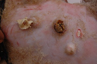
Caseous lymphadenitis (CLA) is an infectious disease caused by the bacterium Corynebacterium pseudotuberculosis, that affects the lymphatic system, resulting in abscesses in the lymph nodes and internal organs. It is found mostly in goats and sheep and at the moment it has no cure.

Guttural pouches are large, auditory-tube diverticula that contain between 300 and 600 ml of air. They are present in odd-toed mammals, some bats, hyraxes, and the American forest mouse. They are paired bilaterally just below the ears, behind the skull and connect to the nasopharynx.
Purpura haemorrhagica is a rare complication of equine strangles and is caused by bleeding from capillaries which results in red spots on the skin and mucous membranes together with oedema (swelling) of the limbs and the head. Purpura hemorrhagica is more common in younger animals.
Stasis papillomatosis is a disease characterized by chronic congestion of the extremities, with blood circulation interrupted in a specific area of the body. A consequence of this congestion and inflammation is long-term lymphatic obstruction. It is also typically characterized by the appearance of numerous papules. Injuries can range from small to large plates composed of brown or pink, smooth or hyperkeratotic papules. The most typical areas where injuries occur are the back of the feet, the toes, the legs, and the area around a venous ulcer formed in the extremities, although the latter is the rarest of all. These injuries include pachydermia, lymphedema, lymphomastic verrucosis and elephantosis verrucosa. The disease can be either localized or generalized; the localized form makes up 78% of cases. Treatment includes surgical and pharmaceutical intervention; indications for partial removal include advanced fibrotic lymphedema and elephantiasis. Despite the existence of these treatments, chronic venous edema, which is a derivation of stasis papillomatosis, is only partially reversible. The skin is also affected and its partial removal may mean that the skin and the subcutaneous tissue are excised. A side effect of the procedure is the destruction of existing cutaneous lymphatic vessels. It also risks papillomatosis, skin necrosis and edema exacerbation.
