Circumesophageal nerve ring

Last updated
A diagram of a hypothetical ancestral mollusc with its nerve ring shown on the left side of the image Archimollusc-en.svg
A diagram of a hypothetical ancestral mollusc with its nerve ring shown on the left side of the image

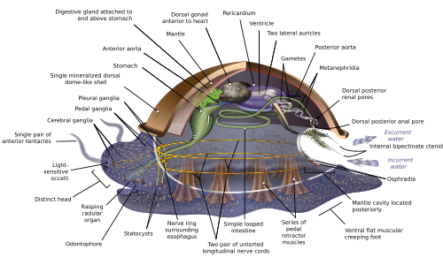
A circumesophageal or circumpharyngeal nerve ring is an arrangement of nerve ganglia around the esophagus/ pharynx of an animal. It is a common feature of nematodes, molluscs, and many other invertebrate animals, though it is absent in all vertebrate animals and is not structurally possible in simpler ones such as water bears.[ citation needed ]

The nerve ring, also called a nerve collar, creates a complete and closed loop around the food-entry parts of the animal's anatomy. In a typical molluscan arrangement, these include the cerebral, pedal, and pleural ganglia, with the esophagus passing through the center of the ring. In nematodes, the ring consists of only two to four large associative cells connected to two paired lateral ganglia, two ventral ganglia, and a single unpaired dorsal ganglion. [1]

Among arthropods, the usual arrangement is a single ganglion (the cerebral) positioned above the esophagus, a single ganglion or nerve mass (the subesophageal) below it, and commissures connecting the two in a ring. [2]

Among the gastropods, the evolutionary torsion event which relocates the anus to near the head of the animal and allowing it to withdraw into its shell has relocated the commissure of the pleural ganglia into a "twist" (the right ganglion has relocated to the left side, and the left ganglion to the right).

Related Research Articles

Central nervous system main information-processing organs of the nervous system, consisting of the brain, spinal cord, and meninges

The central nervous system (CNS) is the part of the nervous system consisting of the brain and spinal cord. The CNS is so named because it integrates the received information and coordinates and influences the activity of all parts of the bodies of bilaterally symmetric animals—that is, all multicellular animals except sponges and radially symmetric animals such as jellyfish—and it contains the majority of the nervous system. Many consider the retina and the optic nerve, as well as the olfactory nerves and olfactory epithelium as parts of the CNS, synapsing directly on brain tissue without intermediate ganglia. As such, the olfactory epithelium is the only central nervous tissue in direct contact with the environment, which opens up for therapeutic treatments. The CNS is contained within the dorsal body cavity, with the brain housed in the cranial cavity and the spinal cord in the spinal canal. In vertebrates, the brain is protected by the skull, while the spinal cord is protected by the vertebrae. The brain and spinal cord are both enclosed in the meninges. Within the CNS, the interneuronal space is filled with a large amount of supporting non-nervous cells called neuroglia or glia from the Greek for "glue".

Ganglion nerve cell cluster or a group of nerve cell bodies located in the autonomic nervous system and sensory system; ganglia house the cells bodies of afferent nerves and efferent nerves

A ganglion is a group of neuron cell bodies in the peripheral nervous system. In the somatic nervous system this includes dorsal root ganglia and trigeminal ganglia among a few others. In the autonomic nervous system there are both sympathetic and parasympathetic ganglia which contain the cell bodies of postganglionic sympathetic and parasympathetic neurons respectively.

Esophagus organ in vertebrates

The esophagus or oesophagus, commonly known as the food pipe or gullet, is an organ in vertebrates through which food passes, aided by peristaltic contractions, from the pharynx to the stomach. The esophagus is a fibromuscular tube, about 25 centimeters long in adults, which travels behind the trachea and heart, passes through the diaphragm and empties into the uppermost region of the stomach. During swallowing, the epiglottis tilts backwards to prevent food from going down the larynx and lungs. The word oesophagus is the Greek word οἰσοφάγος oisophagos, meaning "gullet".

Parasympathetic nervous system one of the three divisions of the autonomic nervous system, the others being the sympathetic nervous system and enteric nervous system.

The parasympathetic nervous system (PSNS) is one of the two divisions of the autonomic nervous system, the other being the sympathetic nervous system. The enteric nervous system (ENS) is now usually referred to as separate from the autonomic nervous system since it has its own independent reflex activity. The autonomic nervous system is responsible for regulating the body's unconscious actions. The parasympathetic system is responsible for stimulation of "rest-and-digest" or "feed and breed" activities that occur when the body is at rest, especially after eating, including sexual arousal, salivation, lacrimation (tears), urination, digestion and defecation. Its action is described as being complementary to that of the sympathetic nervous system, which is responsible for stimulating activities associated with the fight-or-flight response.

Pseudounipolar neuron type of neuron

A pseudounipolar neuron is a type of neuron which has one extension from its cell body. This type of neuron contains an axon that has split into two branches; one branch travels to the periphery branch and the other to the central nervous system.. A single process arises from the cyton and then divides into an axon and a dendrite.They develop embryologically as bipolar in shape and are thus termed pseudounipolar instead of unipolar.

Dorsal root of spinal nerve one of two "roots" which emerge from the spinal cord

The dorsal root of spinal nerve is one of two "roots" which emerge from the spinal cord. It emerges directly from the spinal cord, and travels to the dorsal root ganglion. Nerve fibres with the ventral root then combine to form a spinal nerve. The dorsal root transmits sensory information, forming the afferent sensory root of a spinal nerve.

Posterior cerebral artery

The posterior cerebral artery (PCA) is one of a pair of arteries that supply oxygenated blood to the occipital lobe, part of the back of the human brain. It begins near where the posterior communicating artery and the basilar artery join, and connects with the middle cerebral artery of the same side and internal carotid artery via the posterior communicating artery.

Supraesophageal ganglion

The supraesophageal ganglion is the first part of the arthropod and (especially) insect central nervous system. It receives and processes information from the first, second, and third metameres. The supraesophageal ganglion lies dorsal to the esophagus and consists of three parts, each a pair of ganglia that may be more or less pronounced, reduced, or fused depending on the genus:

Patellogastropoda order of molluscs

The Patellogastropoda, common name true limpets and historically called the Docoglossa, are members of a major phylogenetic group of marine gastropods, treated by experts either as a clade or as a taxonomic order.

The ventral nerve cord (VNC) makes up a part of the central nervous system of some phyla of the bilaterians, particularly within the nematodes, annelids and the arthropods. It usually consists of the segmental ganglia anteriorly with the nerve cords running down the ventral plane of the organism.

Inferior cervical ganglion

The inferior cervical ganglion is situated between the base of the transverse process of the last cervical vertebra and the neck of the first rib, on the medial side of the costocervical artery.

Esophageal plexus

The esophageal plexus is formed by nerve fibers from two sources, branches of the vagus nerve and visceral branches of the sympathetic trunk. The esophageal plexus and the cardiac plexus contain the same types of fibers and are both considered thoracic autonomic plexus(es).

Nervous system of gastropods

The nervous system of gastropods consists of a series of paired ganglia connected by major nerve cords, and a number of smaller branching nerves.

Nerve tract bundle of nerve fibers (axons) connecting nuclei of the central nervous system

A nerve tract is a bundle of nerve fibers (axons) connecting nuclei of the central nervous system. In the peripheral nervous system this is known as a nerve, and has associated connective tissue. The main nerve tracts in the central nervous system are of three types: association fibers, commissural fibers, and projection fibers. A tract may also be referred to as a commissure, fasciculus or decussation. A commissure connects the two cerebral hemispheres at the same levels. Examples are the posterior commissure and the corpus callosum. A decussation is a connection made by fibres that cross at different levels (obliquely), such as the sensory decussation. Examples of a fascicle are the subthalamic fasciculus and the lenticular fasciculus.

<i>Pseudunela cornuta</i> species of mollusc

Pseudunela cornuta is a species of minute sea slug, an acochlidian, a shell-less marine and temporarily brackish gastropod mollusk in the family Pseudunelidae. Adults are about 3 mm long and live in the spaces between sand grains.

The evolution of nervous systems dates back to the first development of nervous systems in animals. Neurons developed as specialized electrical signaling cells in multicellular animals, adapting the mechanism of action potentials present in motile single-celled and colonial eukaryotes. Simple nerve nets seen in animals like Cnidaria (jellyfish) evolved first, consisted of polymodal neurons which serve a dual purpose in motor and sensory functions. Cnidarians can be compared to Ctenophores, which although are both jellyfish, have very different nervous systems. Unlike Cnidarians, Ctenophores have neurons that use electrochemical signaling. This was perplexing because the phylum Ctenophora was considered to be more ancient than that of Porifera (sponges), which have no nervous system at all. This led to the rise of two theories which described how the early nervous system came about. One theory stated that the nervous system came about in an ancestor basal to all of these phylum, however was lost in Porifera. The other theory states that the nervous system arose independently twice, one basal to Cnidarians and one basal to Ctenophores. Bilateral animals – ventral nerve cords in invertebrates and dorsal nerve cords supported by a notochord in chordates-- evolved with a central nervous system that was around a central region, a process known as cephalization.

Bathyacmaea secunda is a species of very small, deep-sea limpet, a marine gastropod mollusk in the family Pectinodontidae. This species inhabits the dark, chemosynthesis-based marine communities of ocean vents and cold seeps near Japan.

Hypoathroid

The term hypoathroid is used to describe the arrangement of ganglia in the nervous system of molluscs. In the hypoathroid state, the pleural ganglia of the "chest" and the pedal ganglia of the "feet" lie close to each other more or less underneath the gut, and they communicate with the cerebral ganglia via long connectives. It is a condition that is characteristic of the Archaeogastropoda clade, and represents one end of a three-part spectrum of such arrangements, the other two being the dystenoid system in which the pleural and cerebral ganglia are closer together but still distinctly separate, and the epiathroid condition in which the pleural, pedal, and cerebral ganglia all lie close together (characteristic, for example, of the Mesogastropoda and Neogastropoda. The centralization of the nervous system generally is considered evidence of an evolutionary advancement among molluscs, while the more diffuse condition is viewed as a sign of evolutionary proximity to the hypothetical "archimollusc" ancestor of all molluscs.

The term epiathroid is used to describe the arrangement of ganglia in the nervous system of molluscs. In the epiathroid state, the pleural ganglia of the "chest" and the pedal ganglia of the "feet" lie close to the cerebral ganglia of the "head" forming a neural cluster which begins to approximate a brain. It is a condition characteristic of the Mesogastropoda and Neogastropoda, and is the obverse of the more-primitive hypoathroid condition in which the pleural and pedal ganglia lie close together under the animal's gut and communicate with the cerebral ganglia via long connectives. The Archaeogastropoda clade is described as "hypoathroid", and is the clade closest to the original hypothetical ancestral mollusc. In between these two extremes lie those animals with a dystenoid nervous system in which the pleural and cerebral ganglia are closer than they are in the hypoathroid condition but still further apart than they are in the epiathroid one.

Bathyhedyle boucheti is a species of panpulmonate slug, a deep-sea dwelling gastropod native to the continental slope off the coast of Mozambique. It is the first ever such panpulmonate slug to be discovered at such depths. It is the only known member of its family group. Its radular formula is 1.1.2.

References

  1. Gregory A. Lewbart (20 September 2011), Invertebrate Medicine, John Wiley & Sons, p. 342, ISBN   978-0-470-96078-3
  2. Fred Punzo (6 December 2012), The Biology of Camel-Spiders: Arachnida, Solifugae, Springer Science & Business Media, p. 45, ISBN   978-1-4615-5727-2