
The cranial vault is the space in the skull within the neurocranium, occupied by the brain.
The cranial vault is the space in the skull within the neurocranium, occupied by the brain.
In humans, the cranial vault is imperfectly composed in newborns, to allow the large human head to pass through the birth canal. During birth, the various bones, connected only by cartilage and ligaments, will move relatively to each other. The open portion between the major bones of the upper part of the vault, called fontanelles, normally remain soft up to two years after birth.
As the fontanelles close, the vault loses some of its plasticity. The sutures between the bones remain until 30 to 40 years of age, allowing for growth of the brain. Cranial vault size is directly proportional to skull size and is developed early. [1]
The size and shape of the brain and the surrounding vault remain quite plastic as the brain grows in childhood. In several ancient societies, head shape was altered for aesthetic or religious reasons by binding cloth or boards tightly around the head during infancy. It is not known whether such artificial cranial deformation has an effect in brain power.

The cranial vault is composed of the endocranium forming the basal parts, topped by the skull roof in land vertebrates. [2]
In fishes, no distinct cranial vault as such exists. Instead, the skull is composed of loosely jointed bones. The cranial vault as a distinct unit arose with the fusion of the skull roof and the endocranium on the early Labyrinthodonts. [2] In amphibians and reptiles, the vault is rather small and inconspicuous, only forming proper vaults in mammals and birds.

Say–Meyer syndrome is a rare X-linked genetic disorder that is mostly characterized as developmental delay. It is one of the rare causes of short stature. It is closely related with trigonocephaly. People with Say–Meyer syndrome have impaired growth, deficits in motor skills development and mental state.

The skull is a bone protective cavity for the brain. The skull is composed of four types of bone i.e., cranial bones, facial bones, ear ossicles and hyoid bone, however two parts are more prominent: the cranium and the mandible. In humans, these two parts are the neurocranium (braincase) and the viscerocranium that includes the mandible as its largest bone. The skull forms the anterior-most portion of the skeleton and is a product of cephalisation—housing the brain, and several sensory structures such as the eyes, ears, nose, and mouth. In humans, these sensory structures are part of the facial skeleton.

A fontanelle is an anatomical feature of the infant human skull comprising soft membranous gaps (sutures) between the cranial bones that make up the calvaria of a fetus or an infant. Fontanelles allow for stretching and deformation of the neurocranium both during birth and later as the brain expands faster than the surrounding bone can grow. Premature complete ossification of the sutures is called craniosynostosis.

The occipital bone is a cranial dermal bone and the main bone of the occiput. It is trapezoidal in shape and curved on itself like a shallow dish. The occipital bone overlies the occipital lobes of the cerebrum. At the base of the skull in the occipital bone, there is a large oval opening called the foramen magnum, which allows the passage of the spinal cord.

The parietal bones are two bones in the skull which, when joined at a fibrous joint, form the sides and roof of the cranium. In humans, each bone is roughly quadrilateral in form, and has two surfaces, four borders, and four angles. It is named from the Latin paries (-ietis), wall.

The axial skeleton is the part of the skeleton that consists of the bones of the head and trunk of a vertebrate. In the human skeleton, it consists of 80 bones and is composed of six parts; the skull, also the ossicles of the middle ear, the hyoid bone, the rib cage, sternum and the vertebral column. The axial skeleton together with the appendicular skeleton form the complete skeleton. Another definition of axial skeleton is the bones including the vertebrae, sacrum, coccyx, skull, ribs, and sternum.
A molera is a "soft spot" on the top of a Chihuahua's skull; it is the equivalent to the bregmatic or anterior fontanelle in human babies, but unlike most mammals Chihuahua's fontanelle persist into maturity. Historically it has been very common amongst Chihuahuas and was regarded as a mark of purity for this miniature dog breed. It is still mentioned in many Chihuahua breed standards, however, it is considered a fault in European countries because of concern that this might reflect underlying malformations such as hydrocephalus and ventriculomegaly, Chiari-like malformation and syringomyelia. Fontanelles are fibrous, membrane-covered gaps that lie between the skull bones and at the intersection of the cranial sutures. The cranial sutures are the junctions between cranial bones. The fontanelles serve as the major sites of bone expansion during post-natal skull growth which accommodates the enlarging brain. The Chihuahua likely has a molera because of neuroparenchymal disproportion i.e. a proportionally big brain for the skull. This is likely because there is premature closure of the skull base cranial sutures. To accommodate the developing brain there is increased growth of the skull bone in a parallel plane giving the dog a characterised domed or "apple-headed" appearance.
In evolutionary developmental biology, heterochrony is any genetically controlled difference in the timing, rate, or duration of a developmental process in an organism compared to its ancestors or other organisms. This leads to changes in the size, shape, characteristics and even presence of certain organs and features. It is contrasted with heterotopy, a change in spatial positioning of some process in the embryo, which can also create morphological innovation. Heterochrony can be divided into intraspecific heterochrony, variation within a species, and interspecific heterochrony, phylogenetic variation, i.e. variation of a descendant species with respect to an ancestral species.

Craniosynostosis is a condition in which one or more of the fibrous sutures in a young infant's skull prematurely fuses by turning into bone (ossification), thereby changing the growth pattern of the skull. Because the skull cannot expand perpendicular to the fused suture, it compensates by growing more in the direction parallel to the closed sutures. Sometimes the resulting growth pattern provides the necessary space for the growing brain, but results in an abnormal head shape and abnormal facial features. In cases in which the compensation does not effectively provide enough space for the growing brain, craniosynostosis results in increased intracranial pressure leading possibly to visual impairment, sleeping impairment, eating difficulties, or an impairment of mental development combined with a significant reduction in IQ.

The crown is the top portion of the head behind the vertex. The anatomy of the crown varies between different organisms. The human crown is made of three layers of the scalp above the skull. The crown also covers a range of bone sutures, and contains blood vessels and branches of the trigeminal nerve.

Saethre–Chotzen syndrome (SCS), also known as acrocephalosyndactyly type III, is a rare congenital disorder associated with craniosynostosis. This affects the shape of the head and face, resulting in a cone-shaped head and an asymmetrical face. Individuals with SCS also have droopy eyelids (ptosis), widely spaced eyes (hypertelorism), and minor abnormalities of the hands and feet (syndactyly). Individuals with more severe cases of SCS may have mild to moderate intellectual or learning disabilities. Depending on the level of severity, some individuals with SCS may require some form of medical or surgical intervention. Most individuals with SCS live fairly normal lives, regardless of whether medical treatment is needed or not.
Craniofacial surgery is a surgical subspecialty that deals with congenital and acquired deformities of the head, skull, face, neck, jaws and associated structures. Although craniofacial treatment often involves manipulation of bone, craniofacial surgery is not tissue-specific; craniofacial surgeons deal with bone, skin, nerve, muscle, teeth, and other related anatomy.

The cranial cavity, also known as intracranial space, is the space within the skull that accommodates the brain. The skull minus the mandible is called the cranium. The cavity is formed by eight cranial bones known as the neurocranium that in humans includes the skull cap and forms the protective case around the brain. The remainder of the skull is called the facial skeleton. Meninges are protective membranes that surround the brain to minimize damage to the brain in the case of head trauma. Meningitis is the inflammation of meninges caused by bacterial or viral infections.

The bregma is the anatomical point on the skull at which the coronal suture is intersected perpendicularly by the sagittal suture.

In human anatomy, the neurocranium, also known as the braincase, brainpan, or brain-pan, is the upper and back part of the skull, which forms a protective case around the brain. In the human skull, the neurocranium includes the calvaria or skullcap. The remainder of the skull is the facial skeleton.

Wadiasaurus is an extinct genus of dicynodont from the family Kannemeyeria, that lived in herds from the early to Middle Triassic. Substantial fossorial evidence of W. indicus was recovered from Yerrapalli Formation of the Pranhita-Godavari valley, India, and it is so far the only Kannemeyeriid known for certain from India. The Kannemeyeriiformes underwent a significant diversification during the middle Triassic, with roughly 40 known species distributed worldwide. All Kannemeyeriiformes were medium to large bodied, graviportal herbivores with relatively erect posture and gait. Wadiasaurus indicus is currently the only known species of Wadiasaurus.

The endocranium in comparative anatomy is a part of the skull base in vertebrates and it represents the basal, inner part of the cranium. The term is also applied to the outer layer of the dura mater in human anatomy.

The skull roof or the roofing bones of the skull are a set of bones covering the brain, eyes and nostrils in bony fishes and all land-living vertebrates. The bones are derived from dermal bone and are part of the dermatocranium.

McGillivray syndrome is a rare syndrome characterized mainly by heart defects, skull and facial abnormalities and ambiguous genitalia. The symptoms of this syndrome are ventricular septal defect, patent ductus arteriosus, small jaw, undescended testes, and webbed fingers. Beside to these symptoms there are more symptoms which is related with bone structure and misshape.
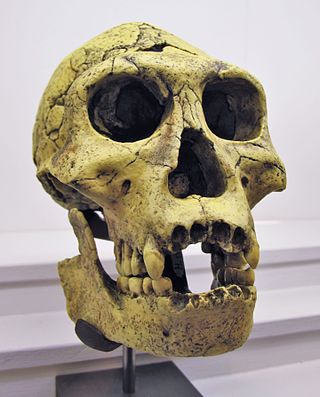
The Dmanisi hominins, Dmanisi people, or Dmanisi man were a population of Early Pleistocene hominins whose fossils have been recovered at Dmanisi, Georgia. The fossils and stone tools recovered at Dmanisi range in age from 1.85 to 1.77 million years old, making the Dmanisi hominins the earliest well-dated hominin fossils in Eurasia and the best preserved fossils of early Homo from a single site so early in time, though earlier fossils and artifacts have been found in Asia. Though their precise classification is controversial and disputed, the Dmanisi fossils are highly significant within research on early hominin migrations out of Africa. The Dmanisi hominins are known from over a hundred postcranial fossils and five famous well-preserved skulls, referred to as Dmanisi Skulls 1–5.