Cystine knot

Last updated
Cystine-knot domain
PDB 1hcn EBI.jpg
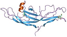
Structure of human chorionic gonadotropin. [1]
Identifiers
SymbolCys_knot
Pfam PF00007
Pfam clan CL0079
InterPro IPR006208
SCOP2 1hcn / SCOPe / SUPFAM
Available protein structures:
Pfam   structures / ECOD  
PDB RCSB PDB; PDBe; PDBj
PDBsum structure summary
PDB 1fl7 , 1hcn , 1hrp , 1qfw , 1xwd

A cystine knot is a protein structural motif containing three disulfide bridges (formed from pairs of cysteine residues). The sections of polypeptide that occur between two of them form a loop through which a third disulfide bond passes, forming a rotaxane substructure. The cystine knot motif stabilizes protein structure and is conserved in proteins across various species. [2] [3] [4] There are three types of cystine knot, which differ in the topology of the disulfide bonds: [5]

The growth factor cystine knot was first observed in the structure of nerve growth factor (NGF), solved by X-ray crystallography and published in 1991 by Tom Blundell in Nature. [6] The GFCK is present in four superfamilies. These include nerve growth factor, transforming growth factor beta (TGF-β), platelet-derived growth factor, and glycoprotein hormones including human chorionic gonadotropin. These are structurally related due to the presence of the cystine knot motif but differ in sequence. [7] All GFCK structures that have been determined are dimeric, but their dimerization modes in different classes are different. [8] The vascular endothelial growth factor subfamily, categorized as part of the platelet-derived growth factor superfamily, includes proteins that are angiogenic factors. [9]

The presence of the cyclic cystine knot (CCK) motif was discovered when cyclotides were isolated from various plant families. The CCK motif has a cyclic backbone, triple stranded beta sheet, and cystine knot conformation. [10]

Novel proteins are being added to the cystine knot motif family, also known as the C-terminal cystine knot (CTCK) proteins. They share approximately 90 amino acid residues in their cysteine-rich C-terminal regions. [9]

Inhibitor cystine knot (ICK) is a structural motif with a triple stranded antiparallel beta sheet linked by three disulfide bonds, forming a knotted core. The ICK motif can be found under the category of phylum, such as animals and plants. It is often found in many venom peptides such as those of snails, spiders, and scorpions. Peptide K-PVIIA, which contains an ICK, can undergo a successful enzymatic backbone cyclization. The disulfide connectivity and the common sequence pattern of the ICK motif provides the stability of the peptides that support cyclization. [11]

Drug implications

The stability and structure of the cystine knot motif implicates possible applications in drug design. The hydrogen bonding between the disulfide bonds of the motif and beta-sheet structures gives rise to highly efficient structure stabilization. In addition, the size of the motif is approximately 30 amino acid residues. [12] These two characteristics make it an attractive biomolecule to be used for drug delivery as it exhibits thermal stability, chemical stability, and proteolytic resistance. The biological activities of these molecules are partially due to the unique interlocking arrangement and cyclized peptide backbone which contains a conserved sequence shared among circulins. [12] Circulins have previously been identified in a screen for anti-HIV activity. [13] Studies have shown that cystine knot proteins can be incubated at temperatures of 65 °C or placed in 1N HCl/1N NaOH without loss of structural and functional integrity. [14] Its resistance from oral and some intestinal proteases suggest possible use for oral delivery. Possible future applications include pain relief as well as antiviral and antibacterial functions. [14]

Related Research Articles

<span class="mw-page-title-main">Alpha helix</span> Type of secondary structure of proteins

An alpha helix is a sequence of amino acids in a protein that are twisted into a coil.

<span class="mw-page-title-main">Beta sheet</span> Protein structural motif

The beta sheet is a common motif of the regular protein secondary structure. Beta sheets consist of beta strands (β-strands) connected laterally by at least two or three backbone hydrogen bonds, forming a generally twisted, pleated sheet. A β-strand is a stretch of polypeptide chain typically 3 to 10 amino acids long with backbone in an extended conformation. The supramolecular association of β-sheets has been implicated in the formation of the fibrils and protein aggregates observed in amyloidosis, Alzheimer's disease and other proteinopathies.

<span class="mw-page-title-main">Protein structure</span> Three-dimensional arrangement of atoms in an amino acid-chain molecule

Protein structure is the three-dimensional arrangement of atoms in an amino acid-chain molecule. Proteins are polymers – specifically polypeptides – formed from sequences of amino acids, which are the monomers of the polymer. A single amino acid monomer may also be called a residue, which indicates a repeating unit of a polymer. Proteins form by amino acids undergoing condensation reactions, in which the amino acids lose one water molecule per reaction in order to attach to one another with a peptide bond. By convention, a chain under 30 amino acids is often identified as a peptide, rather than a protein. To be able to perform their biological function, proteins fold into one or more specific spatial conformations driven by a number of non-covalent interactions, such as hydrogen bonding, ionic interactions, Van der Waals forces, and hydrophobic packing. To understand the functions of proteins at a molecular level, it is often necessary to determine their three-dimensional structure. This is the topic of the scientific field of structural biology, which employs techniques such as X-ray crystallography, NMR spectroscopy, cryo-electron microscopy (cryo-EM) and dual polarisation interferometry, to determine the structure of proteins.

<span class="mw-page-title-main">Trefoil knot fold</span>

The trefoil knot fold is a protein fold in which the protein backbone is twisted into a trefoil knot shape. "Shallow" knots in which the tail of the polypeptide chain only passes through a loop by a few residues are uncommon, but "deep" knots in which many residues are passed through the loop are extremely rare. Deep trefoil knots have been found in the SPOUT superfamily. including methyltransferase proteins involved in posttranscriptional RNA modification in all three domains of life, including bacterium Thermus thermophilus and proteins, in archaea and in eukaryota.

<span class="mw-page-title-main">Cyclotide</span> Disulfide-rich ring peptides found in plants

In biochemistry, cyclotides are small, disulfide-rich peptides isolated from plants. Typically containing 28-37 amino acids, they are characterized by their head-to-tail cyclised peptide backbone and the interlocking arrangement of their three disulfide bonds. These combined features have been termed the cyclic cystine knot (CCK) motif. To date, over 100 cyclotides have been isolated and characterized from species of the families Rubiaceae, Violaceae, and Cucurbitaceae. Cyclotides have also been identified in agriculturally important families such as the Fabaceae and Poaceae.

<span class="mw-page-title-main">Transforming growth factor beta superfamily</span> Protein family

The transforming growth factor beta (TGF-β) superfamily is a large group of structurally related cell regulatory proteins that was named after its first member, TGF-β1, originally described in 1983. They interact with TGF-beta receptors.

<span class="mw-page-title-main">Procollagen-proline dioxygenase</span> Enzyme

Procollagen-proline dioxygenase, commonly known as prolyl hydroxylase, is a member of the class of enzymes known as alpha-ketoglutarate-dependent hydroxylases. These enzymes catalyze the incorporation of oxygen into organic substrates through a mechanism that requires alpha-Ketoglutaric acid, Fe2+, and ascorbate. This particular enzyme catalyzes the formation of (2S, 4R)-4-hydroxyproline, a compound that represents the most prevalent post-translational modification in the human proteome.

<span class="mw-page-title-main">EGF-like domain</span> Protein domain named after the epidermal growth factor protein

The EGF-like domain is an evolutionary conserved protein domain, which derives its name from the epidermal growth factor where it was first described. It comprises about 30 to 40 amino-acid residues and has been found in a large number of mostly animal proteins. Most occurrences of the EGF-like domain are found in the extracellular domain of membrane-bound proteins or in proteins known to be secreted. An exception to this is the prostaglandin-endoperoxide synthase. The EGF-like domain includes 6 cysteine residues which in the epidermal growth factor have been shown to form 3 disulfide bonds. The structures of 4-disulfide EGF-domains have been solved from the laminin and integrin proteins. The main structure of EGF-like domains is a two-stranded β-sheet followed by a loop to a short C-terminal, two-stranded β-sheet. These two β-sheets are usually denoted as the major (N-terminal) and minor (C-terminal) sheets. EGF-like domains frequently occur in numerous tandem copies in proteins: these repeats typically fold together to form a single, linear solenoid domain block as a functional unit.

Potato carboxypeptidase inhibitor (PCI) is a naturally occurring protease inhibitor peptide in potatoes that can form complexes with several metallo-carboxypeptidases, inhibiting them in a strong competitive way with a Ki in the nanomolar range.
PCI consists of 39 amino acids forming a 27-residue globular core stabilized by three disulfide bridges and a C-terminal tail with residues 35–39. PCI contains a small cysteine-rich module, called a T-knot scaffold, that is shared by several different protein families, including the EGF family.

<i>delta</i>-Palutoxin

delta-Palutoxins (δ-palutoxins) consist of a homologous group of four insect-specific toxins from the venom of the spider Pireneitega luctuosa. They show a high toxicity against Spodoptera litura larvae by inhibiting sodium channels, leading to strong paralytic activity and eventually to the death of the insect.

Huwentoxins (HWTX) are a group of neurotoxic peptides found in the venom of the Chinese bird spider Haplopelma schmidti. The species was formerly known as Haplopelma huwenum, Ornithoctonus huwena and Selenocosmia huwena. While structural similarity can be found among several of these toxins, HWTX as a group possess high functional diversity.

<span class="mw-page-title-main">Inhibitor cystine knot</span>

An inhibitor cystine knot is a protein structural motif containing three disulfide bridges. Knottins are one of three folds in the cystine knot motif; the other closely related knots are the growth factor cystine knot (GFCK) and the cyclic cystine knot. Types include a) cyclic mobius, b) cyclic bracelet, c) acyclic inhibitor knottins. Cystine knot motifs are found frequently in nature in a plethora of plants, animals, and fungi and serve diverse functions from appetite suppression to anti-fungal activity.

Ribosomally synthesized and post-translationally modified peptides (RiPPs), also known as ribosomal natural products, are a diverse class of natural products of ribosomal origin. Consisting of more than 20 sub-classes, RiPPs are produced by a variety of organisms, including prokaryotes, eukaryotes, and archaea, and they possess a wide range of biological functions.

GTx1-15 is a toxin from the Chilean tarantula venom that acts as both a voltage-gated calcium channel blocker and a voltage-gated sodium channel blocker.

<span class="mw-page-title-main">Vejocalcin</span> Toxin

Vejocalcin (VjCa, also called Vejocalcine) is a toxin from the venom of the Mexican scorpion Vaejovis mexicanus. Vejocalcin is a member of the calcin family of toxins. It acts as a cell-penetrating peptide (CPP); it binds with high affinity and specificity to skeletal ryanodine receptor 1 (RYR1) of the sarcoplasmic reticulum, thereby triggering calcium release from intracellular Ca2+ stores.

Ptu1 is a toxin that can reversibly bind N-type calcium channels. It is isolated from the assassin bug Peirates turpis. The toxin belongs to the inhibitory cystine knot structural family (ICK) that has a core of disulfide bonds with four loops emerging from it.

Intrepicalcin (ViCaTx1) is a short peptide toxin found in the venom of scorpion Vaejovis intrepidus. It is one of a group of short, basic peptides called calcins, which bind to ryanodine receptors (RyRs) and thereby trigger calcium release from the sarcoplasmic reticulum.

μ-THTX-Cl6a, also known as Cl6a, is a 33-residue peptide toxin extracted from the venom of the spider Cyriopagopus longipes. The toxin acts as an inhibitor of the tetrodotoxin-sensitive (TTX-S) voltage-gated sodium channel (NaV1.7), thereby causing sustained reduction of NaV1.7 currents.

U7-ctenitoxin-Pn1a (or U7-CNTX-Pn1a for short) is a neurotoxin that blocks TRPV1 channels, and can exhibit analgestic effects. It is naturally found in the venom of Phoneutria nigriventer.

Cl6b (μ-THTX-Cl6b) is a peptide toxin from the venom of the spider Cyriopagopus longipes. It acts as a sodium channel blocker: Cl6b significantly and persistently reduces currents through the tetrodotoxin-sensitive sodium channels NaV1.2-1.4, NaV1.6, and NaV1.7.

References

  1. Wu H, Lustbader JW, Liu Y, Canfield RE, Hendrickson WA (June 1994). "Structure of human chorionic gonadotropin at 2.6 A resolution from MAD analysis of the selenomethionyl protein". Structure. 2 (6): 545–58. doi:10.1016/s0969-2126(00)00054-x. PMID   7922031.
  2. "Cystine Knots". The Cyclotide Webpage. Archived from the original on 2015-02-05. Retrieved 2019-04-24.
  3. Sherbet, G.V. (2011), "Growth Factor Families", Growth Factors and Their Receptors in Cell Differentiation, Cancer and Cancer Therapy, Elsevier, pp. 3–5, doi:10.1016/b978-0-12-387819-9.00002-5, ISBN   9780123878199 , retrieved 2019-05-01
  4. Vitt, Ursula A.; Hsu, Sheau Y.; Hsueh, Aaron J. W. (2001-05-01). "Evolution and Classification of Cystine Knot-Containing Hormones and Related Extracellular Signaling Molecules". Molecular Endocrinology. 15 (5): 681–694. doi: 10.1210/mend.15.5.0639 . ISSN   0888-8809. PMID   11328851.
  5. Daly NL, Craik DJ (June 2011). "Bioactive cystine knot proteins". Current Opinion in Chemical Biology. 15 (3): 362–8. doi:10.1016/j.cbpa.2011.02.008. PMID   21362584.
  6. PDB: 1bet ; McDonald NQ, Lapatto R, Murray-Rust J, Gunning J, Wlodawer A, Blundell TL (December 1991). "New protein fold revealed by a 2.3-A resolution crystal structure of nerve growth factor". Nature. 354 (6352): 411–4. Bibcode:1991Natur.354..411M. doi:10.1038/354411a0. PMID   1956407. S2CID   4346788.
  7. Sun PD, Davies DR (1995). "The cystine-knot growth-factor superfamily". Annual Review of Biophysics and Biomolecular Structure. 24 (1): 269–91. doi:10.1146/annurev.bb.24.060195.001413. PMID   7663117.
  8. Jiang X, Dias JA, He X (January 2014). "Structural biology of glycoprotein hormones and their receptors: insights to signaling". Molecular and Cellular Endocrinology. 382 (1): 424–451. doi: 10.1016/j.mce.2013.08.021 . PMID   24001578.
  9. 1 2 Iyer S, Acharya KR (November 2011). "Tying the knot: the cystine signature and molecular-recognition processes of the vascular endothelial growth factor family of angiogenic cytokines". The FEBS Journal. 278 (22): 4304–22. doi:10.1111/j.1742-4658.2011.08350.x. PMC   3328748 . PMID   21917115.
  10. Craik DJ, Daly NL, Bond T, Waine C (December 1999). "Plant cyclotides: A unique family of cyclic and knotted proteins that defines the cyclic cystine knot structural motif". Journal of Molecular Biology. 294 (5): 1327–36. doi:10.1006/jmbi.1999.3383. PMID   10600388.
  11. Kwon, Soohyun; Bosmans, Frank; Kaas, Quentin; Cheneval, Oliver; Cinibear, Anne C; Rosengren, K Johan; Wang, Conan K; Schroeder, Christina I; Craik, David J (19 April 2016). "Efficient enzymatic cyclization of an inhibitory cystine knot‐containing peptide". Biotechnology and Bioengineering. 113 (10): 2202–2212. doi:10.1002/bit.25993. PMC   5526200 . PMID   27093300.
  12. 1 2 Kolmar, Harald. “Biological Diversity and Therapeutic Potential of Natural and Engineered Cystine Knot Miniproteins.” Current Opinion in Pharmacology, vol. 9, no. 5, 2009, pp. 608–614., doi:10.1016/j.coph.2009.05.004.
  13. K.R. Gustafson, R.C. Sowder II, L.E. Henderson, I.C. Parsons, Y. Kashman, J.H. Cardellina II, J.B. McMahon, R.W. Buckheit Jr., L.K. Pannell, M.R. Boyd Circulins A and B: novel HIV-inhibitory macrocyclic peptides from the tropical tree Chassalia parvifolia J. Am. Chem. Soc., 116 (1994), pp. 9337-9338
  14. 1 2 Craik, David J., et al. “The Cystine Knot Motif in Toxins and Implications for Drug Design.” Toxicon, vol. 39, no. 1, 2001, pp. 43–60., doi:10.1016/s0041-0101(00)00160-4.