Related Research Articles

The medulla oblongata or simply medulla is a long stem-like structure which makes up part of the brainstem. It is anterior and partially inferior to the cerebellum. It is a cone-shaped neuronal mass responsible for autonomic (involuntary) functions ranging from vomiting to sneezing. The medulla contains the cardiac, respiratory, vomiting and vasomotor centers and therefore deals with the autonomic functions of breathing, heart rate and blood pressure as well as the sleep wake cycle.
The brainstem is the posterior part of the brain, continuous with the spinal cord. In the human brain the brainstem includes the midbrain, the pons and medulla oblongata of the hindbrain. The midbrain continues with the thalamus of the diencephalon through the tentorial notch, and sometimes the diencephalon is included in the brainstem.

A spinal nerve is a mixed nerve, which carries motor, sensory, and autonomic signals between the spinal cord and the body. In the human body there are 31 pairs of spinal nerves, one on each side of the vertebral column. These are grouped into the corresponding cervical, thoracic, lumbar, sacral and coccygeal regions of the spine. There are eight pairs of cervical nerves, twelve pairs of thoracic nerves, five pairs of lumbar nerves, five pairs of sacral nerves, and one pair of coccygeal nerves. The spinal nerves are part of the peripheral nervous system.

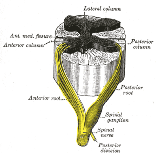
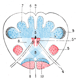
The grey column refers to a somewhat ridge-shaped mass of grey matter in the spinal cord. This presents as three columns: the anterior grey column, the posterior grey column, and the lateral grey column, all of which are visible in cross-section of the spinal cord.

The spinothalamic tract is a sensory pathway to the thalamus. From the ventral posterolateral nucleus in the thalamus, sensory information is relayed upward to the somatosensory cortex of the postcentral gyrus.
The dorsal column–medial lemniscus pathway (DCML) is a sensory pathway of the central nervous system that conveys sensations of fine touch, vibration, two-point discrimination, and proprioception (position) from the skin and joints. It transmits information from the body to the primary somatosensory cortex in the postcentral gyrus of the parietal lobe of the brain. The pathway receives information from sensory receptors throughout the body, and carries this in nerve tracts in the white matter of the dorsal columns of the spinal cord to the medulla, where it is continued in the medial lemniscus, on to the thalamus and relayed from there through the internal capsule and transmitted to the somatosensory cortex. The name dorsal-column medial lemniscus comes from the two structures that carry the sensory information: the dorsal columns of the spinal cord, and the medial lemniscus in the brainstem.

The spinocerebellar tract is a nerve tract originating in the spinal cord and terminating in the same side (ipsilateral) of the cerebellum.
The dorsal root of spinal nerve is one of two "roots" which emerge from the spinal cord. It emerges directly from the spinal cord, and travels to the dorsal root ganglion. Nerve fibres with the ventral root then combine to form a spinal nerve. The dorsal root transmits sensory information, forming the afferent sensory root of a spinal nerve.

The apex of the posterior grey column, one of the three grey columns of the spinal cord, is capped by a V-shaped or crescentic mass of translucent, gelatinous neuroglia, termed the substantia gelatinosa of Rolando, which contains both neuroglia cells, and small nerve cells. The gelatinous appearance is due to a very low concentration of myelinated fibers. It extends the entire length of the spinal cord and into the medulla oblongata where it becomes the spinal nucleus of the trigeminal nerve.
The lateral grey column is one of the three grey columns of the spinal cord ; the others being the anterior and posterior grey columns. The lateral grey column is primarily involved with activity in the sympathetic division of the autonomic motor system. It projects to the side as a triangular field in the thoracic and upper lumbar regions of the postero-lateral part of the anterior grey column.

The posterior thoracic nucleus, is a group of interneurons found in the medial part of lamina VII, also known as the intermediate zone, of the spinal cord. It is mainly located from the cervical vertebra C7 to lumbar L3-L4 levels and is an important structure for proprioception of the lower limb.
The anterior corticospinal tract is a small bundle of descending fibers that connect the cerebral cortex to the spinal cord. Descending tracts are pathways by which motor signals are sent from upper motor neurons in the brain to lower motor neurons which then directly innervate muscle to produce movement. The anterior corticospinal tract is usually small, varying inversely in size with the lateral corticospinal tract, which is the main part of the corticospinal tract.
In neuroanatomy, the dorsal column nuclei are a pair of nuclei in the dorsal columns in the brainstem. The name refers collectively to the cuneate nucleus and gracile nucleus, which are present at the junction between the spinal cord and the medulla oblongata. Both nuclei contain second-order neurons of the dorsal column-medial lemniscus pathway, which carries fine touch and proprioceptive information from the body to the brain. Each nucleus has an associated nerve tract, the gracile fasciculus and the cuneate fasciculus.
The posterolateral tract is a small strand situated in relation to the tip of the posterior column close to the entrance of the posterior nerve roots. It is present throughout the spinal cord, and is most developed in the upper cervical regions.

The medullary pyramids are paired white matter structures of the brainstem's medulla oblongata that contain motor fibers of the corticospinal and corticobulbar tracts – known together as the pyramidal tracts. The lower limit of the pyramids is marked when the fibers cross (decussate).

The spinal cord is a long, thin, tubular structure made up of nervous tissue, which extends from the medulla oblongata in the brainstem to the lumbar region of the vertebral column. It encloses the central canal of the spinal cord, which contains cerebrospinal fluid. The brain and spinal cord together make up the central nervous system (CNS). In humans, the spinal cord begins at the occipital bone, passing through the foramen magnum and entering the spinal canal at the beginning of the cervical vertebrae. The spinal cord extends down to between the first and second lumbar vertebrae, where it ends. The enclosing bony vertebral column protects the relatively shorter spinal cord. It is around 45 cm (18 in) in men and around 43 cm (17 in) long in women. The diameter of the spinal cord ranges from 13 mm in the cervical and lumbar regions to 6.4 mm in the thoracic area.
Ventral propriospinal tract is a collection of nerve fibers, ascending, descending, crossed and uncrossed, that interconnect various levels of the spinal cord. It is a component of the white anterior columns. The fibers are largely myelinated and run close to the spinal central gray for the length of the cord. Shorter fibers are closer to, longer fibers further from the gray. Other prominent components of the anterior columns are the medial longitudinal fasciculus of the spinal cord, the anterior spinothalamic tract of the spinal cord and the lateral vestibulospinal tract of the spinal cord. The ventral propriospinal tract is one of three propriospinal tracts in which most pathways intrinsic to the spinal cord are located. The others are the dorsal propriospinal tract and the lateral propriospinal tract.,
Lateral propriospinal tract is a collection of nerve fibers, ascending, descending, crossed and uncrossed, that interconnect various levels of the spinal cord. Its fibers are largely myelinated. It is a component of the white lateral columns. Most prominent in the cervical and lumbar regions, it is located close to the spinal central gray. Shorter fibers are closer to, longer fibers further from the gray The tract is one of three propriospinal tracts in which most pathways intrinsic to the spinal cord are located. The others are the ventral propriospinal tract and the dorsal propriospinal tract.
Propriospinal tracts are collections of nerve fibers ascending, descending, crossed and uncrossed, that interconnect various levels of the spinal cord. They are located in the white columns of the spinal cord where the columns meet the spinal central gray. Shorter fibers are located closer and longer fibers farther from the gray. The tracts include the ventral propriospinal tract, the lateral propriospinal tract and the dorsal propriospinal tract. Some authors include the semilunar tract in this category. A few other fibers intrinsic to the cord run in the dorsolateral fasciculus of the spinal cord and the septomarginal tract.
The proper fasciculi, or spinospinal fasciculi, or propriospinal tracts, are groups of short fibres, ascending and descending, and crossed and uncrossed, within the spinal cord. These fibres are grouped into anterior, posterior, and lateral regions and make up a spinal pathway. Descending dorsal root collaterals are often included in the pathway.