Dysphagia lusoria

Last updated
Dysphagia lusoria
Gray506.svg
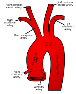
The right subclavian artery is involved in this condition

Dysphagia lusoria (or Bayford-Autenrieth dysphagia) is an abnormal condition characterized by difficulty in swallowing caused by an aberrant right subclavian artery. It was discovered by David Bayford in 1761 and first reported in a paper by the same in 1787. [1]

Contents

Pathophysiology

During development of aortic arch, if the proximal portion of the right fourth arch disappears instead of distal portion, the right subclavian artery will arise as the last branch of aortic arch. It then courses behind the esophagus (or rarely in front of esophagus, or even in front of trachea) to supply blood to right arm. This causes pressure on esophagus and results in dysphagia. It can sometimes result in upper gastrointestinal tract bleeding. [2] Investigation of choice - CT angiography

Treatment

Surgical repair is performed. Reconstruction or ligation of aberrant right subclavian artery by sternotomy/by neck approach.[ citation needed ]

Eponym

David Bayford called it dysphagia lusoria because in Latin, lusus naturæ means sports of nature, freak of nature, or natural anomaly. [3] Bayford-Autenrieth dysphagia is eponym for Bayford and Autenrieth.[ citation needed ]

See also

Related Research Articles

<span class="mw-page-title-main">Esophagus</span> Vertebrate organ through which food passes to the stomach

The esophagus or oesophagus, colloquially known also as the food pipe or gullet, is an organ in vertebrates through which food passes, aided by peristaltic contractions, from the pharynx to the stomach. The esophagus is a fibromuscular tube, about 25 cm (10 in) long in adults, that travels behind the trachea and heart, passes through the diaphragm, and empties into the uppermost region of the stomach. During swallowing, the epiglottis tilts backwards to prevent food from going down the larynx and lungs. The word oesophagus is from Ancient Greek οἰσοφάγος (oisophágos), from οἴσω (oísō), future form of φέρω + ἔφαγον.

<span class="mw-page-title-main">Brachiocephalic artery</span> Artery of the mediastinum

The brachiocephalic artery is an artery of the mediastinum that supplies blood to the right arm and the head and neck. It was previously known as the innominate artery, meaning unnamed artery.

<span class="mw-page-title-main">Circle of Willis</span> Circulatory anastomosis that supplies blood to the brain and surrounding structures

The circle of Willis is a circulatory anastomosis that supplies blood to the brain and surrounding structures in reptiles, birds and mammals, including humans. It is named after Thomas Willis (1621–1675), an English physician.

<span class="mw-page-title-main">Subclavian artery</span> Major arteries of the upper thorax, below the clavicle

In human anatomy, the subclavian arteries are paired major arteries of the upper thorax, below the clavicle. They receive blood from the aortic arch. The left subclavian artery supplies blood to the left arm and the right subclavian artery supplies blood to the right arm, with some branches supplying the head and thorax. On the left side of the body, the subclavian comes directly off the aortic arch, while on the right side it arises from the relatively short brachiocephalic artery when it bifurcates into the subclavian and the right common carotid artery.

<span class="mw-page-title-main">Heyde's syndrome</span> Medical condition

Heyde's syndrome is a syndrome of gastrointestinal bleeding from angiodysplasia in the presence of aortic stenosis.

<span class="mw-page-title-main">Coarctation of the aorta</span> Medical condition

Coarctation of the aorta, also called aortic narrowing, is a congenital condition whereby the aorta is narrow, usually in the area where the ductus arteriosus inserts. The word coarctation means "pressing or drawing together; narrowing". Coarctations are most common in the aortic arch. The arch may be small in babies with coarctations. Other heart defects may also occur when coarctation is present, typically occurring on the left side of the heart. When a patient has a coarctation, the left ventricle has to work harder. Since the aorta is narrowed, the left ventricle must generate a much higher pressure than normal in order to force enough blood through the aorta to deliver blood to the lower part of the body. If the narrowing is severe enough, the left ventricle may not be strong enough to push blood through the coarctation, thus resulting in a lack of blood to the lower half of the body. Physiologically its complete form is manifested as interrupted aortic arch.

<span class="mw-page-title-main">Diverticulum</span> Medical or biological term for an outpouching of a hollow (or a fluid-filled) structure in the body

In medicine or biology, a diverticulum is an outpouching of a hollow structure in the body. Depending upon which layers of the structure are involved, diverticula are described as being either true or false.

<span class="mw-page-title-main">Suspensory muscle of duodenum</span> Muscle between the duodenum and jejunum

The suspensory muscle of duodenum is a thin muscle connecting the junction between the duodenum and jejunum, as well as the duodenojejunal flexure to connective tissue surrounding the superior mesenteric and coeliac arteries. The suspensory muscle most often connects to both the third and fourth parts of the duodenum, as well as the duodenojejunal flexure, although the attachment is quite variable.

<span class="mw-page-title-main">Recurrent laryngeal nerve</span> Nerve in the human body

The recurrent laryngeal nerve (RLN) is a branch of the vagus nerve that supplies all the intrinsic muscles of the larynx, with the exception of the cricothyroid muscles. There are two recurrent laryngeal nerves, right and left. The right and left nerves are not symmetrical, with the left nerve looping under the aortic arch, and the right nerve looping under the right subclavian artery then traveling upwards. They both travel alongside the trachea. Additionally, the nerves are among the few nerves that follow a recurrent course, moving in the opposite direction to the nerve they branch from, a fact from which they gain their name.

<span class="mw-page-title-main">Aortic arch</span> Part of the aorta

The aortic arch, arch of the aorta, or transverse aortic arch is the part of the aorta between the ascending and descending aorta. The arch travels backward, so that it ultimately runs to the left of the trachea.

<span class="mw-page-title-main">Aortic arches</span>

The aortic arches or pharyngeal arch arteries are a series of six paired embryological vascular structures which give rise to the great arteries of the neck and head. They are ventral to the dorsal aorta and arise from the aortic sac.

<span class="mw-page-title-main">Aberrant subclavian artery</span> Medical condition

Aberrant subclavian artery, or aberrant subclavian artery syndrome, is a rare anatomical variant of the origin of the right or left subclavian artery. This abnormality is the most common congenital vascular anomaly of the aortic arch, occurring in approximately 1% of individuals.

<span class="mw-page-title-main">Ortner's syndrome</span> Medical condition

Ortner's syndrome is a rare cardiovocal syndrome and refers to recurrent laryngeal nerve palsy from cardiovascular disease. It was first described by Norbert Ortner (1865–1935), an Austrian physician, in 1897.

Esophageal dysphagia is a form of dysphagia where the underlying cause arises from the body of the esophagus, lower esophageal sphincter, or cardia of the stomach, usually due to mechanical causes or motility problems.

Interrupted aortic arch is a very rare heart defect in which the aorta is not completely developed. There is a gap between the ascending and descending thoracic aorta. In a sense it is the complete form of a coarctation of the aorta. Almost all patients also have other cardiac anomalies, including a ventricular septal defect (VSD), aorto-pulmonary window, and truncus arteriosus. There are three types of interrupted aortic arch, with type B being the most common. Interrupted aortic arch is often associated with DiGeorge syndrome.

A vascular ring is a congenital defect in which there is an abnormal formation of the aorta and/or its surrounding blood vessels. The trachea and esophagus are completely encircled and sometimes compressed by a "ring" formed by these vessels, which can lead to breathing and digestive difficulties.

<span class="mw-page-title-main">Johann Heinrich Ferdinand von Autenrieth</span> German physician (1772–1835)

Johann Heinrich Ferdinand von Autenrieth was a German physician born in Stuttgart.

Double aortic arch is a relatively rare congenital cardiovascular malformation. DAA is an anomaly of the aortic arch in which two aortic arches form a complete vascular ring that can compress the trachea and/or esophagus. Most commonly there is a larger (dominant) right arch behind and a smaller (hypoplastic) left aortic arch in front of the trachea/esophagus. The two arches join to form the descending aorta which is usually on the left side. In some cases the end of the smaller left aortic arch closes and the vascular tissue becomes a fibrous cord. Although in these cases a complete ring of two patent aortic arches is not present, the term ‘vascular ring’ is the accepted generic term even in these anomalies.

<span class="mw-page-title-main">Right-sided aortic arch</span> Medical condition

Right-sided aortic arch is a rare anatomical variant in which the aortic arch is on the right side rather than on the left. During normal embryonic development, the aortic arch is formed by the left fourth aortic arch and the left dorsal aorta. In people with a right-sided aortic arch, instead the right dorsal aorta persists and the distal left aorta disappears.

David Bayford, FRS was a London surgeon, who practised from 1761 to 1782. In later years of his life he practised as a physician.

References

  1. Asherson N (January 1979). "David Bayford. His syndrome and sign of dysphagia lusoria". Ann R Coll Surg Engl. 61 (1): 63–7. PMC   2494476 . PMID   369446.
  2. Vehling-Kaiser, U. (1993). "Lusorian artery lesion as rare cause of severe upper gastrointestinal tract bleeding". Digestive Diseases and Sciences. 38 (1): 178–180. doi:10.1007/BF01296793.
  3. Mahmodlou, Rahim; Sepehrvand, Nariman; Hatami, Sanaz (2014). "Aberrant Right Subclavian Artery: A Life-threatening Anomaly that should be considered during Esophagectomy". Journal of Surgical Technique and Case Report. 6 (2): 61–63. doi:10.4103/2006-8808.147262. PMC   4290042 . PMID   25598945.