The finite volume method (FVM) is a method for representing and evaluating partial differential equations in the form of algebraic equations. In the finite volume method, volume integrals in a partial differential equation that contain a divergence term are converted to surface integrals, using the divergence theorem. These terms are then evaluated as fluxes at the surfaces of each finite volume. Because the flux entering a given volume is identical to that leaving the adjacent volume, these methods are conservative. Another advantage of the finite volume method is that it is easily formulated to allow for unstructured meshes. The method is used in many computational fluid dynamics packages. "Finite volume" refers to the small volume surrounding each node point on a mesh.

Tomography is imaging by sections or sectioning that uses any kind of penetrating wave. The method is used in radiology, archaeology, biology, atmospheric science, geophysics, oceanography, plasma physics, materials science, cosmochemistry, astrophysics, quantum information, and other areas of science. The word tomography is derived from Ancient Greek τόμος tomos, "slice, section" and γράφω graphō, "to write" or, in this context as well, "to describe." A device used in tomography is called a tomograph, while the image produced is a tomogram.

Electrical impedance tomography (EIT) is a noninvasive type of medical imaging in which the electrical conductivity, permittivity, and impedance of a part of the body is inferred from surface electrode measurements and used to form a tomographic image of that part. Electrical conductivity varies considerably among various biological tissues or the movement of fluids and gases within tissues. The majority of EIT systems apply small alternating currents at a single frequency, however, some EIT systems use multiple frequencies to better differentiate between normal and suspected abnormal tissue within the same organ.

Dielectric spectroscopy measures the dielectric properties of a medium as a function of frequency. It is based on the interaction of an external field with the electric dipole moment of the sample, often expressed by permittivity.

A Coulter counter is an apparatus for counting and sizing particles suspended in electrolytes. The Coulter counter is the commercial term for the technique known as resistive pulse sensing or electrical zone sensing. The apparatus is based on the Coulter principle named after its inventor, Wallace H. Coulter.

In chemical processing, a packed bed is a hollow tube, pipe, or other vessel that is filled with a packing material. The packed bed can be randomly filled with small objects like Raschig rings or else it can be a specifically designed structured packing. Packed beds may also contain catalyst particles or adsorbents such as zeolite pellets, granular activated carbon, etc.
In neuroscience, single-unit recordings provide a method of measuring the electro-physiological responses of a single neuron using a microelectrode system. When a neuron generates an action potential, the signal propagates down the neuron as a current which flows in and out of the cell through excitable membrane regions in the soma and axon. A microelectrode is inserted into the brain, where it can record the rate of change in voltage with respect to time. These microelectrodes must be fine-tipped, impedance matching; they are primarily glass micro-pipettes, metal microelectrodes made of platinum, tungsten, iridium or even iridium oxide. Microelectrodes can be carefully placed close to the cell membrane, allowing the ability to record extracellularly.

Electrical resistivity tomography (ERT) or electrical resistivity imaging (ERI) is a geophysical technique for imaging sub-surface structures from electrical resistivity measurements made at the surface, or by electrodes in one or more boreholes. If the electrodes are suspended in the boreholes, deeper sections can be investigated. It is closely related to the medical imaging technique electrical impedance tomography (EIT), and mathematically is the same inverse problem. In contrast to medical EIT, however, ERT is essentially a direct current method. A related geophysical method, induced polarization, measures the transient response and aims to determine the subsurface chargeability properties.
Bioelectrical impedance analysis (BIA) is a method for estimating body composition, in particular body fat and muscle mass, where a weak electric current flows through the body and the voltage is measured in order to calculate impedance of the body. Most body water is stored in muscle. Therefore, if a person is more muscular there is a high chance that the person will also have more body water, which leads to lower impedance. Since the advent of the first commercially available devices in the mid-1980s the method has become popular owing to its ease of use and portability of the equipment. It is familiar in the consumer market as a simple instrument for estimating body fat. BIA actually determines the electrical impedance, or opposition to the flow of an electric current through body tissues which can then be used to estimate total body water (TBW), which can be used to estimate fat-free body mass and, by difference with body weight, body fat.
Process tomography consists of tomographic imaging of systems, such as process pipes in industry. In tomography the 3D distribution of some physical quantity in the object is determined. There is a widespread need to get tomographic information about process. This information can be used, for example, in the design and control of processes.
Industrial process imaging, or industrial process tomography or process tomography are methods used to form an image of a cross-section of vessel or pipe in a chemical engineering or mineral processing, or petroleum extraction or refining plant. Process imaging is used for the development of process equipment such as filters, separators and conveyors, as well as monitoring of production plant including flow rate measurement. As well as conventional tomographic methods widely used in medicine such as X-ray computed tomography, magnetic resonance imaging and gamma ray tomography, and ultra-sound tomography, new and emerging methods such as electrical capacitance tomography and magnetic induction tomography and electrical resistivity tomography are also used.
Focused Impedance Measurement (FIM) is a recent technique for quantifying the electrical resistance in tissues of the human body with improved zone localization compared to conventional methods. This method was proposed and developed by Department of Biomedical Physics and Technology of University of Dhaka under the supervision of Prof. Khondkar Siddique-e-Rabbani; who first introduced the idea. FIM can be considered a bridge between Four Electrode Impedance Measurement (FEIM) and Electrical impedance Tomography (EIT), and provides a middle ground in terms of simplicity and accuracy.
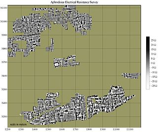
Electrical resistance surveys are one of a number of methods used in archaeological geophysics, as well as in engineering geological investigations. In this type of survey electrical resistance meters are used to detect and map subsurface archaeological features and patterning.
Electrical impedance myography, or EIM, is a non-invasive technique for the assessment of muscle health that is based on the measurement of the electrical impedance characteristics of individual muscles or groups of muscles. The technique has been used for the purpose of evaluating neuromuscular diseases both for their diagnosis and for their ongoing assessment of progression or with therapeutic intervention. Muscle composition and microscopic structure change with disease, and EIM measures alterations in impedance that occur as a result of disease pathology. EIM has been specifically recognized for its potential as an ALS biomarker by Prize4Life, a 501(c)(3) nonprofit organization dedicated to accelerating the discovery of treatments and cures for ALS. The $1M ALS Biomarker Challenge focused on identifying a biomarker precise and reliable enough to cut Phase II drug trials in half. The prize was awarded to Dr. Seward Rutkove, chief, Division of Neuromuscular Disease, in the Department of Neurology at Beth Israel Deaconess Medical Center and Professor of Neurology at Harvard Medical School, for his work in developing the technique of EIM and its specific application to ALS. It is hoped that EIM as a biomarker will result in the more rapid and efficient identification of new treatments for ALS. EIM has shown sensitivity to disease status in a variety of neuromuscular conditions, including radiculopathy, inflammatory myopathy, Duchenne muscular dystrophy, and spinal muscular atrophy.
Richard A. Williams, OBE, FREng, FTSE, FRSE is a British academic and engineer. He is the Principal and Vice-Chancellor of Heriot-Watt University. He took up this position on 1 September 2015. He is also a chemical engineer, Vice President, and a Trustee of the Royal Academy of Engineering.
Boundary estimation in EIT is the term used in the field of electrical impedance tomography, if the inverse problem is the estimation of boundary instead of the conductivity distribution inside an object domain.

Industrial Tomography Systems plc, occasionally abbreviated to ITOMS or simply ITS, is a manufacturer of process visualization systems based upon the principles of tomography. Headquartered in Manchester, UK, the company provides instrumentation to a variety of organisations across a range of sectors; including oil refining, chemical manufacturing, nuclear engineering, dairy manufacturing, and research/academia.
EIDORS is an open-source software tool box written mainly in MATLAB/GNU Octave designed primarily for image reconstruction from electrical impedance tomography (EIT) data, in a biomedical, industrial or geophysical setting. The name was originally an acronym for Electrical Impedance Tomography and Diffuse Optical Reconstruction Software. While the name reflects the original intention to cover image reconstruction of data from the mathematically similar near infra red diffuse optical imaging, to date there has been little development in that area.
Impedance microbiology is a microbiological technique used to measure the microbial number density of a sample by monitoring the electrical parameters of the growth medium. The ability of microbial metabolism to change the electrical conductivity of the growth medium was discovered by Stewart and further studied by other scientists such as Oker-Blom, Parson and Allison in the first half of 20th century. However, it was only in the late 1970s that, thanks to computer-controlled systems used to monitor impedance, the technique showed its full potential, as discussed in the works of Fistenberg-Eden & Eden, Ur & Brown and Cady.
Three-dimensional electrical capacitance tomography also known as electrical capacitance volume tomography (ECVT) is a non-invasive 3D imaging technology applied primarily to multiphase flows. Was introduced in the early 2000s as an extension of the conventional two-dimensional ECT. In conventional electrical capacitance tomography, sensor plates are distributed around a surface of interest. Measured capacitance between plate combinations is used to reconstruct 2D images (tomograms) of material distribution. Because the ECT sensor plates are required to have lengths on the order of the domain cross-section, 2D ECT does not provide the required resolution in the axial dimension. In ECT, the fringing field from the edges of the plates is viewed as a source of distortion to the final reconstructed image and is thus mitigated by guard electrodes. 3D ECT exploits this fringing field and expands it through 3D sensor designs that deliberately establish an electric field variation in all three dimensions. In 3D tomography, the data are acquired in 3D geometry, and the reconstruction algorithm produces the three-dimensional image directly, in contrast to 2D tomography, where 3D information might be obtained by stacking 2D slices reconstructed individually.

