Computer vision tasks include methods for acquiring, processing, analyzing and understanding digital images, and extraction of high-dimensional data from the real world in order to produce numerical or symbolic information, e.g. in the forms of decisions. Understanding in this context means the transformation of visual images into descriptions of the world that make sense to thought processes and can elicit appropriate action. This image understanding can be seen as the disentangling of symbolic information from image data using models constructed with the aid of geometry, physics, statistics, and learning theory.

Medical imaging is the technique and process of imaging the interior of a body for clinical analysis and medical intervention, as well as visual representation of the function of some organs or tissues (physiology). Medical imaging seeks to reveal internal structures hidden by the skin and bones, as well as to diagnose and treat disease. Medical imaging also establishes a database of normal anatomy and physiology to make it possible to identify abnormalities. Although imaging of removed organs and tissues can be performed for medical reasons, such procedures are usually considered part of pathology instead of medical imaging.

Tomography is imaging by sections or sectioning that uses any kind of penetrating wave. The method is used in radiology, archaeology, biology, atmospheric science, geophysics, oceanography, plasma physics, materials science, cosmochemistry, astrophysics, quantum information, and other areas of science. The word tomography is derived from Ancient Greek τόμος tomos, "slice, section" and γράφω graphō, "to write" or, in this context as well, "to describe." A device used in tomography is called a tomograph, while the image produced is a tomogram.

Electrical impedance tomography (EIT) is a noninvasive type of medical imaging in which the electrical conductivity, permittivity, and impedance of a part of the body is inferred from surface electrode measurements and used to form a tomographic image of that part. Electrical conductivity varies considerably among various biological tissues or the movement of fluids and gases within tissues. The majority of EIT systems apply small alternating currents at a single frequency, however, some EIT systems use multiple frequencies to better differentiate between normal and suspected abnormal tissue within the same organ.
Tomographic reconstruction is a type of multidimensional inverse problem where the challenge is to yield an estimate of a specific system from a finite number of projections. The mathematical basis for tomographic imaging was laid down by Johann Radon. A notable example of applications is the reconstruction of computed tomography (CT) where cross-sectional images of patients are obtained in non-invasive manner. Recent developments have seen the Radon transform and its inverse used for tasks related to realistic object insertion required for testing and evaluating computed tomography use in airport security.
Iterative reconstruction refers to iterative algorithms used to reconstruct 2D and 3D images in certain imaging techniques. For example, in computed tomography an image must be reconstructed from projections of an object. Here, iterative reconstruction techniques are usually a better, but computationally more expensive alternative to the common filtered back projection (FBP) method, which directly calculates the image in a single reconstruction step. In recent research works, scientists have shown that extremely fast computations and massive parallelism is possible for iterative reconstruction, which makes iterative reconstruction practical for commercialization.

In chemical processing, a packed bed is a hollow tube, pipe, or other vessel that is filled with a packing material. The packed bed can be randomly filled with small objects like Raschig rings or else it can be a specifically designed structured packing. Packed beds may also contain catalyst particles or adsorbents such as zeolite pellets, granular activated carbon, etc.

Electrical resistivity tomography (ERT) or electrical resistivity imaging (ERI) is a geophysical technique for imaging sub-surface structures from electrical resistivity measurements made at the surface, or by electrodes in one or more boreholes. If the electrodes are suspended in the boreholes, deeper sections can be investigated. It is closely related to the medical imaging technique electrical impedance tomography (EIT), and mathematically is the same inverse problem. In contrast to medical EIT, however, ERT is essentially a direct current method. A related geophysical method, induced polarization, measures the transient response and aims to determine the subsurface chargeability properties.

Electrical capacitance tomography (ECT) is a method for determination of the dielectric permittivity distribution in the interior of an object from external capacitance measurements. It is a close relative of electrical impedance tomography and is proposed as a method for industrial process monitoring.
Terahertz tomography is a class of tomography where sectional imaging is done by terahertz radiation. Terahertz radiation is electromagnetic radiation with a frequency between 0.1 and 10 THz; it falls between radio waves and light waves on the spectrum; it encompasses portions of the millimeter waves and infrared wavelengths. Because of its high frequency and short wavelength, terahertz wave has a high signal-to-noise ratio in the time domain spectrum. Tomography using terahertz radiation can image samples that are opaque in the visible and near-infrared regions of the spectrum. Terahertz wave three-dimensional (3D) imaging technology has developed rapidly since its first successful application in 1997, and a series of new 3D imaging technologies have been proposed successively.
Industrial process imaging, or industrial process tomography or process tomography are methods used to form an image of a cross-section of vessel or pipe in a chemical engineering or mineral processing, or petroleum extraction or refining plant. Process imaging is used for the development of process equipment such as filters, separators and conveyors, as well as monitoring of production plant including flow rate measurement. As well as conventional tomographic methods widely used in medicine such as X-ray computed tomography, magnetic resonance imaging and gamma ray tomography, and ultra-sound tomography, new and emerging methods such as electrical capacitance tomography and magnetic induction tomography and electrical resistivity tomography are also used.
Richard A. Williams, OBE, FREng, FTSE, FRSE is a British academic and engineer. He is the Principal and Vice-Chancellor of Heriot-Watt University. He took up this position on 1 September 2015. He is also a chemical engineer, Vice President, and a Trustee of the Royal Academy of Engineering.

Industrial computed tomography (CT) scanning is any computer-aided tomographic process, usually X-ray computed tomography, that uses irradiation to produce three-dimensional internal and external representations of a scanned object. Industrial CT scanning has been used in many areas of industry for internal inspection of components. Some of the key uses for industrial CT scanning have been flaw detection, failure analysis, metrology, assembly analysis and reverse engineering applications. Just as in medical imaging, industrial imaging includes both nontomographic radiography and computed tomographic radiography.
Boundary estimation in EIT is the term used in the field of electrical impedance tomography, if the inverse problem is the estimation of boundary instead of the conductivity distribution inside an object domain.

Industrial Tomography Systems plc, occasionally abbreviated to ITOMS or simply ITS, is a manufacturer of process visualization systems based upon the principles of tomography. Headquartered in Manchester, UK, the company provides instrumentation to a variety of organisations across a range of sectors; including oil refining, chemical manufacturing, nuclear engineering, dairy manufacturing, and research/academia.
EIDORS is an open-source software tool box written mainly in MATLAB/GNU Octave designed primarily for image reconstruction from electrical impedance tomography (EIT) data, in a biomedical, industrial or geophysical setting. The name was originally an acronym for Electrical Impedance Tomography and Diffuse Optical Reconstruction Software. While the name reflects the original intention to cover image reconstruction of data from the mathematically similar near infra red diffuse optical imaging, to date there has been little development in that area.
Ultrasound computer tomography (USCT), sometimes also Ultrasound computed tomography, Ultrasound computerized tomography or just Ultrasound tomography, is a form of medical ultrasound tomography utilizing ultrasound waves as physical phenomenon for imaging. It is mostly in use for soft tissue medical imaging, especially breast imaging.
Impedance microbiology is a microbiological technique used to measure the microbial number density of a sample by monitoring the electrical parameters of the growth medium. The ability of microbial metabolism to change the electrical conductivity of the growth medium was discovered by Stewart and further studied by other scientists such as Oker-Blom, Parson and Allison in the first half of 20th century. However, it was only in the late 1970s that, thanks to computer-controlled systems used to monitor impedance, the technique showed its full potential, as discussed in the works of Fistenberg-Eden & Eden, Ur & Brown and Cady.
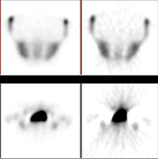
Three-dimensional electrical capacitance tomography also known as electrical capacitance volume tomography (ECVT) is a non-invasive 3D imaging technology applied primarily to multiphase flows. Was introduced in the early 2000s as an extension of the conventional two-dimensional ECT. In conventional electrical capacitance tomography, sensor plates are distributed around a surface of interest. Measured capacitance between plate combinations is used to reconstruct 2D images (tomograms) of material distribution. Because the ECT sensor plates are required to have lengths on the order of the domain cross-section, 2D ECT does not provide the required resolution in the axial dimension. In ECT, the fringing field from the edges of the plates is viewed as a source of distortion to the final reconstructed image and is thus mitigated by guard electrodes. 3D ECT exploits this fringing field and expands it through 3D sensor designs that deliberately establish an electric field variation in all three dimensions. In 3D tomography, the data are acquired in 3D geometry, and the reconstruction algorithm produces the three-dimensional image directly, in contrast to 2D tomography, where 3D information might be obtained by stacking 2D slices reconstructed individually.
Jaakko A. Malmivuo is a Finnish engineer, academic, author, and opera singer. He was a professor of Bioelectromagnetism at Tampere University of Technology (TUT) from 1976 to 2010, an adjunct professor in the Faculty of Medicine at the University of Tampere as well as a visiting professor in the Faculty of Electrical Engineering and Computer Science, Electronics, and Medical Signal Processing at Technische Universität Berlin. Moreover, he was a director of the Ragnar Granit Institute at TUT from 1992.

