Electromyoneurography

Last updated
Electromyoneurography
Purposemeasurement of a peripheral nerve’s conduction velocity

Electromyoneurography (EMNG) is the combined use of electromyography and electroneurography [1] This technique allows for the measurement of a peripheral nerve's conduction velocity upon stimulation (electroneurography) alongside electrical recording of muscular activity (electromyography). Their combined use proves to be clinically relevant by allowing for both the source and location of a particular neuromuscular disease to be known, and for more accurate diagnoses.

Contents

Characteristics

Electromyoneurography is a technique that uses surface electrical probes to obtain electrophysiological readings from nerve and muscle cells. The nerve activity is generally recorded using surface electrodes, stimulating the nerve at one site and recording from another with a minimum distance between the two. The time difference of the potential is a measure of the time taken for the potential to travel the distance across the two sites and is a measure of the conduction velocity along the nerve. The amplitude of the potential, measured baseline to peak, or peak to peak, is a measure of the number of fibers conducting the response. Abnormality in data obtained from nerve measurements, such as absent or low amplitude, indicates potential nerve damage. [2]

This technique is used in many medical fields today. One example of its use is to detect neuropathy due to diseases like diabetes mellitus. [3] It can also be used to detect muscle weakness or paralysis due to sepsis or multi-organ failure in comatose patients. [4] This method remains a largely used medical technique due to its efficiency and relative simplicity. It is especially attractive due to the lack of special precautions or preparation involved with this procedure. There is minimal pain and no significant risks except those associated with needle use. [5]

History

The technique of electromyoneurography was first practiced in the late 1970s by the American Academy of General Practice. The use of this technique enhances diagnostic capability by defining and localizing the target site. In 1978, Milton B. Spiegel, research physician with The Rehabilitation Institute of South Florida, wrote one of the first major academic papers surrounding the uses and benefits of electromyoneurography. It was in this paper that Dr. Spiegel suggested that pre-examination of the patients' range of motion and reflexes would eliminate time and exploration of nerve entrapments during the electromyoneurographic procedure. [1]

In the early 1980s, the practice of utilizing electromyoneurography became more widely accepted in the medical community, specifically aiding in the diagnoses of neuropathy, radiculopathy, and axonopathy. As to more recent use, electromyoneurography has been employed throughout the 21st century, aiding in the diagnosis of carpal tunnel syndrome, abnormal glucose levels, and many other myopathies. This procedure now analyzes the nerve conduction and muscle potentials through the use of H-Reflex and F-Wave studies. Combined with a pre-examination, electromyoneurography is utilized to detect neuromuscular abnormalities. [6]

Modern Application

This is sample data table showing what data obtained from electromyoneurography looks like. Findings are measured in the form of amplitude (mV), latency (ms), and velocity (m/s) of the injured radial nerve, before and after surgery. Electrophysiological findings sample.png
This is sample data table showing what data obtained from electromyoneurography looks like. Findings are measured in the form of amplitude (mV), latency (ms), and velocity (m/s) of the injured radial nerve, before and after surgery.

Electromyoneurography has a variety of modern applications. The high level of sensitivity that electromyoneurography employs makes it ideal for detecting peripheral nerve damage as well as a variety of myopathies in their early stages. This electrophysiological data obtaining technique has been able to heighten diagnostic capabilities when looking at peripheral neuropathy disorders like radiculopathy, and axonopathy in addition to myopathies such as muscular dystrophy, myotonia, and myasthenia gravis. [1] Electromyoneurography was the main technique used in a study to detect diabetic polyneuropathy, a serious condition that is progressive in nature. [7]

Electromyoneurography can also be used to measure patient recovery from surgical procedures, such as nerve repair. A study conducted on patients with proximal radial nerve injuries used the procedure to indicate the degree of both pre- and postoperative nerve damage. [8] In this particular study, electromyoneurography was the preferred method of measuring recovery, chosen over magnetic resonance imaging (MRI) and computed tomography (CT) scans. When looking at the sample data table, one can see that postoperative patients generally see an increase in mean radial nerve amplitude, a decrease in mean radial nerve latency and increases in nerve motor conduction velocity. These results are all general trends that would be expected when operating on damaged nerves in effort to increase their performance.[ citation needed ]

Electromyoneurography's unique combination of recording in muscle and nerve simultaneously typically results in a higher level of diagnostic ability in the field of medicine. This heightened utility often results in a lesser demand for more invase techniques for acquiring electrophysiological data, such as myelography, [1] a procedure where complications are not uncommon and the amount of attention required for post-operative care is more involved.

Conditions Diagnosed with Electromyoneurography

Electromyoneurography has been found to be particularly useful in diagnosing the following neuromuscular conditions, though it is not an exhaustive list:

I. Myopathy (disease or disturbance of striated muscle fibers or cell membrane) II. Neuropathy (disease or disorder of the lower motor neuron)
Primary (muscle fiber): muscular dystrophy
Duchenne muscular dystrophy
Facioscapulohumeral muscular dystrophy
Limb-girdle muscular dystrophy
Myelopathy (lesion involving motor neuron in anterior horn of the spinal cord)
Spinal muscular atrophy
Progressive muscular atrophy
Poliomyelitis
Amyotrophic lateral sclerosis
Charcot–Marie–Tooth disease
Cell membrane hyper-irritability (attributed to spindle cell hyperactivity)
Myotonic dystrophy
Myotonia congenita
Paramyotonia congenita
Radiculopathy (lesion involving the nerve root)
Spinal disc herniation
Spinal stenosis
Guillain–Barré syndrome
Myasthenia
Myasthenia gravis
Lambert–Eaton myasthenic syndrome
Hypokalemia
Glycogen storage disease type V
Cushing's syndrome
Axonopathy (disease or damage to the axon or peripheral nerve)
Carpal tunnel syndrome
Radial neuropathy
Meralgia paraesthetica
Hypothyroidism

Procedure Outline

In an electromyoneurography procedure, recording of the muscle is done by insertion of a needle. The recordings are taken when the muscle is at rest and when the muscle is contracting; the muscle will contract based on the directions of the one performing the test (instructing the patient to move certain body parts in certain directions forming muscle contractions). Various regions of muscle on the body are examined in an electromyoneurography test and the procedure lasts anywhere between 30 and 60 minutes (2–5 minutes per muscle). In addition to examining the muscles, the conduction velocity of nerve signals are measured. The nerve's ability to transmit signals is tested by inserting recording electrodes to capture the data and signal electrodes to initiate signals down a nerve by applying a small shock. Self-generated potentials also occur naturally for recording, in addition to the artificial "shock". Evaluating a nerve's conduction velocity, together with testing potentials, allows for a beneficial diagnosis that can detect pain and sensory problems at the neuromuscular level. [5]

Expected Test Results

Action potential propagation animation Action potential propagation animation.gif
Action potential propagation animation

The needle is normally attached to a recording device known as an electromyography machine. The results show the appearance of action potential or graded potential spikes. While interpretation of the results requires background knowledge, irregular data can be used to diagnose many diseases. If the activity of the nerves at rest is abnormal, this may indicate nerve lesion, radiculopathy, or lower motor nerve degeneration. The amplitude or duration of the potential spike may also be used to gather information. A decreased amplitude or duration may indicate nerve damage due to a muscle diseases, whereas an increase in these demonstrates reinervation, or repair by new nerve connections to the muscles, has occurred. [5]

Related Research Articles

In neuroscience, an F wave is one of several motor responses which may follow the direct motor response (M) evoked by electrical stimulation of peripheral motor or mixed nerves. F-waves are the second of two late voltage changes observed after stimulation is applied to the skin surface above the distal region of a nerve, in addition to the H-reflex which is a muscle reaction in response to electrical stimulation of innervating sensory fibers. Traversal of F-waves along the entire length of peripheral nerves between the spinal cord and muscle, allows for assessment of motor nerve conduction between distal stimulation sites in the arm and leg, and related motoneurons (MN's) in the cervical and lumbosacral cord. F-waves are able to assess both afferent and efferent loops of the alpha motor neuron in its entirety. As such, various properties of F-wave motor nerve conduction are analyzed in nerve conduction studies (NCS), and often used to assess polyneuropathies, resulting from states of neuronal demyelination and loss of peripheral axonal integrity.

<span class="mw-page-title-main">Lambert–Eaton myasthenic syndrome</span> Medical condition

Lambert–Eaton myasthenic syndrome (LEMS) is a rare autoimmune disorder characterized by muscle weakness of the limbs.

Diabetic neuropathy is various types of nerve damage associated with diabetes mellitus. Symptoms depend on the site of nerve damage and can include motor changes such as weakness; sensory symptoms such as numbness, tingling, or pain; or autonomic changes such as urinary symptoms. These changes are thought to result from a microvascular injury involving small blood vessels that supply nerves. Relatively common conditions which may be associated with diabetic neuropathy include distal symmetric polyneuropathy; third, fourth, or sixth cranial nerve palsy; mononeuropathy; mononeuropathy multiplex; diabetic amyotrophy; and autonomic neuropathy.

<span class="mw-page-title-main">Polyneuropathy</span> Medical condition

Polyneuropathy is damage or disease affecting peripheral nerves in roughly the same areas on both sides of the body, featuring weakness, numbness, and burning pain. It usually begins in the hands and feet and may progress to the arms and legs and sometimes to other parts of the body where it may affect the autonomic nervous system. It may be acute or chronic. A number of different disorders may cause polyneuropathy, including diabetes and some types of Guillain–Barré syndrome.

<span class="mw-page-title-main">Electromyography</span> Electrodiagnostic medicine technique

Electromyography (EMG) is a technique for evaluating and recording the electrical activity produced by skeletal muscles. EMG is performed using an instrument called an electromyograph to produce a record called an electromyogram. An electromyograph detects the electric potential generated by muscle cells when these cells are electrically or neurologically activated. The signals can be analyzed to detect abnormalities, activation level, or recruitment order, or to analyze the biomechanics of human or animal movement. Needle EMG is an electrodiagnostic medicine technique commonly used by neurologists. Surface EMG is a non-medical procedure used to assess muscle activation by several professionals, including physiotherapists, kinesiologists and biomedical engineers. In computer science, EMG is also used as middleware in gesture recognition towards allowing the input of physical action to a computer as a form of human-computer interaction.

<span class="mw-page-title-main">Alcoholic polyneuropathy</span> Medical condition

Alcoholic polyneuropathy is a neurological disorder in which peripheral nerves throughout the body malfunction simultaneously. It is defined by axonal degeneration in neurons of both the sensory and motor systems and initially occurs at the distal ends of the longest axons in the body. This nerve damage causes an individual to experience pain and motor weakness, first in the feet and hands and then progressing centrally. Alcoholic polyneuropathy is caused primarily by chronic alcoholism; however, vitamin deficiencies are also known to contribute to its development. This disease typically occurs in chronic alcoholics who have some sort of nutritional deficiency. Treatment may involve nutritional supplementation, pain management, and abstaining from alcohol.

<span class="mw-page-title-main">Nerve conduction study</span> Diagnostic test for nerve function

A nerve conduction study (NCS) is a medical diagnostic test commonly used to evaluate the function, especially the ability of electrical conduction, of the motor and sensory nerves of the human body. These tests may be performed by medical specialists such as clinical neurophysiologists, physical therapists, physiatrists, and neurologists who subspecialize in electrodiagnostic medicine. In the United States, neurologists and physiatrists receive training in electrodiagnostic medicine as part of residency training and in some cases acquire additional expertise during a fellowship in clinical neurophysiology, electrodiagnostic medicine, or neuromuscular medicine. Outside the US, clinical neurophysiologists learn needle EMG and NCS testing.

<span class="mw-page-title-main">Nerve conduction velocity</span> Speed at which an electrochemical impulse propagates down a neural pathway

In neuroscience, nerve conduction velocity (CV) is the speed at which an electrochemical impulse propagates down a neural pathway. Conduction velocities are affected by a wide array of factors, which include age, sex, and various medical conditions. Studies allow for better diagnoses of various neuropathies, especially demyelinating diseases as these conditions result in reduced or non-existent conduction velocities. CV is an important aspect of nerve conduction studies.

Proximal diabetic neuropathy, also known as diabetic amyotrophy, is a complication of diabetes mellitus that affects the nerves that supply the thighs, hips, buttocks and/or lower legs. Proximal diabetic neuropathy is a type of diabetic neuropathy characterized by muscle wasting, weakness, pain, or changes in sensation/numbness of the leg. It is caused by damage to the nerves of the lumbosacral plexus.

<span class="mw-page-title-main">Chronic inflammatory demyelinating polyneuropathy</span> Medical condition

Chronic inflammatory demyelinating polyneuropathy (CIDP) is an acquired autoimmune disease of the peripheral nervous system characterized by progressive weakness and impaired sensory function in the legs and arms. The disorder is sometimes called chronic relapsing polyneuropathy (CRP) or chronic inflammatory demyelinating polyradiculoneuropathy. CIDP is closely related to Guillain–Barré syndrome and it is considered the chronic counterpart of that acute disease. Its symptoms are also similar to progressive inflammatory neuropathy. It is one of several types of neuropathy.

<span class="mw-page-title-main">Centronuclear myopathy</span> Medical condition

Centronuclear myopathies (CNM) are a group of congenital myopathies where cell nuclei are abnormally located in the center of muscle cells instead of their normal location at the periphery.

<span class="mw-page-title-main">Rheobase</span>

Rheobase is a measure of membrane potential excitability. In neuroscience, rheobase is the minimal current amplitude of infinite duration that results in the depolarization threshold of the cell membranes being reached, such as an action potential or the contraction of a muscle. In Greek, the root rhe translates to "current or flow", and basi means "bottom or foundation": thus the rheobase is the minimum current that will produce an action potential or muscle contraction.

<span class="mw-page-title-main">Radiculopathy</span> Medical condition

Radiculopathy, also commonly referred to as pinched nerve, refers to a set of conditions in which one or more nerves are affected and do not work properly. Radiculopathy can result in pain, weakness, altered sensation (paresthesia) or difficulty controlling specific muscles. Pinched nerves arise when surrounding bone or tissue, such as cartilage, muscles or tendons, put pressure on the nerve and disrupt its function.

Sudomotor function refers to the autonomic nervous system control of sweat gland activity in response to various environmental and individual factors. Sweat production is a vital thermoregulatory mechanism used by the body to prevent heat-related illness as the evaporation of sweat is the body’s most effective method of heat reduction and the only cooling method available when the air temperature rises above skin temperature. In addition, sweat plays key roles in grip, microbial defense, and wound healing.

Critical illness polyneuropathy (CIP) and critical illness myopathy (CIM) are overlapping syndromes of diffuse, symmetric, flaccid muscle weakness occurring in critically ill patients and involving all extremities and the diaphragm with relative sparing of the cranial nerves. CIP and CIM have similar symptoms and presentations and are often distinguished largely on the basis of specialized electrophysiologic testing or muscle and nerve biopsy. The causes of CIP and CIM are unknown, though they are thought to be a possible neurological manifestation of systemic inflammatory response syndrome. Corticosteroids and neuromuscular blocking agents, which are widely used in intensive care, may contribute to the development of CIP and CIM, as may elevations in blood sugar, which frequently occur in critically ill patients.

<span class="mw-page-title-main">Hereditary motor and sensory neuropathy</span> Medical condition

Hereditary motor and sensory neuropathies (HMSN) is a name sometimes given to a group of different neuropathies which are all characterized by their impact upon both afferent and efferent neural communication. HMSN are characterised by atypical neural development and degradation of neural tissue. The two common forms of HMSN are either hypertrophic demyelinated nerves or complete atrophy of neural tissue. Hypertrophic condition causes neural stiffness and a demyelination of nerves in the peripheral nervous system, and atrophy causes the breakdown of axons and neural cell bodies. In these disorders, a patient experiences progressive muscle atrophy and sensory neuropathy of the extremities.

Repetitive nerve stimulation is a variant of the nerve conduction study where electrical stimulation is delivered to a motor nerve repeatedly several times per second. By observing the change in the muscle electrical response (CMAP) after several stimulations, a physician can assess for the presence of a neuromuscular junction disease, and differentiate between presynaptic and postsynaptic conditions. The test was first described by German neurologist Friedrich Jolly in 1895, and is also known as Jolly's test.

Clinical Electrophysiological Testing is based on techniques derived from electrophysiology used for the clinical diagnosis of patients. There are many processes that occur in the body which produce electrical signals that can be detected. Depending on the location and the source of these signals, distinct methods and techniques have been developed to properly target them.

Electrodiagnosis (EDX) is a method of medical diagnosis that obtains information about diseases by passively recording the electrical activity of body parts or by measuring their response to external electrical stimuli. The most widely used methods of recording spontaneous electrical activity are various forms of electrodiagnostic testing (electrography) such as electrocardiography (ECG), electroencephalography (EEG), and electromyography (EMG). Electrodiagnostic medicine is a medical subspecialty of neurology, clinical neurophysiology, cardiology, and physical medicine and rehabilitation. Electrodiagnostic physicians apply electrophysiologic techniques, including needle electromyography and nerve conduction studies to diagnose, evaluate, and treat people with impairments of the neurologic, neuromuscular, and/or muscular systems. The provision of a quality electrodiagnostic medical evaluation requires extensive scientific knowledge that includes anatomy and physiology of the peripheral nerves and muscles, the physics and biology of the electrical signals generated by muscle and nerve, the instrumentation used to process these signals, and techniques for clinical evaluation of diseases of the peripheral nerves and sensory pathways.

<span class="mw-page-title-main">Shin Joong Oh</span>

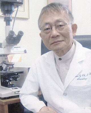
Shin Joong Oh is a Korean physician who is Distinguished Professor of Neurology Emeritus at The University of Alabama at Birmingham in the United States. Oh is a clinician, researcher, and educator known for his contributions to the fields of neurology and electrodiagnostic medicine, particularly electromyography. He retired in 2014.

References

  1. 1 2 3 4 M. B. Spiegel (November 1978). "Electromyoneurography". American Family Physician . 18 (5): 119–130. PMID   717221.
  2. Khushnuma A. Mansukhani & Bhavna H. Doshi (July–September 2008). "Interpretation of electroneuromyographic studies in diseases of neuromuscular junction and myopathies". Neurology India . 56 (3): 339–347. doi: 10.4103/0028-3886.43453 . PMID   18974561. S2CID   19873087.
  3. N. Ovayolu, E. Akarsu, E. Madenci, S. Torun, O. Ucan & M. Yilmaz (July 2008). "Clinical characteristics of patients with diabetic polyneuropathy: the role of clinical and electromyographic evaluation and the effect of the various types on the quality of life". International Journal of Clinical Practice . 62 (7): 1019–1025. doi:10.1111/j.1742-1241.2008.01730.x. PMC   2658015 . PMID   18410351.{{cite journal}}: CS1 maint: multiple names: authors list (link)
  4. N. Latronico, F. Fenzi, D. Recupero, B. Guarneri, G. Tomelleri, P. Tonin, G. De Maria, L. Antonini, N. Rizzuto & A. Candiani (June 1996). "Critical illness myopathy and neuropathy". Lancet . 347 (9015): 1579–1582. doi:10.1016/s0140-6736(96)91074-0. PMID   8667865. S2CID   701091.{{cite journal}}: CS1 maint: multiple names: authors list (link)
  5. 1 2 3 "Electroneuromyography." MedInstitute Y&C Institute of medical rehabilitation. MedInstitute, n.d. Web. 26 Apr 2013. <http://www.medinstitute.net/index.php5?&page_id=19&path=5,19 Archived 2008-12-19 at the Wayback Machine >.
  6. "MedicAid Services Manual" (PDF). Division of Health Care Financing and Policy. Retrieved 21 March 2013.[ permanent dead link ]
  7. Milan Cvijanovic, Miroslav Ilin, Petar Slankamenac, Sofija Banic Horvat & Zita Jovin (January–February 2011). "The sensitivity of electromyoneurography in the diagnosis of diabetic polyneuropathy". Medicinski Pregled . 64 (1–2): 11–14. doi:10.2298/mpns1102011c. PMID   21545063.{{cite journal}}: CS1 maint: multiple names: authors list (link)
  8. Bulent Duz, Ilker Solmaz, Erdinc Civelek, M. Bulent Onal, Serhat Pusat & Mehmet Daneyemez (March–April 2010). "Analysis of proximal radial nerve injury in the arm". Neurology India . 58 (2): 230–234. doi: 10.4103/0028-3886.63802 . PMID   20508341.{{cite journal}}: CS1 maint: multiple names: authors list (link)