Equine protozoal myeloencephalitis

Last updated
Equine protozoal myeloencephalitis in various stages Equine Protozoal Myeloencephalitis.jpg
Equine protozoal myeloencephalitis in various stages

Equine protozoal myeloencephalitis (EPM) is a disease that affects the central nervous system of horses. It is caused by a protozoal infection that is brought about by the apicomplexan parasites Sarcocystis neurona or Neospora hughesi . Most cases are caused by S. neurona. The lifecycle and transmission of N. hughesi is not well understood. [1] The parasites create lesions in both the brain and spinal cord of the affected horses leading to neurological issues. [2] Most horses infected with S. neurona do not exhibit neurological symptoms consistent with EPM.

Contents

Causes

Lifecycle of S. neurona Equine Protozoal Myeloencephalitis life cycle.jpg
Lifecycle of S. neurona

EPM is caused primarily by the parasite Sarcocystis neurona. To complete its lifecycle, S. neurona requires two hosts, a definitive host, and an intermediate host. The definitive host is the opossum. In North America, the definitive host is the Virginia opossum (Didelphis virginiana); South American opossums can also act as the definitive host. [3]

A number of mammals can serve as intermediate hosts. In the laboratory, raccoons, cats, armadillos, skunks, and sea otters have been shown to be intermediate hosts of S. neurona.

The opossum passes the parasite through feces. Horses contract EPM from contaminated feed or water. Horses cannot pass the disease among themselves; that is, one horse cannot contract the disease from another infected horse. The horse is a dead-end, or aberrant, host of the parasite. Although all horses are believed to be susceptible to EPM the disease is usually found in younger horses typically around three to six years of age. [2] EPM does not typically correlate with other factors such as poor nutrition or other diseases. A study conducted in the US found there was no correlation to the genders of the horses infected nor was there a correlation with the time of year. [4]

Six subspecies of S. neurona can be identified by surface antigens (SAG). Equine EPM is caused by the parasites that exhibit SAG1, SAG5, and SAG6. SAG1 and SAG5 are responsible for the majority of EPM cases in horses. Horses produce antibodies to these surface antigens. Serum antibody testing is available that measures levels of these antibodies in the blood of horses, which is helpful in diagnosing EPM in an ataxic horse. Serial blood levels are helpful in guiding treatment. In experimentally infected horses it takes 14 days from infection to positive antibody tests. 80% of horses with EPM have positive antibody tests. A negative antibody test in the presence of EPM results if testing is done before 17 days or if the horse has been treated with antiprotozoal drugs which delays antibody production.

Symptoms

The onset of EPM can come on suddenly or gradually however once the lesion is created it is extremely hard to reverse the damage. The most common symptoms of EPM are ataxia, general weakness with muscle spasticity. However, this is not specific to EPM and is common in many other neurological disorders. Clinical signs among horses with EPM include a wide array of symptoms that may result from primary or secondary problems. Apparent lameness, particularly atypical lameness or slight gait asymmetry of the rear limbs are commonly caused by EPM. Focal muscle atrophy, or even generalized muscle atrophy or loss of condition may occur. Secondary signs also occur with neurologic disease. Airway abnormalities, such as laryngeal hemiplegia, snoring, or airway noise of undetermined origin may result from damage to the nerves which control the throat, although this is quite uncommon.

In experimentally infected horses, very early signs included loss of appetite, decreased tongue tone, facial paresis, altered mental status, generalized weakness, and lameness.

Sarcocystis neurona is thought to not need to enter the CNS to cause disease, in some cases S. neurona has been found in the CNS, but usually not. In cases where S. neurona is found in the CNS, white blood cells probably play a role in the parasite's penetration of the blood-brain barrier.

Diagnosis

Diagnosing EPM can be challenging because the signs can vary significantly from horse to horse, and the symptoms can be similar to those of other CNS diseases. The only way to definitively diagnose EPM is through post-mortem testing, but there are ways to exclude other diseases and establish a basis for the EPM diagnosis. One method is a neurological examination that assesses the CNS function of the horse. Another option is a cervical radiograph, which can identify any compression in the spinal cord that may cause a horse to lose coordination. [5] A veterinarian can also draw blood and spinal fluid and send it to a lab to confirm if a horse has been exposed to opossum feces. Labs can perform immunodiagnostic testing on spinal fluid to evaluate the production of antibodies that fight against these parasites. [1]

Treatment and prevention

EPM is treatable, but irreversible damage to the nervous system is possible. Identifying the disease as early as possible and beginning treatment with antiprotozoal drugs is important. Currently, three FDA approved treatments are available in the US: ReBalance (sulfadiazine and pyrimethamine), Marquis (ponazuril), and Protazil (diclazuril). These drugs minimize the infection, but do not kill the parasite. The use of anti-inflammatory agents such as banamine, corticosteroids, or phenylbutazone are often used to help reduce inflammation and limit further damage to the CNS. Antioxidants, such as vitamin E, may help promote the restoration of nervous tissue. Response to treatment is often variable, and treatment may be expensive. Recently, antiprotozoal treatments that kill the parasite and clear the infection have shown promise. The inflammatory component is thought responsible for the symptoms of EPM;  anti-inflammatory drugs that target the IL-6 pathway have been particularly effective at reversing symptoms. However it is important to note that there is no vaccine to cure EPM. There are several ways that horse owners can potentially lessen the chances of their horse(s) getting EPM. Some of these preventative measure include proper storage of hay and feed, if possible, not feeding on the ground, reducing wildlife access to stalls and pastures, thoroughly cleaning the horse’s water source often, and prompt disposal of animal carcasses. [1]

History

In the 1960s, a syndrome dubbed "segmental myelitis" was first reported in horses in the US by Rooney and colleagues. [3] Later, in the 1970s, it was recognized by Mayhew and colleagues that the syndrome was associated with an infection by protozoa, and the current name of the disease, "equine protozoal myeloencephalitis", was coined. [3] In 1991, a morphologically distinct parasitic agent was identified in samples taken from horses across the US which had EPM; the parasite was tentatively named Sarcocystis neurona. [6] That same year, S. neurona was isolated from tissue taken from three infected horses. [6] Following identification of the cause of EPM, in 1993 the first test was developed which could identify antibodies to S. neurona in the serum of horses. [6] In 1995, the opossum Didelphis virginiana was first associated with EPM when genetic sequences from S. neurona were found in the opossum's intestinal contents. [6]

The term EPM refers to the clinical neurologic symptoms caused by the parasite, not infection itself.

Related Research Articles

<span class="mw-page-title-main">Toxoplasmosis</span> Protozoan parasitic disease

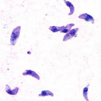
Toxoplasmosis is a parasitic disease caused by Toxoplasma gondii, an apicomplexan. Infections with toxoplasmosis are associated with a variety of neuropsychiatric and behavioral conditions. Occasionally, people may have a few weeks or months of mild, flu-like illness such as muscle aches and tender lymph nodes. In a small number of people, eye problems may develop. In those with a weak immune system, severe symptoms such as seizures and poor coordination may occur. If a person becomes infected during pregnancy, a condition known as congenital toxoplasmosis may affect the child.

<i>Dirofilaria immitis</i> Species of worm that causes parasitic disease in animals

Dirofilaria immitis, also known as heartworm or dog heartworm, is a parasitic roundworm that is a type of filarial worm, a small thread-like worm, that causes dirofilariasis. It is spread from host to host through the bites of mosquitoes. There are four genera of mosquitoes that transmit dirofilariasis, Aedes, Culex, Anopheles, and Mansonia. The definitive host is the dog, but it can also infect cats, wolves, coyotes, jackals, foxes, ferrets, bears, seals, sea lions and, under rare circumstances, humans.

Parelaphostrongylus tenuis is a neurotropic nematode parasite common to white-tailed deer, Odocoileus virginianus, which causes damage to the central nervous system. Moose, elk, caribou, mule deer, and others are also susceptible to the parasite, but are aberrant hosts and are infected in neurological instead of meningeal tissue. The frequency of infection in these species increases dramatically when their ranges overlap high densities of white-tailed deer.

Coccidiosis is a parasitic disease of the intestinal tract of animals caused by coccidian protozoa. The disease spreads from one animal to another by contact with infected feces or ingestion of infected tissue. Diarrhea, which may become bloody in severe cases, is the primary symptom. Most animals infected with coccidia are asymptomatic, but young or immunocompromised animals may suffer severe symptoms and death.

<i>Neospora caninum</i> Species of Conoidasida in the apicomplex phylum

Neospora caninum is a coccidian parasite that was identified as a species in 1988. Prior to this, it was misclassified as Toxoplasma gondii due to structural similarities. The genome sequence of Neospora caninum has been determined by the Wellcome Trust Sanger Institute and the University of Liverpool. Neospora caninum is an important cause of spontaneous abortion in infected livestock.

<span class="mw-page-title-main">Viral encephalitis</span> Medical condition

Viral encephalitis is inflammation of the brain parenchyma, called encephalitis, by a virus. The different forms of viral encephalitis are called viral encephalitides. It is the most common type of encephalitis and often occurs with viral meningitis. Encephalitic viruses first cause infection and replicate outside of the central nervous system (CNS), most reaching the CNS through the circulatory system and a minority from nerve endings toward the CNS. Once in the brain, the virus and the host's inflammatory response disrupt neural function, leading to illness and complications, many of which frequently are neurological in nature, such as impaired motor skills and altered behavior.

<span class="mw-page-title-main">Neurosyphilis</span> Infection of the central nervous system in a patient with syphilis

Neurosyphilis is the infection of the central nervous system in a patient with syphilis. In the era of modern antibiotics, the majority of neurosyphilis cases have been reported in HIV-infected patients. Meningitis is the most common neurological presentation in early syphilis. Tertiary syphilis symptoms are exclusively neurosyphilis, though neurosyphilis may occur at any stage of infection.

Wobbler disease is a catchall term referring to several possible malformations of the cervical vertebrae that cause an unsteady (wobbly) gait and weakness in dogs and horses. A number of different conditions of the cervical (neck) spinal column cause similar clinical signs. These conditions may include malformation of the vertebrae, intervertebral disc protrusion, and disease of the interspinal ligaments, ligamenta flava, and articular facets of the vertebrae. Wobbler disease is also known as cervical vertebral instability (CVI), cervical spondylomyelopathy (CSM), and cervical vertebral malformation (CVM). In dogs, the disease is most common in large breeds, especially Great Danes and Doberman Pinschers. In horses, it is not linked to a particular breed, though it is most often seen in tall, race-bred horses of Thoroughbred or Standardbred ancestry. It is most likely inherited to at least some extent in dogs and horses.

<i>Sarcocystis</i> Genus of protists in the apicomplex phylum

Sarcocystis is a genus of protozoan parasites, with many species infecting mammals, reptiles and birds. Its name is dervived from Greek sarx = flesh and kystis = bladder.

<i>Neospora</i> Genus of single-celled organisms

Neospora is a single celled parasite of livestock and companion animals. It was not discovered until 1984 in Norway, where it was found in dogs. Neosporosis, the disease that affects cattle and companion animals, has a worldwide distribution. Neosporosis causes abortions in cattle and paralysis in companion animals. It is highly transmissible and some herds can have up to a 90% prevalence. Up to 33% of pregnancies can result in aborted fetuses on one dairy farm. In many countries this organism is the main cause of abortion in cattle. Neosporosis is now considered as a major cause of abortion in cattle worldwide. Many reliable diagnostic tests are commercially available. Neospora caninum does not appear to be infectious to humans. In dogs, Neospora caninum can cause neurological signs, especially in congenitally infected puppies, where it can form cysts in the central nervous system.

<span class="mw-page-title-main">Blastocystosis</span> Medical condition

Blastocystosis refers to a medical condition caused by infection with Blastocystis. Blastocystis is a protozoal, single-celled parasite that inhabits the gastrointestinal tracts of humans and other animals. Many different types of Blastocystis exist, and they can infect humans, farm animals, birds, rodents, amphibians, reptiles, fish, and even cockroaches. Blastocystosis has been found to be a possible risk factor for development of irritable bowel syndrome.

<span class="mw-page-title-main">Ponazuril</span> Chemical compound

Ponazuril (INN), sold by Merial, Inc., now part of Boehringer Ingelheim, under the trade name Marquis® , is a drug currently approved for the treatment of equine protozoal myeloencephalitis (EPM) in horses, caused by coccidia Sarcocystis neurona. More recently, veterinarians have been preparing a formulary version of the medication for use in small animals such as cats, dogs, and rabbits against coccidia as an intestinal parasite. Coccidia treatment in small animals is far shorter than treatment for EPM.

The central nervous system (CNS) controls most of the functions of the body and mind. It comprises the brain, spinal cord and the nerve fibers that branch off to all parts of the body. The CNS viral diseases are caused by viruses that attack the CNS. Existing and emerging viral CNS infections are major sources of human morbidity and mortality.

<i>Encephalitozoon cuniculi</i> Microsporidial pathogen

Encephalitozoon cuniculi is a microsporidial pathogen of mammals with world-wide distribution. An important cause of neurologic and renal disease in rabbits, E. cuniculi can also cause disease in immunocompromised people.

<span class="mw-page-title-main">Coenurosis in humans</span> Medical condition

Coenurosis is a parasitic infection that results when humans ingest the eggs of dog tapeworm species Taenia multiceps, T. serialis, T. brauni, or T. glomerata.

<i>Anoplocephala perfoliata</i> Species of flatworm

Anoplocephala perfoliata is the most common intestinal tapeworm of horses, and an agent responsible for some cases of equine colic.

<i>Neorickettsia risticii</i> Species of bacterium

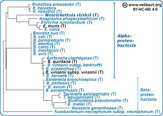
Neorickettsia risticii, formerly Ehrlichia risticii, is an obligate intracellular gram negative bacteria that typically lives as an endosymbiont to parasitic flatworms, specifically flukes. N. risticii is the known causative agent of equine neorickettsiosis, which gets its name from its discovery near the Potomac River in Maryland and Virginia. N. risticii was first recovered from horses in this region in 1984 but was not recognized as the causative agent of PHF until 1979. Potomac horse fever is currently endemic in the United States but has also been reported with lower frequency in other regions, including Canada, Brazil, Uruguay, and Europe. PHF is a condition that is clinically important for horses since it can cause serious signs such as fever, diarrhea, colic, and laminitis. PHF has a fatality rate of approximately 30%, making this condition one of the concerns for horse owners in endemic regions N. risticii is typically acquired in the middle to late summer near freshwater streams or rivers, as well as on irrigated pastures. This is a seasonal infection because it relies on the ingestion of an arthropod vector, which are more commonly found on pasture in the summer months. Although N. risticii is a well known causative agent for PHF in horses, it may act as a potential pathogen in cats and dogs as well. Not only has N. risticii been successfully cultured from monocytes of dogs and cats, but cats have become clinically ill after experimental infection with the bacteria. In addition, N. risticii has been isolated and cultured from human histiocytic lymphoma cells.

<i>Taenia hydatigena</i> Species of flatworm

Taenia hydatigena is one of the adult forms of the canine and feline tapeworm. This infection has a worldwide geographic distribution. Humans with taeniasis can infect other humans or animal intermediate hosts by eggs and gravid proglottids passed in the feces.

Sarcocystis calchasi is an apicomplexan parasite. It has been identified to be the cause of Pigeon protozoal encephalitis (PPE) in the intermediate hosts, domestic pigeons. PPE is a central-nervous disease of domestic pigeons. Initially there have been reports of this parasite in Germany, with an outbreak in 2008 and in 2011 in the United States. Sarcocystis calchasi is transmitted by the definitive host Accipter hawks.

Sarcocystis neurona is primarily a neural parasite of horses and its management is of concern in veterinarian medicine. The protozoan Sarcocystis neurona is a protozoan of single celled character and belongs to the family, Sarcocystidae, a group called coccidia. The protozoan, S. neurona, is a member of the genus Sarcocystis, and is most commonly associated with equine protozoal myeloencephalitis (EPM). The protozoan, S. neurona, can be easily cultivated and genetically manipulated, hence its common use as a model to study numerous aspects of cell biology.

References

  1. 1 2 3 Reed, S.M.; Furr, M.; Howe, D.K.; Johnson, A.L.; MacKay, R.J.; Morrow, J.K.; Pusterla, N.; Witonsky, S. (March 2016). "Equine protozoal myeloencephalitis: an updated consensus statement with a focus on parasite biology, diagnosis, treatment, and prevention". Journal of Veterinary Internal Medicine. 30 (2): 491–502. doi:10.1111/jvim.13834. ISSN   0891-6640. PMC   4913613 . PMID   26857902.
  2. 1 2 Dubey, J. P. (May 2022). "Sarcocystis neurona, Neospora spp. and Toxoplasma gondii infections in horses and equine protozoal myeloencephalitis (EPM): five decades of personal experience, perspectives and update". Parasitology. 149 (6): 717–728. doi:10.1017/S0031182021002055. ISSN   0031-1820. PMID   35260209.
  3. 1 2 3 MacKay, RJ; Howe, DK (August 2022). "Equine protozoal myeloencephalitis". The Veterinary clinics of North America. Equine practice. 38 (2): 249–268. doi:10.1016/j.cveq.2022.05.003. PMID   35810151.
  4. Fayer, R.; Mayhew, I. G.; Baird, J. D.; Dill, S. G.; Foreman, J. H.; Fox, J. C.; Higgins, R. J.; Reed, S. M.; Ruoff, W. W.; Sweeney, R. W.; Tuttle, P. (March 1990). "Epidemiology of Equine Protozoal Myeloencephalitis in North America Based on Histologically Confirmed Cases: A Report". Journal of Veterinary Internal Medicine. 4 (2): 54–57. doi:10.1111/j.1939-1676.1990.tb03103.x. ISSN   0891-6640.
  5. "Equine Protozoal Myeloencephalitis | AAEP". aaep.org. Retrieved 2023-12-01.
  6. 1 2 3 4 Dubey, JP; Lindsay, DS; Saville, WJ; Reed, SM; Granstrom, DE; Speer, CA (26 February 2001). "A review of Sarcocystis neurona and equine protozoal myeloencephalitis (EPM)". Veterinary Parasitology . 95 (2–4): 89–131. doi:10.1016/s0304-4017(00)00384-8. PMID   11223193.

Further reading