Sarcocystis neurona

Last updated

Sarcocystis neurona
Equine Protozoal Myeloencephalitis.jpg
Sporulated oocyst
Scientific classification OOjs UI icon edit-ltr.svg
Domain: Eukaryota
Clade: Diaphoretickes
Clade: SAR
Clade: Alveolata
Phylum: Apicomplexa
Class: Conoidasida
Subclass: Coccidia
Order: Eucoccidiorida
Suborder: Eimeriorina
Family: Sarcocystidae
Genus: Sarcocystis
Species:
S. neurona
Binomial name
Sarcocystis neurona
Dubey et al., 1991

Sarcocystis neurona is primarily a neural parasite of horses and its management is of concern in veterinary medicine. The protozoan Sarcocystis neurona is a protozoan of single celled character and belongs to the family Sarcocystidae, in a group called coccidia. [1] The protozoan, S. neurona, is a member of the genus Sarcocystis , and is most commonly associated with equine protozoal myeloencephalitis (EPM). [2] The protozoan, S. neurona, can be easily cultivated and genetically manipulated, hence its common use as a model to study numerous aspects of cell biology. [3]

Contents

Life cycle

The life cycle of Sarcocystis neurona Equine Protozoal Myeloencephalitis life cycle.jpg
The life cycle of Sarcocystis neurona

Sarcocystis neurona has a wide range of potential intermediate hosts, making it one of the most unique life cycle of the genus Sarcocystis. The life cycle of this parasite requires two hosts, a definitive and an intermediate host. The definitive host of S. neurona are opossums, and more specifically opossums of species Didelphis virginiana and D. albiventris . [4] The full life cycle of S. neurona has yet to be understood, but many advances have been made in fully understanding it after the publication of this subject in 2001 by Dubey. [2] Environmentally resistant oocysts and sporocysts are released into the environment by the definitive hosts' (opossums) feces. Numerous mammals primarily including armadillos, raccoons, sea otters, skunks, and cats serve as intermediate hosts for S. neurona and ingest many sporocysts. After multiple asexual cycles, the sporocysts develop into a resting stage in their muscles called sarcocysts. The sarcocysts produced from the muscles of the intermediate host are ingested by the definitive host and bradyzoites are released in the intestine of the definitive host. The bradyzoites result in sporulated oocysts by undergoing a sexual cycle. The sporulated oocysts are excreted in the feces of the opossum. Horses are aberrant or dead-end hosts, because only schizonts and merozoites have been identified and confined to the brain in spinal cord after a horse has ingested sporocysts in contaminated water and feed. This disease cannot be passed from horse to horse. [3]

Pathogenesis

There are many unanswered questions concerning the development of the disease once this protozoan infects the horse by ingestion of sporocysts in contaminated water and feed. It is presumed that sporocysts release sporozoites that are able to penetrate the intestinal wall and enter arterial endothelial cells within the horse. It is assumed that schizonts develop in these cells until the cell ruptures and merozoites are released into the bloodstream, repeating this stage numerous times to produce large numbers of merozoites. [5] During this stage, no clinical signs may be present, and the infection can be cleared with seropositivity indicating the past infection, or the protozoan can progress to the central nervous system. How S. neurona enters the central nervous system of horses is unknown, but it has been hypothesized that merozoites enter the central nervous system through cytoplasm of endothelial cells or by infected leukocytes. When the merozoite accesses the central nervous system, it is suggested that schizonts form in one or more areas of the CNS including the cerebrum, brainstem, cranial nerves, and/or the spinal cord of the horse. Transmission of the protozoan from the infected horse to other animals is not possible based on the schizonts and daughter merozoites remaining uninfective in the neural tissue. [5] Recent studies suggest that approximately 22–65% of horses in the United States, depending on the geographic location, are seropositive for S. neurona antibodies. Although theories are often developed, the reason explaining why only some horses develop clinical signs of S. neurona and some do not is unknown. Theoretical factors including stress and further unrelated health events are thought to contribute to the onset of this disease. Little is known about the incubation period between exposure to the protozoan and development of clinical disease of Sarcocystis neurona. [5]

Clinical disease

Sarcocystis neurona can parasitize all regions of the central nervous system, including the anterior cerebrum to the end of the spinal cord. Clinical signs of EPM rely on the parasitzing of the CNS. As discussed above, S. neurona schizonts and merozoites are found in the neurons, mononuclear cells, glial cells, and possibly other neural cells. Studies in immune deficient, interferon gamma gene knockout (KO) mice fed S. neurona sporocysts, indicate the Sarcocystis neurona multiplies to a maximum extent in visceral tissue before being transported to the CNS via vehicles discussed above. According to the study, three weeks after infection, S. neuronais primarily confined to the CNS prior to the third week of infection. [6] Early clinical signs of EPM include stumbling and frequent interference, which can often be mistaken for lameness of thoracic and/or pelvic limbs in horses. In horses, the disease often progresses gradually and includes clinical signs such as ataxia. In some horses mild clinical signs may herald a rapidly progressive disease course. The vital signs of infected horses are usually normal during physical examination, although thinning and mild depression may be present. Following a neurological examination asymmetric weakness, spasms and lack of muscle control involving the limbs is revealed. Brain or cranial nerve deficits most frequently observed in horses includes, but are not limited to, depression, head tilt, difficulty swallowing, and facial nerve paralysis. Depending on the location and severity of the lesion, severity of spinal cord damage may lead to an abnormality in walking. [7]

Diagnosis

Although many neurological disorders affect horses, EPM remains the most commonly diagnosed infectious neurological disease in the United States. [8] It is suggested to begin with a complete neurological examination of potentially infected horses to rule out alternative diagnoses. Several serological diagnostic tests have been used to detect antibodies against S. neurona in animals. The first assay developed for detection of antibodies against S. neurona and the diagnosis of EPM was the immunoblot test, also called the Western Blot (WB) test. [9] Development of the WB test over the years has benefited greatly in EPM diagnosis, however the WB technique is mainly a research tool that requires high levels of precision and accuracy. "Second generation" serological assays that are more informative and provide greater throughput have been developed. [10] However, due to EPM only occurring in a small proportion of horses infected with S. neurona, the detection of antibodies against this parasite offers minimal diagnostic value. Antibody detection in cerebrospinal fluid (CSF) offers more insight, but tends to be cofounded by flood contamination during the collection of the sample. [11]

Enzyme-linked immunosorbent assays (ELISAs) are easy to perform, provide a more objective interpretation of the results, and allow for increased throughput testing. ELISAs have been developed based on S. neurona antigens being expressed as recombinant proteins in E. coli. [12] The ELISAs are based on the S. neurona merozoite surface antigens (SnSAGs). The SnSAGs are good targets due to being abundant and immunogenic. All of the SnSAGs are not equally useful in serological assays due to antigenic diversity found in the different strains of S. neurona. Validative studies have proved that the SnSAG ELISAs are specific and do not cross-react with serum from horses infected with other species of Sarcocystis. [13] Surface proteins S. neurona accurately based on their vulnerability as immunologic markers.

Treatment and prevention

Treatment of horses with suspected equine protozoal myeloencephalitis should begin immediately after clinical signs and symptoms of the disease have been recognized and confirmed. Treatment has been confined to dihydrofolate reductase inhibitors including sulfonamides and pyrimethamine over the years. According to the USDA, usual treatment consists of an oral dose of 20 mg/kg sulfadiazine once or twice in a day. Affected horses should additionally be placed on a dose of 1.0 mg/kg pyrimethamine orally for 120 days or longer. Other coccidiostats are currently being evaluated to treat EPM. Recommended measures of prevention include proper hygiene and preventing opossums to horse feed and horse grazing pastures. [1]

Related Research Articles

<span class="mw-page-title-main">Toxoplasmosis</span> Protozoan parasitic disease

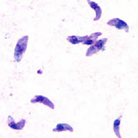
Toxoplasmosis is a parasitic disease caused by Toxoplasma gondii, an apicomplexan. Infections with toxoplasmosis are associated with a variety of neuropsychiatric and behavioral conditions. Occasionally, people may have a few weeks or months of mild, flu-like illness such as muscle aches and tender lymph nodes. In a small number of people, eye problems may develop. In those with a weak immune system, severe symptoms such as seizures and poor coordination may occur. If a woman becomes infected during pregnancy, a condition known as congenital toxoplasmosis may affect the child.

<span class="mw-page-title-main">Isosporiasis</span> Human intestinal disease

Isosporiasis, also known as cystoisosporiasis, is a human intestinal disease caused by the parasite Cystoisospora belli. It is found worldwide, especially in tropical and subtropical areas. Infection often occurs in immuno-compromised individuals, notably AIDS patients, and outbreaks have been reported in institutionalized groups in the United States. The first documented case was in 1915. It is usually spread indirectly, normally through contaminated food or water (CDC.gov).

<span class="mw-page-title-main">Coccidia</span> Subclass of protists

Coccidia (Coccidiasina) are a subclass of microscopic, spore-forming, single-celled obligate intracellular parasites belonging to the apicomplexan class Conoidasida. As obligate intracellular parasites, they must live and reproduce within an animal cell. Coccidian parasites infect the intestinal tracts of animals, and are the largest group of apicomplexan protozoa.

<span class="mw-page-title-main">Gametocyte</span> Eukaryotic germ stem cell

A gametocyte is a eukaryotic germ cell that divides by mitosis into other gametocytes or by meiosis into gametids during gametogenesis. Male gametocytes are called spermatocytes, and female gametocytes are called oocytes.

<i>Plasmodium malariae</i> Species of single-celled organism

Plasmodium malariae is a parasitic protozoan that causes malaria in humans. It is one of several species of Plasmodium parasites that infect other organisms as pathogens, also including Plasmodium falciparum and Plasmodium vivax, responsible for most malarial infection. Found worldwide, it causes a so-called "benign malaria", not nearly as dangerous as that produced by P. falciparum or P. vivax. The signs include fevers that recur at approximately three-day intervals – a quartan fever or quartan malaria – longer than the two-day (tertian) intervals of the other malarial parasite.

<i>Eimeria</i> Genus of single-celled organisms

Eimeria is a genus of apicomplexan parasites that includes various species capable of causing the disease coccidiosis in animals such as cattle, poultry and smaller ruminants including sheep and goats. Eimeria species are considered to be monoxenous because the life cycle is completed within a single host, and stenoxenous because they tend to be host specific, although a number of exceptions have been identified. Species of this genus infect a wide variety of hosts. Thirty-one species are known to occur in bats (Chiroptera), two in turtles, and 130 named species infect fish. Two species infect seals. Five species infect llamas and alpacas: E. alpacae, E. ivitaensis, E. lamae, E. macusaniensis, and E. punonensis. A number of species infect rodents, including E. couesii, E. kinsellai, E. palustris, E. ojastii and E. oryzomysi. Others infect poultry, rabbits and cattle. For full species list, see below.

Cytauxzoon felis is a protozoal organism transmitted to domestic cats by tick bites, and whose natural reservoir host is the bobcat. C. felis has been found in other wild felid species such as the cougar, as well as a white tiger in captivity. C. felis infection is limited to the family Felidae which means that C. felis poses no zoonotic risk or agricultural risk. Until recently it was believed that after infection with C. felis, pet cats almost always died. As awareness of C. felis has increased it has been found that treatment is not always futile. More cats have been shown to survive the infection than was previously thought. New treatments offer as much as 60% survival rate.

<i>Sarcocystis</i> Genus of protists in the apicomplex phylum

Sarcocystis is a genus of protozoan parasites, with many species infecting mammals, reptiles and birds. Its name is dervived from Greek sarx = flesh and kystis = bladder.

<span class="mw-page-title-main">Equine protozoal myeloencephalitis</span> Central nervous system disease of horses

Equine protozoal myeloencephalitis (EPM) is a disease that affects the central nervous system of horses. It is caused by a protozoal infection that is brought about by the apicomplexan parasites Sarcocystis neurona or Neospora hughesi. Most cases are caused by S. neurona. The lifecycle and transmission of N. hughesi is not well understood. The parasites create lesions in both the brain and spinal cord of the affected horses leading to neurological issues. Most horses infected with S. neurona do not exhibit neurological symptoms consistent with EPM.

Megaloschizonts are large schizonts that produce extremely high numbers of merozoites. They are found in various species of the Phylum Apicomplexa. The Apicomplexa phylum contains several parasitic protozoans. They have a very complex life cycle that includes several stages. Megaloschizonts and the smaller schizonts are the part of the life cycle that takes place inside the infected host organism and operates as an asexually reproductive cell. Megaloschizonts appear as grey-white nodules found in the smooth muscle of major organs, such as the heart, liver, lung or spleen.

<span class="mw-page-title-main">Ponazuril</span> Chemical compound

Ponazuril (INN), sold by Merial, Inc., now part of Boehringer Ingelheim, under the trade name Marquis® , is a drug currently approved for the treatment of equine protozoal myeloencephalitis (EPM) in horses, caused by coccidia Sarcocystis neurona. More recently, veterinarians have been preparing a formulary version of the medication for use in small animals such as cats, dogs, and rabbits against coccidia as an intestinal parasite. Coccidia treatment in small animals is far shorter than treatment for EPM.

<span class="mw-page-title-main">Apicomplexan life cycle</span> Apicomplexa life cycle

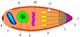
Apicomplexans, a group of intracellular parasites, have life cycle stages that allow them to survive the wide variety of environments they are exposed to during their complex life cycle. Each stage in the life cycle of an apicomplexan organism is typified by a cellular variety with a distinct morphology and biochemistry.

Virus quantification is counting or calculating the number of virus particles (virions) in a sample to determine the virus concentration. It is used in both research and development (R&D) in academic and commercial laboratories as well as in production situations where the quantity of virus at various steps is an important variable that must be monitored. For example, the production of virus-based vaccines, recombinant proteins using viral vectors, and viral antigens all require virus quantification to continually monitor and/or modify the process in order to optimize product quality and production yields and to respond to ever changing demands and applications. Other examples of specific instances where viruses need to be quantified include clone screening, multiplicity of infection (MOI) optimization, and adaptation of methods to cell culture.

<i>Theileria parva</i> Species of single-celled organism

Theileria parva is a species of parasites, named in honour of Arnold Theiler, that causes East Coast fever (theileriosis) in cattle, a costly disease in Africa. The main vector for T. parva is the tick Rhipicephalus appendiculatus. Theiler found that East Coast fever was not the same as redwater, but caused by a different protozoan.

The plaque reduction neutralization test is used to quantify the titer of neutralizing antibody for a virus.

Leucocytozoon caulleryi is a species of the genus Leucocytozoon, a genus of parasitic alveolates.

<i>Neorickettsia risticii</i> Species of bacterium

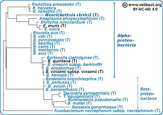
Neorickettsia risticii, formerly Ehrlichia risticii, is an obligate intracellular gram negative bacteria that typically lives as an endosymbiont to parasitic flatworms, specifically flukes. N. risticii is the known causative agent of equine neorickettsiosis, which gets its name from its discovery near the Potomac River in Maryland and Virginia. N. risticii was first recovered from horses in this region in 1984 but was not recognized as the causative agent of PHF until 1979. Potomac horse fever is currently endemic in the United States but has also been reported with lower frequency in other regions, including Canada, Brazil, Uruguay, and Europe. PHF is a condition that is clinically important for horses since it can cause serious signs such as fever, diarrhea, colic, and laminitis. PHF has a fatality rate of approximately 30%, making this condition one of the concerns for horse owners in endemic regions N. risticii is typically acquired in the middle to late summer near freshwater streams or rivers, as well as on irrigated pastures. This is a seasonal infection because it relies on the ingestion of an arthropod vector, which are more commonly found on pasture in the summer months. Although N. risticii is a well known causative agent for PHF in horses, it may act as a potential pathogen in cats and dogs as well. Not only has N. risticii been successfully cultured from monocytes of dogs and cats, but cats have become clinically ill after experimental infection with the bacteria. In addition, N. risticii has been isolated and cultured from human histiocytic lymphoma cells.

<i>Cystoisospora belli</i> Species of single-celled organism

Cystoisospora belli, previously known as Isospora belli, is a parasite that causes an intestinal disease known as cystoisosporiasis. This protozoan parasite is opportunistic in immune suppressed human hosts. It primarily exists in the epithelial cells of the small intestine, and develops in the cell cytoplasm. The distribution of this coccidian parasite is cosmopolitan, but is mainly found in tropical and subtropical areas of the world such as the Caribbean, Central and S. America, India, Africa, and S.E. Asia. In the U.S., it is usually associated with HIV infection and institutional living.

Sarcocystis calchasi is an apicomplexan parasite. It has been identified to be the cause of Pigeon protozoal encephalitis (PPE) in the intermediate hosts, domestic pigeons. PPE is a central-nervous disease of domestic pigeons. Initially there have been reports of this parasite in Germany, with an outbreak in 2008 and in 2011 in the United States. Sarcocystis calchasi is transmitted by the definitive host Accipter hawks.

<span class="mw-page-title-main">Quartan fever</span> Medical condition

Quartan fever is one of the four types of malaria which can be contracted by humans.

References

  1. 1 2 Beltsville MD,"EPM/Sarcocystis neurona" United States Department of Agriculture
  2. 1 2 Dubey, J.P.; Lindsay, D.S.; Saville, W.J.A.; Reed, S.M.; Granstrom, D.E.; Speer, C.A. (February 2001). "A review of Sarcocystis neurona and equine protozoal myeloencephalitis (EPM)". Veterinary Parasitology. 95 (2–4): 89–131. doi:10.1016/S0304-4017(00)00384-8. PMID   11223193.
  3. 1 2 Dubey, J.P.; Howe, D.K.; Furr, M.; Saville, W.J.; Marsh, A.E.; Reed, S.M.; Grigg, M.E. (April 2015). "An update on Sarcocystis neurona infections in animals and equine protozoal myeloencephalitis (EPM)". Veterinary Parasitology. 209 (1–2): 1–42. doi:10.1016/j.vetpar.2015.01.026. PMC   4461864 . PMID   25737052.
  4. "EPM/Sarcocystis neurona: USDA ARS". www.ars.usda.gov. United States Department of Agriculture Agricultural Research Service. Retrieved 20 April 2018.
  5. 1 2 3 Gilmor, Katherine (Summer 2004) Boulineau, Theresa, ed. "Equine Protozoal Myeloencephalitis", Purdue Newsletter, retrieved 2024-03-09.
  6. Lindsay DS, Dubey JP "Determination of the activity of pyrantel tartrate against Sarcocystis neurona in gamma-interferon gene knockout mice". US National Library of Medicine NAtional Institutes of Health 22 May 2001.
  7. Beltsville MD, "EPM/Sarcocystis neurona". United States Department of Agriculture.
  8. Morley S, Traub-Dargatz J, Saville W, Wagner B, Garber L, Hillberg-Seitzinger A. Equine protozoal myeloencephalitis. J Equine Vet Sci. 2001; 21: 262–270.
  9. Granstrom DE, Dubey JP, Davis SW, Fayer R, Fox JC, Poonacha KB, Giles RC, Comer PF. Equine protozoal myeloencephalitis: antigen analysis of cultured Sarcocystis neurona merozoites. J Vet Diagn Invest. 1993;5:88–90
  10. Lindsay DS, Thomas NJ, Rosypal AC, Dubey JP. Dual Sarcocystis neurona and Toxoplasma gondii infection in a Northern sea otter from Washington state, USA. Vet Parasitol. 2001a;97:319–327
  11. Lymphocyte phenotype subsets in the cerebrospinal fluid of normal horses and horses with equine protozoal myeloencephalitis. Furr M, Pontzer C, Gasper P Vet Ther. 2001 Fall; 2(4):317-24
  12. Ellison SP, Kennedy T, Brown KK. Development of an ELISA to detect antibodies to rSAG1 in the horse. Int J Appl Res Vet Med. 2003a;1:318–327
  13. Enzyme-linked immunosorbent assays for detection of equine antibodies specific to Sarcocystis neurona surface antigens. Hoane JS, Morrow JK, Saville WJ, Dubey JP, Granstrom DE, Howe DK Clin Diagn Lab Immunol. 2005 Sep; 12(9):1050-6