Related Research Articles

The femur, or thigh bone is the only bone in the thigh. The thigh is the region of the lower limb between the hip and the knee. In many four-legged animals the femur is the upper bone of the hindleg.

In humans and other primates, the knee joins the thigh with the leg and consists of two joints: one between the femur and tibia, and one between the femur and patella. It is the largest joint in the human body. The knee is a modified hinge joint, which permits flexion and extension as well as slight internal and external rotation. The knee is vulnerable to injury and to the development of osteoarthritis.
Teratology is the study of abnormalities of physiological development in organisms during their life span. It is a sub-discipline in medical genetics which focuses on the classification of congenital abnormalities in dysmorphology caused by teratogens. Teratogens are substances that may cause non-heritable birth defects via a toxic effect on an embryo or fetus. Defects include malformations, disruptions, deformations, and dysplasia that may cause stunted growth, delayed mental development, or other congenital disorders that lack structural malformations. The related term developmental toxicity includes all manifestations of abnormal development that are caused by environmental insult. The extent to which teratogens will impact an embryo is dependent on several factors, such as how long the embryo has been exposed, the stage of development the embryo was in when exposed, the genetic makeup of the embryo, and the transfer rate of the teratogen.

An epiphysis is one of the rounded ends or tips of a long bone that ossify from a secondary center of ossification. Between the epiphysis and diaphysis lies the metaphysis, including the epiphyseal plate. At the joint, the epiphysis is covered with articular cartilage; below that covering is a zone similar to the epiphyseal plate, known as subchondral bone.
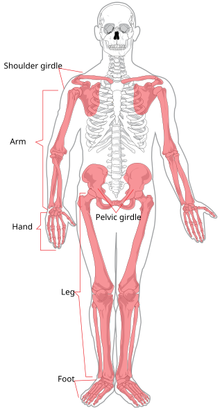
The appendicular skeleton is the portion of the skeleton of vertebrates consisting of the bones that support the appendages. There are 126 bones. The appendicular skeleton includes the skeletal elements within the limbs, as well as supporting shoulder girdle and pelvic girdle. The word appendicular is the adjective of the noun appendage, which itself means a part that is joined to something larger.

Coxa vara is a deformity of the hip, whereby the angle between the head and the shaft of the femur is reduced to less than 120 degrees. This results in the leg being shortened and the development of a limp. It may be congenital and is commonly caused by injury, such as a fracture. It can also occur when the bone tissue in the neck of the femur is softer than normal, causing it to bend under the weight of the body. This may either be congenital or the result of a bone disorder. The most common cause of coxa vara is either congenital or developmental. Other common causes include metabolic bone diseases, post-Perthes deformity, osteomyelitis, and post traumatic. Shepherd's Crook deformity is a severe form of coxa vara where the proximal femur is severely deformed with a reduction in the neck shaft angle beyond 90 degrees. It is most commonly a sequela of osteogenesis imperfecta, Paget's disease, osteomyelitis, tumour and tumour-like conditions.

In vertebrate anatomy, hip refers to either an anatomical region or a joint.

Proximal femoral focal deficiency (PFFD), also known as Congenital Femoral Deficiency (CFD), is a rare, non-hereditary birth defect that affects the pelvis, particularly the hip bone, and the proximal femur. The disorder may affect one side or both, with the hip being deformed and the leg shortened.
Synostosis is fusion of two or more bones. It can be normal in puberty, fusion of the epiphyseal plate to become the epiphyseal line, or abnormal. When synostosis is abnormal it is a type of dysostosis. Examples of synostoses include:

Unequal leg length is where the legs are either different lengths or appear to be different lengths because of misalignment. It has been estimated that at least 0.1% of the population have a difference greater than 20 mm (0.79 in).

Fibular hemimelia or longitudinal fibular deficiency is "the congenital absence of the fibula and it is the most common congenital absence of long bone of the extremities." It is the shortening of the fibula at birth, or the complete lack thereof. Fibular hemimelia often causes severe knee instability due to deficiencies of the ligaments. Severe forms of fibula hemimelia can result in a malformed ankle with limited motion and stability. Fusion or absence of two or more toes are also common. In humans, the disorder can be noted by ultrasound in utero to prepare for amputation after birth or complex bone lengthening surgery. The amputation usually takes place at six months with removal of portions of the legs to prepare them for prosthetic use. The other treatments, which include repeated corrective osteotomies and leg-lengthening surgery, are costly and associated with residual deformity.

Aardonyx is a genus of basal sauropodomorph dinosaur. It is known from the type species Aardonyx celestae found from the Early Jurassic Elliot Formation of South Africa. A. celestae was named after Celeste Yates, who prepared much of the first known fossil material of the species. It has arm features that are intermediate between prosauropods and sauropods.

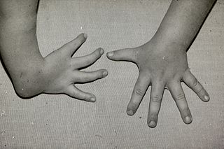
Ectrodactyly, split hand, or cleft hand involves the deficiency or absence of one or more central digits of the hand or foot and is also known as split hand/split foot malformation (SHFM). The hands and feet of people with ectrodactyly (ectrodactyls) are often described as "claw-like" and may include only the thumb and one finger with similar abnormalities of the feet.
Radial dysplasia, also known as radial club hand or radial longitudinal deficiency, is a congenital difference occurring in a longitudinal direction resulting in radial deviation of the wrist and shortening of the forearm. It can occur in different ways, from a minor anomaly to complete absence of the radius, radial side of the carpal bones and thumb. Hypoplasia of the distal humerus may be present as well and can lead to stiffness of the elbow. Radial deviation of the wrist is caused by lack of support to the carpus, radial deviation may be reinforced if forearm muscles are functioning poorly or have abnormal insertions. Although radial longitudinal deficiency is often bilateral, the extent of involvement is most often asymmetric.
Facial femoral syndrome is a rare congenital disorder. It is also known as femoral dysgenesis, bilateral femoral dysgenesis, bilateral-Robin anomaly and femoral hypoplasia-unusual facies syndrome. The main features of this disorder are underdeveloped thigh bones (femurs) and unusual facial features.
Hecht Scott syndrome is a rare genetic disease that causes congenital limb formation. The main characterisation is the aplasia or hypoplasia of bones of the limb. It is currently presenting in less than 1 in 1,000,000 newborns. It has been known to be more commonly present in males. It was first diagnosed in 2005 by Courtens et al. who recognised the malformations with his present case and four others that were similarly described in literature.

Van Den Berghe Dequeker syndrome, also known as ulnar hypoplasia-split foot syndrome is a very rare congenital limb malformation syndrome which is characterized by severe ulnar hypoplasia, absence of the index to pinky finger in both hands, and split-foot.
Gollop-Wolfgang complex is a very rare genetic disorder which is characterized by skeletal and digital anomalies.
References
- ↑ "Orphanet: Femur fibula ulna complex". Orpha.net. Retrieved 2011-10-26.
- ↑ Holmes, Lewis B. (2011), Common Malformations, Oxford University Press, p. 178, ISBN 978-0-19-513602-9