Related Research Articles

Keratitis is a condition in which the eye's cornea, the clear dome on the front surface of the eye, becomes inflamed. The condition is often marked by moderate to intense pain and usually involves any of the following symptoms: pain, impaired eyesight, photophobia, red eye and a 'gritty' sensation.

Dry eye syndrome (DES), also known as keratoconjunctivitis sicca (KCS), is the condition of having dry eyes. Other associated symptoms include irritation, redness, discharge, and easily fatigued eyes. Blurred vision may also occur. Symptoms range from mild and occasional to severe and continuous. Scarring of the cornea may occur in untreated cases.
Trichiasis is a medical term for abnormally positioned eyelashes that grow back toward the eye, touching the cornea or conjunctiva. This can be caused by infection, inflammation, autoimmune conditions, congenital defects, eyelid agenesis and trauma such as burns or eyelid injury. It is the leading cause of infectious blindness in the world.

Exophthalmos is a bulging of the eye anteriorly out of the orbit. Exophthalmos can be either bilateral or unilateral. Complete or partial dislocation from the orbit is also possible from trauma or swelling of surrounding tissue resulting from trauma.

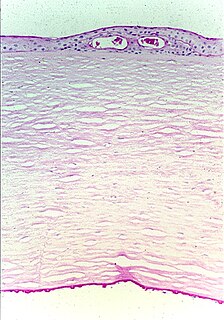
Fuchs dystrophy, also referred to as Fuchs endothelial corneal dystrophy (FECD) and Fuchs endothelial dystrophy (FED), is a slowly progressing corneal dystrophy that usually affects both eyes and is slightly more common in women than in men. Although early signs of Fuchs dystrophy are sometimes seen in people in their 30s and 40s, the disease rarely affects vision until people reach their 50s and 60s.

Entropion is a medical condition in which the eyelid folds inward. It is very uncomfortable, as the eyelashes continuously rub against the cornea causing irritation. Entropion is usually caused by genetic factors. This is different from when an extra fold of skin on the lower eyelid causes lashes to turn in towards the eye (epiblepharon). In epiblepharons, the eyelid margin itself is in the correct position, but the extra fold of skin causes the lashes to be misdirected. Entropion can also create secondary pain of the eye. The upper or lower eyelid can be involved, and one or both eyes may be affected. When entropion occurs in both eyes, this is known as "bilateral entropion". Repeated cases of trachoma infection may cause scarring of the inner eyelid, which may cause entropion. In human cases, this condition is most common to people over 60 years of age.
Ocular melanosis (OM) is a blue-gray and/or brown lesion of the conjunctiva that can be separated into benign conjunctival epithelial melanosis (BCEM) and primary acquired melanosis (PAM), of which the latter is considered a risk factor for uveal melanoma. The disease is caused by an increase of melanocytes in the iris, choroid, and surrounding structures. Overproduction of pigment by these cells can block the trabecular meshwork through which fluid drains from the eye. The increased fluid in the eye leads to increased pressure, which can lead to glaucoma. In humans, this is sometimes known as pigment dispersion syndrome.
Granulomatous meningoencephalitis (GME) is an inflammatory disease of the central nervous system (CNS) of dogs and, rarely, cats. It is a form of meningoencephalitis. GME is likely second only to encephalitis caused by canine distemper virus as the most common cause of inflammatory disease of the canine CNS. The disease is more common in female dogs of young and middle age. It has a rapid onset. The lesions of GME exist mainly in the white matter of the cerebrum, brainstem, cerebellum, and spinal cord. The cause is only known to be noninfectious and is considered at this time to be idiopathic. Because lesions resemble those seen in allergic meningoencephalitis, GME is thought to have an immune-mediated cause, but it is also thought that the disease may be based on an abnormal response to an infectious agent. One study searched for viral DNA from canine herpesvirus, canine adenovirus, and canine parvovirus in brain tissue from dogs with GME, necrotizing meningoencephalitis, and necrotizing leukoencephalitis, but failed to find any.

A distichia is an eyelash that arises from an abnormal part of the eyelid. This abnormality, attributed to a genetic mutation, is known to affect dogs and humans. Distichiae usually exit from the duct of the meibomian gland at the eyelid margin. They are usually multiple, and sometimes more than one arises from a duct. They can affect either the upper or lower eyelid and are usually bilateral. The lower eyelids of dogs usually have no eyelashes.

Ectopia lentis is a displacement or malposition of the eye's crystalline lens from its normal location. A partial dislocation of a lens is termed lens subluxation or subluxated lens; a complete dislocation of a lens is termed lens luxation or luxated lens.

A corneal ulcer, or ulcerative keratitis, is an inflammatory condition of the cornea involving loss of its outer layer. It is very common in dogs and is sometimes seen in cats. In veterinary medicine, the term corneal ulcer is a generic name for any condition involving the loss of the outer layer of the cornea, and as such is used to describe conditions with both inflammatory and traumatic causes.

Nuclear sclerosis is an age-related change in the density of the crystalline lens nucleus that occurs in all older animals. It is caused by compression of older lens fibers in the nucleus by new fiber formation. The denser construction of the nucleus causes it to scatter light. Although nuclear sclerosis may describe a type of early cataract in human medicine, in veterinary medicine the term is also known as lenticular sclerosis and describes a bluish-grey haziness at the nucleus that usually does not affect vision, except for unusually dense cases. Immature senile cataract has to be differentiated with nuclear sclerosis while making its diagnosis.
Retinal dysplasia is an eye disease affecting the retina of animals and, less commonly, humans. It is usually a nonprogressive disease and can be caused by viral infections, drugs, vitamin A deficiency, or genetic defects. Retinal dysplasia is characterized by folds or rosettes of the retinal tissue.
Collie eye anomaly (CEA) is a congenital, inherited, bilateral eye disease of dogs, which affects the retina, choroid, and sclera. It can be a mild disease or cause blindness. CEA is caused by a simple autosomal recessive gene defect. There is no treatment.

Persistent pupillary membrane (PPM) is a condition of the eye involving remnants of a fetal membrane that persist as strands of tissue crossing the pupil. The pupillary membrane in mammals exists in the fetus as a source of blood supply for the lens. It normally atrophies from the time of birth to the age of four to eight weeks. PPM occurs when this atrophy is incomplete. It generally does not cause any symptoms. The strands can connect to the cornea or lens, but most commonly to other parts of the iris. Attachment to the cornea can cause small corneal opacities, while attachment to the lens can cause small cataracts. Using topical atropine to dilate the pupil may help break down PPMs.

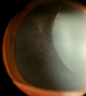
Synchysis scintillans is a degenerative condition of the eye resulting in liquefied vitreous humor and the accumulation of cholesterol crystals within the vitreous. It is also known as cholesterolosis bulbi. The vitreous liquifies in a process known as syneresis. Synchysis scintillans appears as small white floaters that freely move in the posterior part of the eye, giving a snow globe effect. It is most commonly seen in eyes that have suffered from a degenerative disease and are end-stage.

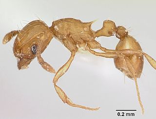
The electric ant, also known as the little fire ant, is a small, light to golden brown (ginger) social ant native to Central and South America, now spread to parts of Africa, Taiwan, North America, Puerto Rico, Israel, Cuba, and six Pacific Island groups plus north-eastern Australia (Cairns).

Corneal dystrophies are a group of diseases that affect the cornea in dogs.
Feline corneal sequestrum is the development of dark areas of dead tissue in the cornea of domestic cats. This disease is painful to the cat, although it develops slowly over a longer period of time. Cats will usually demonstrate teary eye(s), squinting or closing of the eye(s), and covering of the third eyelid.
Exposure keratopathy is medical condition affecting the cornea of eyes. It can lead to corneal ulceration and permanent loss of vision due to corneal opacity.
References
- 1 2 3 Roze, Maurice (2005). "Corneal Diseases in Cats". Proceedings of the 30th World Congress of the World Small Animal Veterinary Association. Retrieved 2007-03-23.
- 1 2 Gelatt, Kirk N.; Gilger, Brian C.; Kern, Thomas J. (2013). "Section IV: Special ophthalmology". Veterinary ophthalmology (5 ed.). Ames, Iowa: Wiley-Blackwell. pp. 1499–1500. ISBN 9780470960400.
- 1 2 Gelatt, Kirk N. (ed.) (1999). Veterinary Ophthalmology (3rd ed.). Lippincott, Williams & Wilkins. ISBN 0-683-30076-8.
{{cite book}}:|author=has generic name (help) - ↑ Theron, Leonard (2005). Wasmannia auropunctata linked keratopathy Hypothesis - The Polynesian Case. Doctorate in Veterinary Medicine Master (Thesis). hdl:2268/652.