Fluoro-jade stain

Last updated
Fluoro-jade B positive neurons in the rat cortex after injection of kainic acid KA fljb.jpg
Fluoro-jade B positive neurons in the rat cortex after injection of kainic acid

Fluoro-jade stain is a fluorochrome derived from fluorescein, and is commonly used in neuroscience disciplines to label degenerating neurons in ex vivo tissue of the central nervous system. The first fluoro-jade derivative was reported by Larry Schmued in 1997 as an alternative method from traditional methods for labeling degenerating neurons such as silver nitrate staining, H&E stain, or Nissl stain. [1] Fluoro-jade may be preferred to other degenerative stains due to simplicity of staining procedures and visual interpretation, which are common drawbacks of conventional degenerative stains. However, the mechanism by which fluoro-jade labels degenerating neurons is unknown thus creating some controversy to the actual physiological condition of the labeled cells.

Contents

Chemical properties

Currently, there are three fluoro-jade dyes (fluoro-jade, [1] fluoro-jade B, [2] and fluoro-jade C [3] ), all of which are anionic derivatives of fluorescein and highly acidic. [3] Specifically, fluoro-jade is a mixture of 5-carboxyfluorescein and 6-carboxyfluorescein disodium salts, whereas fluoro-jade B is a mixture of (1) trisodium 5-(6-hydroxy-3-oxo-3H-xanthen-9yl)benzene, 1,2,4 tricarboxylic acid, (2) disodium 2-(6-hydroxy-3-oxo-3H-xanthen-9yl)-5-(2,4-dihydroxybenzol)terepthalic acid, and (3) disodium 2,5-bis(6-hydroxy-3-oxo-3H-xanthen-9yl)terepthalic acid. [4] All three fluoro-jade species have similar excitation and emission profiles as fluorescein (excitation: 495 nm; emission:521 nm) and thus can be visualized using a fluorescein/FITC filter. The newer dyes, fluoro-jade B and fluoro-jade C, were developed to improve signal to noise ratio, therefore creating superior compounds for visualizing finer neuronal morphology including dendrites, axons and nerve terminals.

General staining techniques

Nearly all processed tissue is compatible with fluoro-jade stain including tissue from rodents (mice and rats), non-human primates and humans. Mounted tissue is stepwise rehydrated with decreasing concentrations of alcohol. Potassium permanganate may be used to decrease background staining and protect tissue from fading and photo bleaching. Fluoro-jade is highly soluble in water and is therefore first dissolved in distilled water. In order to be specific for degenerating neurons, fluoro-jade must be used in an acidic environment, therefore fluoro-jade is further diluted in glacial acetic acid. Additional water washes should be used to rinse tissue before drying and coverslipping. [1]

Fluoro-jade staining procedures are flexible and therefore can be adapted to be compatible with other staining techniques such as immunohistochemistry. Several modifications to the general procedures can be made such as reducing potassium permanganate incubations to avoid disrupting immunofluorescent labeling. Background can be decreased by alternative methods such as lowering staining temperature or decreasing fluoro-jade concentration, which may be more compatible with other labeling techniques. [1] Such alterations, however should be determined empirically to optimize specific experimental conditions.

In addition to staining tissue from treated subjects, positive and negative controls should be included to ensure method specificity and validity. Typically tissue from untreated control subjects is included to show specificity for degenerating neurons as fluoro-jade should not stain non-degenerating tissue. Additionally, a positive control is included to ensure validity of staining procedures; to show that degenerating neurons will be stained with fluoro-jade. An acceptable positive control includes neurodegenerative tissue from subjects where fluoro-jade has already been validated, such as kainic acid treated animals.

Analysis

Fluoro-jade–stained tissue can be visualized under an epifluorescent microscope using a filter system designed for fluorescein or fluorescein isothiocyanate (FITC) (excitation: 495 nm; emission:521 nm). Multiple morphological features can be detected using fluoro-jade stain including cell bodies, dendrites, axons, and axon terminals. [1] Even though all fluoro-jade derivatives can detect these specific morphological features, the newer derivatives (fluoro-jade B and fluoro-jade C) have greater specificity and resolution and therefore are superior in detecting finer morphological features. [3] Fluoro-jade is typically quantified in every 6th–12th 40 nm section within the region of interest and expressed as cells/section. Alternatively, stereological procedures may be used to estimate total fluoro-jade positive cells within the defined region.

Technique validation

As the mechanism of fluoro-jade labeling is unknown, correlative analysis with traditional neuronal degeneration stains was used to validate this technique. Initially fluoro-jade staining was compared with H&E and de Olmos's cupric-staining methodologies in a variety of neurotoxic models of neurodegeneration such as injection of kainic acid, MPTP, or multivalent metals. Each of these neurotoxic insults produce brain-region specific neuronal degeneration and thus could be used to the determine specificity of fluoro-jade. Indeed, these studies demonstrated that fluoro-jade consistently reproduced insult specific staining patterns of neuronal degeneration that were identical to H&E and de Olmos's cupric staining patterns after the same neurotoxic insults. These results suggest that fluoro-jade is a reliable marker of neurodegeneration. [1]

Larry Schmued suggests that a basic "death molecule" is expressed by damaged cells and that the highly anionic and acidic fluoro-jade may be specific for this target. [1] Further supporting the validity of fluoro-jade stain and this hypothesis is the work of Auer et al., [5] who demonstrated that another anionic dye, fuchsine acid, could successfully bind to damaged neurons after a hyperglycemic insult presumably by the same electrostatic mechanism as fluoro-jade. These neurons were characterized by cell death morphology including condensed chromatin, a disrupted plasma membrane, and a disrupted nuclear membrane.

Although fluoro-jade and de Olmos's silver stain have the same pattern of staining in models of neurotoxicity, there are inherent differences between the two methodologies that may have physiological implications. For example, a time course analysis of neuronal degeneration between the two techniques shows that silver stain is evident earlier after a neurotoxic insult which may suggest that fluoro-jade is specific for a later, more committed stage of the degenerative process. [6]

Applications

Traumatic brain injury [7] [8]

Spinal cord injury [9]

Alzheimer's disease [10]

Aging [11]

Stroke [12] [13]

Epilepsy [6]

Alcoholism [14]

Drug abuse [15]

Other

Fluoro-jade may also be useful for other applications other than labeling degeneration neurons of the brain. Several reports have demonstrated that fluoro-jade is also useful in detecting glia, specifically reactive astroglia [16] and microglia. [17] Thus fluoro-jade may be used to assess glial responses associated with neurotoxicity. Additionally, other studies demonstrate that fluoro-jade can also label neurons outside the CNS such as neurons of the dorsal root ganglia. [18] Finally, fluoro-jade may find use in non-neuronal systems as investigators have reported its use to assess cell death in renal tubular epithelial cells, in vitro [19] and in vivo. [20]

Related Research Articles

<span class="mw-page-title-main">Neurotoxin</span> Toxin harmful to nervous tissue

Neurotoxins are toxins that are destructive to nerve tissue. Neurotoxins are an extensive class of exogenous chemical neurological insults that can adversely affect function in both developing and mature nervous tissue. The term can also be used to classify endogenous compounds, which, when abnormally contacted, can prove neurologically toxic. Though neurotoxins are often neurologically destructive, their ability to specifically target neural components is important in the study of nervous systems. Common examples of neurotoxins include lead, ethanol, glutamate, nitric oxide, botulinum toxin, tetanus toxin, and tetrodotoxin. Some substances such as nitric oxide and glutamate are in fact essential for proper function of the body and only exert neurotoxic effects at excessive concentrations.

<span class="mw-page-title-main">Glia</span> Support cells in the nervous system

Glia, also called glial cells(gliocytes) or neuroglia, are non-neuronal cells in the central nervous system (brain and spinal cord) and the peripheral nervous system that do not produce electrical impulses. The neuroglia make up more than one half the volume of neural tissue in our body. They maintain homeostasis, form myelin in the peripheral nervous system, and provide support and protection for neurons. In the central nervous system, glial cells include oligodendrocytes, astrocytes, ependymal cells, and microglia, and in the peripheral nervous system they include Schwann cells and satellite cells.

<span class="mw-page-title-main">Haemodynamic response</span>

In haemodynamics, the body must respond to physical activities, external temperature, and other factors by homeostatically adjusting its blood flow to deliver nutrients such as oxygen and glucose to stressed tissues and allow them to function. Haemodynamic response (HR) allows the rapid delivery of blood to active neuronal tissues. The brain consumes large amounts of energy but does not have a reservoir of stored energy substrates. Since higher processes in the brain occur almost constantly, cerebral blood flow is essential for the maintenance of neurons, astrocytes, and other cells of the brain. This coupling between neuronal activity and blood flow is also referred to as neurovascular coupling.

<span class="mw-page-title-main">Astrocyte</span> Type of brain cell

Astrocytes, also known collectively as astroglia, are characteristic star-shaped glial cells in the brain and spinal cord. They perform many functions, including biochemical control of endothelial cells that form the blood–brain barrier, provision of nutrients to the nervous tissue, maintenance of extracellular ion balance, regulation of cerebral blood flow, and a role in the repair and scarring process of the brain and spinal cord following infection and traumatic injuries. The proportion of astrocytes in the brain is not well defined; depending on the counting technique used, studies have found that the astrocyte proportion varies by region and ranges from 20% to around 40% of all glia. Another study reports that astrocytes are the most numerous cell type in the brain. Astrocytes are the major source of cholesterol in the central nervous system. Apolipoprotein E transports cholesterol from astrocytes to neurons and other glial cells, regulating cell signaling in the brain. Astrocytes in humans are more than twenty times larger than in rodent brains, and make contact with more than ten times the number of synapses.

<span class="mw-page-title-main">Astrogliosis</span> Increase in astrocytes in response to brain injury

Astrogliosis is an abnormal increase in the number of astrocytes due to the destruction of nearby neurons from central nervous system (CNS) trauma, infection, ischemia, stroke, autoimmune responses or neurodegenerative disease. In healthy neural tissue, astrocytes play critical roles in energy provision, regulation of blood flow, homeostasis of extracellular fluid, homeostasis of ions and transmitters, regulation of synapse function and synaptic remodeling. Astrogliosis changes the molecular expression and morphology of astrocytes, in response to infection for example, in severe cases causing glial scar formation that may inhibit axon regeneration.

<span class="mw-page-title-main">Glial fibrillary acidic protein</span> Type III intermediate filament protein

Glial fibrillary acidic protein (GFAP) is a protein that is encoded by the GFAP gene in humans. It is a type III intermediate filament (IF) protein that is expressed by numerous cell types of the central nervous system (CNS), including astrocytes and ependymal cells during development. GFAP has also been found to be expressed in glomeruli and peritubular fibroblasts taken from rat kidneys, Leydig cells of the testis in both hamsters and humans, human keratinocytes, human osteocytes and chondrocytes and stellate cells of the pancreas and liver in rats.

<span class="mw-page-title-main">Microglia</span> Glial cell located throughout the brain and spinal cord

Microglia are a type of neuroglia located throughout the brain and spinal cord. Microglia account for about 10-15% of cells found within the brain. As the resident macrophage cells, they act as the first and main form of active immune defense in the central nervous system (CNS). Microglia are distributed in large non-overlapping regions throughout the CNS. Microglia are key cells in overall brain maintenance—they are constantly scavenging the CNS for plaques, damaged or unnecessary neurons and synapses, and infectious agents. Since these processes must be efficient to prevent potentially fatal damage, microglia are extremely sensitive to even small pathological changes in the CNS. This sensitivity is achieved in part by the presence of unique potassium channels that respond to even small changes in extracellular potassium. Recent evidence shows that microglia are also key players in the sustainment of normal brain functions under healthy conditions. Microglia also constantly monitor neuronal functions through direct somatic contacts and exert neuroprotective effects when needed.

Neuroprotection refers to the relative preservation of neuronal structure and/or function. In the case of an ongoing insult the relative preservation of neuronal integrity implies a reduction in the rate of neuronal loss over time, which can be expressed as a differential equation. It is a widely explored treatment option for many central nervous system (CNS) disorders including neurodegenerative diseases, stroke, traumatic brain injury, spinal cord injury, and acute management of neurotoxin consumption. Neuroprotection aims to prevent or slow disease progression and secondary injuries by halting or at least slowing the loss of neurons. Despite differences in symptoms or injuries associated with CNS disorders, many of the mechanisms behind neurodegeneration are the same. Common mechanisms of neuronal injury include decreased delivery of oxygen and glucose to the brain, energy failure, increased levels in oxidative stress, mitochondrial dysfunction, excitotoxicity, inflammatory changes, iron accumulation, and protein aggregation. Of these mechanisms, neuroprotective treatments often target oxidative stress and excitotoxicity—both of which are highly associated with CNS disorders. Not only can oxidative stress and excitotoxicity trigger neuron cell death but when combined they have synergistic effects that cause even more degradation than on their own. Thus limiting excitotoxicity and oxidative stress is a very important aspect of neuroprotection. Common neuroprotective treatments are glutamate antagonists and antioxidants, which aim to limit excitotoxicity and oxidative stress respectively.

<span class="mw-page-title-main">Rostral migratory stream</span> One path neural stem cells take to reach the olfactory bulb


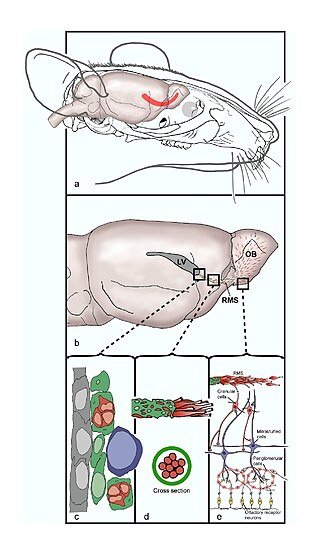
The rostral migratory stream (RMS) is a specialized migratory route found in the brain of some animals along which neuronal precursors that originated in the subventricular zone (SVZ) of the brain migrate to reach the main olfactory bulb (OB). The importance of the RMS lies in its ability to refine and even change an animal's sensitivity to smells, which explains its importance and larger size in the rodent brain as compared to the human brain, as our olfactory sense is not as developed. This pathway has been studied in the rodent, rabbit, and both the squirrel monkey and rhesus monkey. When the neurons reach the OB they differentiate into GABAergic interneurons as they are integrated into either the granule cell layer or periglomerular layer.

<span class="mw-page-title-main">Neuroimmune system</span>

The neuroimmune system is a system of structures and processes involving the biochemical and electrophysiological interactions between the nervous system and immune system which protect neurons from pathogens. It serves to protect neurons against disease by maintaining selectively permeable barriers, mediating neuroinflammation and wound healing in damaged neurons, and mobilizing host defenses against pathogens.

Glutamate transporters are a family of neurotransmitter transporter proteins that move glutamate – the principal excitatory neurotransmitter – across a membrane. The family of glutamate transporters is composed of two primary subclasses: the excitatory amino acid transporter (EAAT) family and vesicular glutamate transporter (VGLUT) family. In the brain, EAATs remove glutamate from the synaptic cleft and extrasynaptic sites via glutamate reuptake into glial cells and neurons, while VGLUTs move glutamate from the cell cytoplasm into synaptic vesicles. Glutamate transporters also transport aspartate and are present in virtually all peripheral tissues, including the heart, liver, testes, and bone. They exhibit stereoselectivity for L-glutamate but transport both L-aspartate and D-aspartate.

Gliosis is a nonspecific reactive change of glial cells in response to damage to the central nervous system (CNS). In most cases, gliosis involves the proliferation or hypertrophy of several different types of glial cells, including astrocytes, microglia, and oligodendrocytes. In its most extreme form, the proliferation associated with gliosis leads to the formation of a glial scar.

Neural stem cells (NSCs) are self-renewing, multipotent cells that firstly generate the radial glial progenitor cells that generate the neurons and glia of the nervous system of all animals during embryonic development. Some neural progenitor stem cells persist in highly restricted regions in the adult vertebrate brain and continue to produce neurons throughout life. Differences in the size of the central nervous system are among the most important distinctions between the species and thus mutations in the genes that regulate the size of the neural stem cell compartment are among the most important drivers of vertebrate evolution.

<span class="mw-page-title-main">Radial glial cell</span> Bipolar-shaped progenitor cells of all neurons in the cerebral cortex and some glia

Radial glial cells, or radial glial progenitor cells (RGPs), are bipolar-shaped progenitor cells that are responsible for producing all of the neurons in the cerebral cortex. RGPs also produce certain lineages of glia, including astrocytes and oligodendrocytes. Their cell bodies (somata) reside in the embryonic ventricular zone, which lies next to the developing ventricular system.

<span class="mw-page-title-main">Subventricular zone</span> Region outside each lateral ventricle of the brain

The subventricular zone (SVZ) is a region situated on the outside wall of each lateral ventricle of the vertebrate brain. It is present in both the embryonic and adult brain. In embryonic life, the SVZ refers to a secondary proliferative zone containing neural progenitor cells, which divide to produce neurons in the process of neurogenesis. The primary neural stem cells of the brain and spinal cord, termed radial glial cells, instead reside in the ventricular zone (VZ).

Neuroregeneration refers to the regrowth or repair of nervous tissues, cells or cell products. Such mechanisms may include generation of new neurons, glia, axons, myelin, or synapses. Neuroregeneration differs between the peripheral nervous system (PNS) and the central nervous system (CNS) by the functional mechanisms involved, especially in the extent and speed of repair. When an axon is damaged, the distal segment undergoes Wallerian degeneration, losing its myelin sheath. The proximal segment can either die by apoptosis or undergo the chromatolytic reaction, which is an attempt at repair. In the CNS, synaptic stripping occurs as glial foot processes invade the dead synapse.

<span class="mw-page-title-main">Quinolinic acid</span> Dicarboxylic acid with pyridine backbone

Quinolinic acid, also known as pyridine-2,3-dicarboxylic acid, is a dicarboxylic acid with a pyridine backbone. It is a colorless solid. It is the biosynthetic precursor to niacin.

Transneuronal degeneration is the death of neurons resulting from the disruption of input from or output to other nearby neurons. It is an active excitotoxic process when a neuron is overstimulated by a neurotransmitter causing the dysfunction of that neuron which drives neighboring neurons into metabolic deficit, resulting in rapid, widespread loss of neurons. This can be either anterograde or retrograde, indicating the direction of the degeneration relative to the original site of damage. There are varying causes for transneuronal degeneration such as brain lesions, disconnection syndromes, respiratory chain deficient neuron interaction, and lobectomies. Although there are different causes, transneuronal degeneration generally results in the same effects to varying degrees. Transneuronal degeneration is thought to be linked to a number of diseases, most notably Huntington's disease and Alzheimer's disease, and researchers recently have been performing experiments with monkeys and rats, monitoring lesions in different parts of the body to study more closely how exactly the process works.

Endogenous regeneration in the brain is the ability of cells to engage in the repair and regeneration process. While the brain has a limited capacity for regeneration, endogenous neural stem cells, as well as numerous pro-regenerative molecules, can participate in replacing and repairing damaged or diseased neurons and glial cells. Another benefit that can be achieved by using endogenous regeneration could be avoiding an immune response from the host.

<span class="mw-page-title-main">Neuronal lineage marker</span> Endogenous tag expressed in different cells along neurogenesis and differentiated cells

A neuronal lineage marker is an endogenous tag that is expressed in different cells along neurogenesis and differentiated cells such as neurons. It allows detection and identification of cells by using different techniques. A neuronal lineage marker can be either DNA, mRNA or RNA expressed in a cell of interest. It can also be a protein tag, as a partial protein, a protein or an epitope that discriminates between different cell types or different states of a common cell. An ideal marker is specific to a given cell type in normal conditions and/or during injury. Cell markers are very valuable tools for examining the function of cells in normal conditions as well as during disease. The discovery of various proteins specific to certain cells led to the production of cell-type-specific antibodies that have been used to identify cells.

References

  1. 1 2 3 4 5 6 7 Schmued LC; Albertson C; Slikker Jr. W (1997). "Fluoro-Jade: a novel fluorochrome for the sensitive and reliable histochemical localization of neuronal degeneration". Brain Research . 751 (1): 37–46. doi:10.1016/S0006-8993(96)01387-X. PMID   9098566.
  2. Schmued LC, Hopkins KJ (August 2000). "Fluoro-Jade B: a high affinity fluorescent marker for the localization of neuronal degeneration". Brain Research . 874 (2): 123–30. doi:10.1016/S0006-8993(00)02513-0. PMID   10960596.
  3. 1 2 3 Schmued LC, Stowers CC, Scallet AC, Xu L (February 2005). "Fluoro-Jade C results in ultra high resolution and contrast labeling of degenerating neurons". Brain Research . 1035 (1): 24–31. doi:10.1016/j.brainres.2004.11.054. PMID   15713273.
  4. Sarkar, Sumit; Larry Schmued (2011). "11". In Brad Bolon; Mark Butt (eds.). Fundamental Neuropathology for Pathologists and Toxicologists: Principles and Techniques. John Wiley & Sons. p. Chapter 11. ISBN   9781118002230.
  5. Auer R. N.; Kalimo H.; Olsson Y.; Siesjo¨ B. K. (1985). "The temporal evolution of hypoglycemic brain damage. I. Light- and electron-microscopic findings in the rat cerebral cortex". Acta Neuropathol. 67 (1–2): 13–24. doi:10.1007/bf00688120. PMID   4024866.
  6. 1 2 Poirier JL, Capek R, De Koninck Y (2000). "Differential progression of Dark Neuron and Fluoro-Jade labelling in the rat hippocampus following pilocarpine-induced status epilepticus". Neuroscience . 97 (1): 59–68. doi:10.1016/S0306-4522(00)00026-9. PMID   10771339.
  7. Anderson, K.J.; Miller, K.M.; Fugaccia, I.; Scheff, S.W. (2005). "Regional distribution of Fluoro-Jade B staining in the hippocampus following traumatic brain injury". Experimental Neurology . 193 (1): 125–130. doi:10.1016/j.expneurol.2004.11.025. PMID   15817271.
  8. Hellmich, H.L.; Capra, B.; Eidson, K.; Garcia, J.; Kennedy, D.; Uchida, T.; Parsley, M.; Cowar, J.; DeWitt, D.S.; Prough, D.S. (2005). "Dose-dependent neuronal injury after traumatic brain injury". Brain Research . 1044 (2): 144–154. doi:10.1016/j.brainres.2005.02.054. PMID   15885213.
  9. Anderson, K.J.; Fugaccia, I.; Scheff, S.W. (2003). "Fluoro-Jade B stains quiescent and reactive astrocytes in the rodent spinal cord". J. Neurotrauma . 20 (11): 1223–1231. doi:10.1089/089771503770802899. PMID   14651809.
  10. Damjanac, M.; Rioux Bilan, A.; Barrier, L.; Pontcharraud, R.; Anne, C.; Hugon, J.; Page, G. (2007). "Fluoro-Jade B staining as useful tool to identify activated microglia and astrocytes in a mouse transgenic model of Alzheimer's Disease". Brain Research . 1128 (1): 40–49. doi:10.1016/j.brainres.2006.05.050. PMID   17125750.
  11. Colombo, J. A.; Puissant, V.I. (2002). "Fluoro Jade Stains Early and Reactive Astroglia in the Primate Cerebral Cortex". J Histochem Cytochem. 50 (8): 1135–1137. doi: 10.1177/002215540205000815 .
  12. Duckworth, E.A.M.; Butler, T.; Collier, L.; Collier, S.; Pennypacker, K. R. (2006). "NF-kappaB protects neurons from ischemia injury after middle cerebral artery occlusion in mice". Brain Research. 1088 (1): 167–175. doi:10.1016/j.brainres.2006.02.103. PMID   16630592.
  13. Duckworth, E.A.M.; Bulter, T.L.; De Mesquita, D.; Collier, S.N.; Collier, L.; Pennypacker, K. R. (2005). "Temporary focal ischemia in the mouse: Technical aspects and patterns of Fluoro-Jade evident neurodegeneration". Brain Research . 1042 (1): 29–36. doi:10.1016/j.brainres.2005.02.021. PMID   15823250.
  14. Obernier, J.A.; Bouldin, J.A.; Crews, F.T. (2002). "Binge ethanol exposure in adult rats causes necrotic cell death". Alcohol Clin Exp Res . 26 (4): 547–557. doi:10.1111/j.1530-0277.2002.tb02573.x. PMID   11981132.
  15. Eisch, A.J.; Schmued, L.C.; Marshall, J.F. (1998). "Characterizing cortical neuron injury with fluoro-jade labeling after a neurotoxic regimen of methamphetamine". Synapse . 30 (3): 329–333. doi:10.1002/(SICI)1098-2396(199811)30:3<329::AID-SYN10>3.0.CO;2-V. PMID   9776136.
  16. Anderson, K.J.; Isabella, F.; Scheff, S.W. (2003). "Fluoro-jade stains quiescent and reactive astrocytes in the rodent spinal cord". J. Neurotrauma . 20 (11): 1223–1231. doi:10.1089/089771503770802899. PMID   14651809.
  17. Damjanac, M.; Rioux Bilan, A.; Barrier, L.; Pontcharraud, R.; Anne, C.; Hugon, J.; Page, G. (2007). "Fluro-jade B staining as useful tool to identify activated microglia and astrocytes in a mouse transgenic model of Alzheimer's disease". Brain Research . 1128 (1): 40–49. doi:10.1016/j.brainres.2006.05.050. PMID   17125750.
  18. Hornfelt, M.; Edstrom, A.; Ekstrom, P.A.R. (1999). "Upregulation of cytosolic phospholipase A2 correlates with apoptosis in mouse superior cervical and dorsal root ganglia neurons". Neuroscience Letters . 265 (2): 87–90. doi:10.1016/S0304-3940(99)00046-4. PMID   10327175.
  19. Gu, Qiang; Lantz, Susan; Rosas-Hernandez, Hector; Cuevas, Elvis; Ali, Syed F.; Paule, Merle G.; Sarkar, Sumit (June 2014). "In vitro detection of cytotoxicity using FluoroJade-C". Toxicology in Vitro. 28 (4): 469–472. doi:10.1016/j.tiv.2014.01.007. PMID   24462471.
  20. Ikeda, Mizuko; Wakasaki, Rumie; Schenning, Katie J.; Swide, Thomas; Lee, Jeong Heon; Miller, M. Bernie; Choi, Hak Soo; Anderson, Sharon; Hutchens, Michael P. (April 2017). "Determination of renal function and injury using near-infrared fluorimetry in experimental cardiorenal syndrome". American Journal of Physiology. Renal Physiology. 312 (4): F629–F639. doi:10.1152/ajprenal.00573.2016. PMC   5407071 . PMID   28077373.