Related Research Articles

A finger is a prominent digit on the forelimbs of most tetrapod vertebrate animals, especially those with prehensile extremities such as humans and other primates. Most tetrapods have five digits (pentadactyly), and short digits are typically referred to as toes, while those that are notably elongated are called fingers. In humans, the fingers are flexibly articulated and opposable, serving as an important organ of tactile sensation and fine movements, which are crucial to the dexterity of the hands and the ability to grasp and manipulate objects.
The flexor digitorum profundus is a muscle in the forearm of humans that flexes the fingers. It is considered an extrinsic hand muscle because it acts on the hand while its muscle belly is located in the forearm.

Psoriatic arthritis (PsA) is a long-term inflammatory arthritis that occurs in people affected by the autoimmune disease psoriasis. The classic feature of psoriatic arthritis is swelling of entire fingers and toes with a sausage-like appearance. This often happens in association with changes to the nails such as small depressions in the nail (pitting), thickening of the nails, and detachment of the nail from the nailbed. Skin changes consistent with psoriasis frequently occur before the onset of psoriatic arthritis but psoriatic arthritis can precede the rash in 15% of affected individuals. It is classified as a type of seronegative spondyloarthropathy.

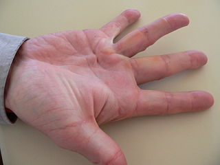
Dupuytren's contracture is a condition in which one or more fingers become permanently bent in a flexed position. It is named after Guillaume Dupuytren, who first described the underlying mechanism of action, followed by the first successful operation in 1831 and publication of the results in The Lancet in 1834. It usually begins as small, hard nodules just under the skin of the palm, then worsens over time until the fingers can no longer be fully straightened. While typically not painful, some aching or itching may be present. The ring finger followed by the little and middle fingers are most commonly affected. It can affect one or both hands. The condition can interfere with activities such as preparing food, writing, putting the hand in a tight pocket, putting on gloves, or shaking hands.
Sclerodactyly is a localized thickening and tightness of the skin of the fingers or toes that yields a characteristic claw-like appearance and spindle shape of the affected digits, and renders them immobile or of limited mobility. The thickened, discolored patches of skin are called morphea, and may involve connective tissue below the skin, as well as muscle and other tissues. Sclerodactyly is often preceded by months or even years by Raynaud's phenomenon when it is part of systemic scleroderma.

The extensor digitorum muscle is a muscle of the posterior forearm present in humans and other animals. It extends the medial four digits of the hand. Extensor digitorum is innervated by the posterior interosseous nerve, which is a branch of the radial nerve.
In human anatomy, the extensor pollicis longus muscle (EPL) is a skeletal muscle located dorsally on the forearm. It is much larger than the extensor pollicis brevis, the origin of which it partly covers and acts to stretch the thumb together with this muscle.

A mallet finger, also known as hammer finger or PLF finger or Hannan finger, is an extensor tendon injury at the farthest away finger joint. This results in the inability to extend the finger tip without pushing it. There is generally pain and bruising at the back side of the farthest away finger joint.

In human anatomy, the dorsal interossei (DI) are four muscles in the back of the hand that act to abduct (spread) the index, middle, and ring fingers away from hand's midline and assist in flexion at the metacarpophalangeal joints and extension at the interphalangeal joints of the index, middle and ring fingers.

In human anatomy, the abductor digiti minimi is a skeletal muscle situated on the ulnar border of the palm of the hand. It forms the ulnar border of the palm and its spindle-like shape defines the hypothenar eminence of the palm together with the skin, connective tissue, and fat surrounding it. Its main function is to pull the little finger away from the other fingers.

The interphalangeal joints of the hand are the hinge joints between the phalanges of the fingers that provide flexion towards the palm of the hand.

Boutonniere deformity is a deformed position of the fingers or toes, in which the joint nearest the knuckle is permanently bent toward the palm while the farthest joint is bent back away. Causes include injury, inflammatory conditions like rheumatoid arthritis, and genetic conditions like Ehlers-Danlos syndrome.

The interphalangeal joints of the foot are between the phalanx bones of the toes in the feet.

An ulnar claw, also known as claw hand or ‘Spinster’s Claw’, is a deformity or an abnormal attitude of the hand that develops due to ulnar nerve damage causing paralysis of the lumbricals. A claw hand presents with a hyperextension at the metacarpophalangeal joints and flexion at the proximal and distal interphalangeal joints of the 4th and 5th fingers. The patients with this condition can make a full fist but when they extend their fingers, the hand posture is referred to as claw hand. The ring- and little finger can usually not fully extend at the proximal interphalangeal joint (PIP).

A hand is a prehensile, multi-fingered appendage located at the end of the forearm or forelimb of primates such as humans, chimpanzees, monkeys, and lemurs. A few other vertebrates such as the koala are often described as having "hands" instead of paws on their front limbs. The raccoon is usually described as having "hands" though opposable thumbs are lacking.
Knuckle pads are circumscribed, keratotic, fibrous growths over the dorsa of the interphalangeal joints. They are described as well-defined, round, plaque-like, fibrous thickening that may develop at any age, and grow to be 10 to 15mm in diameter in the course of a few weeks or months, then go away over time.
Diabetic cheiroarthropathy, also known as Diabetic stiff hand syndrome or limited joint mobility syndrome, is a cutaneous condition characterized by waxy, thickened skin and limited joint mobility of the hands and fingers, leading to flexion contractures, a condition associated with diabetes mellitus and it is observed in roughly 30% of diabetic patients with longstanding disease. It can be a predictor for other diabetes-related complications and was one of the earliest known complications of diabetes, first documented in 1974.

The extrinsic extensor muscles of the hand are located in the back of the forearm and have long tendons connecting them to bones in the hand, where they exert their action. Extrinsic denotes their location outside the hand. Extensor denotes their action which is to extend, or open flat, joints in the hand. They include the extensor carpi radialis longus (ECRL), extensor carpi radialis brevis (ECRB), extensor digitorum (ED), extensor digiti minimi (EDM), extensor carpi ulnaris (ECU), abductor pollicis longus (APL), extensor pollicis brevis (EPB), extensor pollicis longus (EPL), and extensor indicis (EI).
Fiddler's neck is an occupational disease that affects violin and viola players.
The intense contact between a musical instrument and skin may exaggerate existing skin conditions or cause new skin conditions. Skin conditions like hyperhidrosis, lichen planus, psoriasis, eczema, and urticaria may be caused in instrumental musicians due to occupational exposure and stress. Allergic contact dermatitis and irritant contact dermatitis are the most common skin conditions seen in string musicians.
References
- ↑ Rapini, Ronald P.; Bolognia, Jean L.; Jorizzo, Joseph L. (2007). Dermatology: 2-Volume Set. St. Louis: Mosby. ISBN 978-1-4160-2999-1.
- 1 2 3 Rimmer, S.; Spielvogel, R. L. (1990). "Dermatologic problems of musicians". Journal of the American Academy of Dermatology. 22 (4): 657–663. doi:10.1016/0190-9622(90)70093-W. PMID 2138638.
- ↑ Garrod, A. E. (1904). "Concerning Pads upon the Finger Joints and their Clinical Relationships". British Medical Journal. 2 (2270): 8. doi:10.1136/bmj.2.2270.8. PMC 2354178 . PMID 20761632.
- 1 2 Bird, H. A. (1987). "Development of Garrod's pads in the fingers of a professional violinist". Annals of the Rheumatic Diseases. 46 (2): 169–170. doi:10.1136/ard.46.2.169. PMC 1002087 . PMID 3827341.
- ↑ Cutis. Technical Publishing Company. 1994. p. 160. Retrieved 13 November 2017.
- ↑ Young, Christopher J.; Gladman, Marc A. (2013). Examination Surgery: A Guide to Passing the Fellowship Examination in General Surgery. Elsevier Health Sciences. p. 213. ISBN 9780729541480 . Retrieved 13 November 2017.