
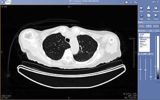
Radiography is an imaging technique using X-rays, gamma rays, or similar ionizing radiation and non-ionizing radiation to view the internal form of an object. Applications of radiography include medical and industrial radiography. Similar techniques are used in airport security,. To create an image in conventional radiography, a beam of X-rays is produced by an X-ray generator and it is projected towards the object. A certain amount of the X-rays or other radiation are absorbed by the object, dependent on the object's density and structural composition. The X-rays that pass through the object are captured behind the object by a detector. The generation of flat two-dimensional images by this technique is called projectional radiography. In computed tomography, an X-ray source and its associated detectors rotate around the subject, which itself moves through the conical X-ray beam produced. Any given point within the subject is crossed from many directions by many different beams at different times. Information regarding the attenuation of these beams is collated and subjected to computation to generate two-dimensional images on three planes which can be further processed to produce a three-dimensional image.

Radiology is the medical specialty that uses medical imaging to diagnose diseases and guide their treatment, within the bodies of humans and other animals. It began with radiography, but today it includes all imaging modalities, including those that use no ionizing electromagnetic radiation, as well as others that do, such as computed tomography (CT), fluoroscopy, and nuclear medicine including positron emission tomography (PET). Interventional radiology is the performance of usually minimally invasive medical procedures with the guidance of imaging technologies such as those mentioned above.

Medical imaging is the technique and process of imaging the interior of a body for clinical analysis and medical intervention, as well as visual representation of the function of some organs or tissues (physiology). Medical imaging seeks to reveal internal structures hidden by the skin and bones, as well as to diagnose and treat disease. Medical imaging also establishes a database of normal anatomy and physiology to make it possible to identify abnormalities. Although imaging of removed organs and tissues can be performed for medical reasons, such procedures are usually considered part of pathology instead of medical imaging.

Fluoroscopy, informally referred to as "fluoro", is an imaging technique that uses X-rays to obtain real-time moving images of the interior of an object. In its primary application of medical imaging, a fluoroscope allows a surgeon to see the internal structure and function of a patient, so that the pumping action of the heart or the motion of swallowing, for example, can be watched. This is useful for both diagnosis and therapy and occurs in general radiology, interventional radiology, and image-guided surgery.
Teleradiology is the transmission of radiological patient images from procedures such as x-rays photographs, Computed tomography (CT), and MRI imaging, from one location to another for the purposes of sharing studies with other radiologists and physicians. Teleradiology allows radiologists to provide services without actually having to be at the location of the patient. This is particularly important when a sub-specialist such as an MRI radiologist, neuroradiologist, pediatric radiologist, or musculoskeletal radiologist is needed, since these professionals are generally only located in large metropolitan areas working during daytime hours. Teleradiology allows for specialists to be available at all times.

Radiographers, also known as radiologic technologists, diagnostic radiographers and medical radiation technologists are healthcare professionals who specialise in the imaging of human anatomy for the diagnosis and treatment of pathology. Radiographers are infrequently, and almost always erroneously, known as x-ray technicians. In countries that use the title radiologic technologist they are often informally referred to as techs in the clinical environment; this phrase has emerged in popular culture such as television programmes. The term radiographer can also refer to a therapeutic radiographer, also known as a radiation therapist.

A timeline of United States inventions encompasses the ingenuity and innovative advancements of the United States within a historical context, dating from the Contemporary era to the present day, which have been achieved by inventors who are either native-born or naturalized citizens of the United States. Patent protection secures a person's right to his or her first-to-invent claim of the original invention in question, highlighted in Article I, Section 8, Clause 8 of the United States Constitution which gives the following enumerated power to the United States Congress:
To promote the Progress of Science and useful Arts, by securing for limited Times to Authors and Inventors the exclusive Right to their respective Writings and Discoveries.

Projectional radiography, also known as conventional radiography, is a form of radiography and medical imaging that produces two-dimensional images by X-ray radiation. The image acquisition is generally performed by radiographers, and the images are often examined by radiologists. Both the procedure and any resultant images are often simply called 'X-ray'. Plain radiography or roentgenography generally refers to projectional radiography. Plain radiography can also refer to radiography without a radiocontrast agent or radiography that generates single static images, as contrasted to fluoroscopy, which are technically also projectional.

An abdominal x-ray is an x-ray of the abdomen. It is sometimes abbreviated to AXR, or KUB.
Kakarla Subba Rao was an Indian radiologist who served as the first director of Nizam's Institute of Medical Sciences, Hyderabad. For his contributions to the field of medicine, Rao was conferred Padma Shri in 2000, the fourth highest civilian award by the Government of India. He was also the founder and president of the Telugu Association of North America.
Physician self-referral is a term describing the practice of a physician ordering tests on a patient that are performed by either the referring physician himself or a fellow faculty member from whom he receives financial compensation in return for the referral. Examples of self-referral include an internist performing an EKG, a surgeon suggesting an operation that he himself would perform, and a physician ordering imaging tests that would be done at a facility he owns or leases.

In medical imaging, an anti-scatter grid is a device for limiting the amount of scattered radiation reaching the detector, thereby improving the quality of diagnostic medical x-ray images. The grid is positioned on the opposite side of the patient from the x-ray source, and between the patient and the X-ray detector or film. Reducing the amount of scattered x-rays increases the image's contrast resolution, and consequently the visibility of soft tissues.
The International Day of Radiology (IDoR) is an annual event promoting the role of medical imaging in modern healthcare. It is celebrated on November 8 each year and coincides with the anniversary of the discovery of x-rays. It was first introduced in 2012, as a joint initiative of the European Society of Radiology (ESR), the Radiological Society of North America (RSNA), and the American College of Radiology (ACR). The International Day of Radiology is acknowledged and celebrated by nearly 200 national, sub-speciality, and related societies around the world. 'Radiographers Association of Madhya Pradesh(India)''' has celebrated this day since 1996 and the theme for this day was raised by '''Mr.Shivakant Vajpai''', Secretary of Madhya Pradesh Radiographers Association, also holding a designation of Radiation Safety Officer and Senior Radiographer in government of Madhya Pradesh, India.

Rudolph Nissen was a German surgeon who chaired surgery departments in Turkey, the United States and Switzerland. The Nissen fundoplication, a surgical procedure for the treatment of gastroesophageal reflux disease, is named after him.
Ronald J. Ross is a Cleveland, Ohio radiologist known for research on brain injury in professional and amateur boxers and for the first clinical use of nuclear magnetic resonance imaging on human patients. Ross is also credited with the first use of head and whole body computed tomography imaging (CT) in a private clinical setting in the United States.
Alice Ettinger was a prominent radiologist and professor of medicine. A native of Germany, Ettinger trained there before coming to the Tufts University School of Medicine. She had come for a visit to Boston to demonstrate the spot film imaging technique, and she decided to stay at Tufts permanently.
Cesare Gianturco was an Italian-American physician and one of the earliest contributors to the specialty of interventional radiology. After many years as the radiology chief at the Carle Clinic in Illinois and a faculty member at the University of Illinois College of Medicine, Gianturco moved to Houston, where he conducted research at MD Anderson Hospital.

In radiography, focal plane tomography is tomography by simultaneously moving the X-ray generator and X-ray detector so as to keep a consistent exposure of only the plane of interest during image acquisition. This was the main method of obtaining tomographs in medical imaging until the late-1970s. It has since been largely replaced by more advanced imaging techniques such as CT and MRI. It remains in use today in a few specialized applications, such as for acquiring orthopantomographs of the jaw in dental radiography.
Judy Yee is an American radiologist. She is the University Chair of Radiology at Montefiore and professor of radiology at Albert Einstein College of Medicine.
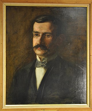
Charles Lester Leonard (1861–1913) was an American physician and X-ray pioneer. Leonard was the first radiologist at the Hospital of the University of Pennsylvania, founded the Philadelphia Roentgen Ray Society, and served as president of the American Roentgen Ray Society in 1904–1905. He was known as one of the foremost experts in urological X-ray diagnosis, and he was the first American physician to demonstrate kidney stone disease with X-rays.
