Related Research Articles

In mammalian oral anatomy, the canine teeth, also called cuspids, dogteeth, eye teeth, vampire teeth, or fangs, are the relatively long, pointed teeth. In the context of the upper jaw, they are also known as fangs. They can appear more flattened, however, causing them to resemble incisors and leading them to be called incisiform. They developed and are used primarily for firmly holding food in order to tear it apart, and occasionally as weapons. They are often the largest teeth in a mammal's mouth. Individuals of most species that develop them normally have four, two in the upper jaw and two in the lower, separated within each jaw by incisors; humans and dogs are examples. In most species, canines are the anterior-most teeth in the maxillary bone. The four canines in humans are the two upper maxillary canines and the two lower mandibular canines. They are specially prominent in dogs (Canidae), hence the name.

The gums or gingiva consist of the mucosal tissue that lies over the mandible and maxilla inside the mouth. Gum health and disease can have an effect on general health.

Cementoenamel junction (CEJ) is defined as the area of the union of cementum and enamel at the cervical region of the tooth. It is a slightly visible anatomical border identified on a tooth. It is the location where the enamel, which covers the anatomical crown of a tooth, and the cementum, which covers the anatomical root of a tooth, meet. Informally it is known as the neck of the tooth. The border created by these two dental tissues has much significance as it is usually the location where the gingiva attaches to a healthy tooth by fibers called the gingival fibers.
Dental explorers, also known as sickle probes, are tools found in the dental arsenal that are frequently utilised. The explorer is designed with a sharp tip at the end to improve tactile perception.
Periodontology or periodontics is the specialty of dentistry that studies supporting structures of teeth, as well as diseases and conditions that affect them. The supporting tissues are known as the periodontium, which includes the gingiva (gums), alveolar bone, cementum, and the periodontal ligament. A periodontist is a dentist that specializes in the prevention, diagnosis and treatment of periodontal disease and in the placement of dental implants.

The maxillary central incisor is a human tooth in the front upper jaw, or maxilla, and is usually the most visible of all teeth in the mouth. It is located mesial to the maxillary lateral incisor. As with all incisors, their function is for shearing or cutting food during mastication (chewing). There is typically a single cusp on each tooth, called an incisal ridge or incisal edge. Formation of these teeth begins at 14 weeks in utero for the deciduous (baby) set and 3–4 months of age for the permanent set.

The maxillary first molar is the human tooth located laterally from both the maxillary second premolars of the mouth but mesial from both maxillary second molars.

The mandibular central incisor is the tooth located on the jaw, adjacent to the midline of the face. It is mesial from both mandibular lateral incisors. As with all incisors, its function includes shearing or cutting food during mastication, commonly known as chewing. There are no cusps on the tooth. Instead, the surface area of the tooth used in eating is called an incisal ridge or incisal edge. Though the two are similar, there are some minor differences between the deciduous (baby) mandibular central incisor and that of the permanent mandibular central incisor. The mandibular central incisors are usually the first teeth to appear in the mouth, typically around the age of 6–8 months.

The mandibular second premolar is the tooth located distally from both the mandibular first premolars of the mouth but mesial from both mandibular first molars. The function of this premolar is assist the mandibular first molar during mastication, commonly known as chewing. Mandibular second premolars have three cusps. There is one large cusp on the buccal side of the tooth. The lingual cusps are well developed and functional. Therefore, whereas the mandibular first premolar resembles a small canine, the mandibular second premolar is more alike to the first molar. There are no deciduous (baby) mandibular premolars. Instead, the teeth that precede the permanent mandibular premolars are the deciduous mandibular molars.
The mandibular first molar or six-year molar is the tooth located distally from both the mandibular second premolars of the mouth but mesial from both mandibular second molars. It is located on the mandibular (lower) arch of the mouth, and generally opposes the maxillary (upper) first molars and the maxillary 2nd premolar in normal class I occlusion. The function of this molar is similar to that of all molars in regard to grinding being the principal action during mastication, commonly known as chewing. There are usually five well-developed cusps on mandibular first molars: two on the buccal, two lingual, and one distal. The shape of the developmental and supplementary grooves, on the occlusal surface, are described as being M-shaped. There are great differences between the deciduous (baby) mandibular molars and those of the permanent mandibular molars, even though their function are similar. The permanent mandibular molars are not considered to have any teeth that precede it. Despite being named molars, the deciduous molars are followed by permanent premolars.
Dental anatomy is a field of anatomy dedicated to the study of human tooth structures. The development, appearance, and classification of teeth fall within its purview. Tooth formation begins before birth, and the teeth's eventual morphology is dictated during this time. Dental anatomy is also a taxonomical science: it is concerned with the naming of teeth and the structures of which they are made, this information serving a practical purpose in dental treatment.
This is a list of definitions of commonly used terms of location and direction in dentistry. This set of terms provides orientation within the oral cavity, much as anatomical terms of location provide orientation throughout the body.
The gingival fibers are the connective tissue fibers that inhabit the gingival tissue adjacent to teeth and help hold the tissue firmly against the teeth. They are primarily composed of type I collagen, although type III fibers are also involved.
The interdental papilla, also known as the interdental gingiva, is the part of the gums (gingiva) that exists coronal to the free gingival margin on the mesial and distal surfaces of the teeth. The interdental papillae fill in the area between the teeth apical to their contact areas to prevent food impaction; they assume a conical shape for the anterior teeth and a blunted shape buccolingually for the posterior teeth.

Periodontal scalers are dental instruments used in the prophylactic and periodontal care of teeth, including scaling and root planing. The working ends come in a variety of shapes and sizes, but they are always narrow at the tip, so as to allow for access to narrow embrasure spaces between teeth. They differ from periodontal curettes, which possess a blunt tip.

Gingivitis is a non-destructive disease that causes inflammation of the gums; ulitis is an alternative term. The most common form of gingivitis, and the most common form of periodontal disease overall, is in response to bacterial biofilms that are attached to tooth surfaces, termed plaque-induced gingivitis. Most forms of gingivitis are plaque-induced.

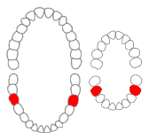
In dentistry, a furcation defect is bone loss, usually a result of periodontal disease, affecting the base of the root trunk of a tooth where two or more roots meet. The extent and configuration of the defect are factors in both diagnosis and treatment planning.
The syntette combines the benefits of a universal curette and a Gracey curette in one. It is a dental instrument used by dentists and dental hygienists to remove calculus subgingivally on the mesial and distal surfaces on all teeth throughout the mouth.

In human anatomy, the mouth is the first portion of the alimentary canal that receives food and produces saliva. The oral mucosa is the mucous membrane epithelium lining the inside of the mouth.
Periodontal surgery is a form of dental surgery that prevents or corrects anatomical, traumatic, developmental, or plaque-induced defects in the bone, gingiva, or alveolar mucosa. The objectives of this surgery include accessibility of instruments to the root surface, elimination of inflammation, creation of an oral environment for plaque control, periodontal disease control, oral hygiene maintenance, maintaining proper embrasure space, addressing gingiva–alveolar mucosa problems, and esthetic improvement. Surgical procedures include crown lengthening, frenectomy, and mucogingival flap surgery.
References
- 1 2 Brand RW, Isselhard DE (2014-03-12). Anatomy of Orofacial Structures - Enhanced 7th Edition - E-Book: A Comprehensive Approach. Elsevier Health Sciences. ISBN 9780323227728.
- ↑ Vandana KL, Haneet RK (September 2014). "Cementoenamel junction: An insight". Journal of Indian Society of Periodontology. 18 (5): 549–54. doi: 10.4103/0972-124X.142437 . PMC 4239741 . PMID 25425813.
- ↑ Yuodelis RA, Weaver JD, Sapkos S (January 1973). "Facial and lingual contours of artificial complete crown restorations and their effects on the periodontium". The Journal of Prosthetic Dentistry. 29 (1): 61–6. doi:10.1016/0022-3913(73)90140-6. PMID 4564800.