Hydrogel dressing

Last updated

Hydrogels are three-dimensional networks consisting of chemically or physically cross-linked hydrophilic polymers. [1] The insoluble hydrophilic structures absorb polar wound exudates and allow oxygen diffusion at the wound bed to accelerate healing. [2] Hydrogel dressings can be designed to prevent bacterial infection, retain moisture, promote optimum adhesion to tissues, and satisfy the basic requirements of biocompatibility. [1] [2] Hydrogel dressings can also be designed to respond to changes in the microenvironment at the wound bed. [3] Hydrogel dressings should promote an appropriate microenvironment for angiogenesis, recruitment of fibroblasts, and cellular proliferation. [2] [4]

Contents

Hydrogels respond elastically to applied stress; gels made from materials like collagen exhibit high toughness and low sliding friction, reducing damage from mechanical stress. [1] [5] Hydrogel dressings should possess mechanical and physical properties similar to the 3D microenvironment of the extracellular matrix of human skin. [6] Hydrogel wound dressings are designed to have a mechanism for application and removal which minimizes further trauma to tissues. [1]

Hydrogel dressings can be sorted into the categories: synthetic, natural, and hybrid. [1] Synthetic hydrogel dressings have been produced using biomimetic extracellular matrix nanofibers such as polyvinyl alcohol (PVA). [7] Self-assembling designer peptide hydrogels are another type of synthetic hydrogel in development. [8] Natural hydrogel dressings are further subdivided into either polysaccharide-based (e.g. alginates) or proteoglycan- and/or protein-based (e.g. collagen). [7] Hybrid hydrogel dressings incorporate synthetic nanoparticles and natural materials. [2]

Characteristics

Chemical characteristics

Hydrogel dressings exhibit chemical or physical cross-linking. Chemical cross-linking involved formation of covalent bonds between polymer chains. Chemically cross-linked hydrogel dressings are synthesized by chain-growth polymerization, step-growth polymerization, enzymes, or irradiation polymerization.[ citation needed ] Synthetic dressings incorporating nanoparticles such as PVA and polyethylene glycol (PEG) are assembled using chemical cross-linking mechanisms. [9] [10] Physically cross-linked hydrogel dressings are assembled via ionic interaction, hydrogen bonding, hydrophobic interactions, or crystallization.[ citation needed ] Physically cross-linked hydrogels disintegrate due to local changes in pH, ionic strength, and temperature. [3] Natural dressings incorporating polysaccharides and proteoglycans/proteins form a 3D network using physical cross-linking. [11] Hydrogel dressings mimic the cross-linked 3D network of extracellular matrix fibers in human skin. [1]

Hydrogels can be formed through a self-assembly process in which monomers diffuse in solution then form non covalent interactions.[ citation needed ] Hydrogels used in wound dressings can be self-assembled upon addition of divalent metal cations or electrically charged polysaccharides due to electrostatic interactions. [12] [13] Self-assembly via hydrophobic interactions can be induced in amphiphilic polysaccharides-based gels by addition of water; it can also be induced in non amphiphilic polysaccharide-based hydrogels by addition of hydrophobic grafts. [8] [12]

Cross-linking of soluble hydrophilic monomers forms a 3D insoluble netted structure which can incorporate a large amount of water. [14] The 3D polymeric network of hydrogels is highly hydrated with 90-99% water w/w; it is capable of binding many times more water molecules when assembled than in the uncross-linked state. [2] [3] Hydrogel dressings can absorb up to 600 times their initial amount of water, including fluid-based wound exudates. [2] [14] Hydrogels are effective biomaterials for wound dressings and tissue engineering because they exchange fluid, hydrating necrotic tissues. [2] [6] The absorption of secretions causes the hydrogel dressing to swell, expanding the cross links in the polymer chains. [6] The expanded 3D cross-linked network can irreversibly incorporate pathogens and detritus, thereby removing them from the wound. [6]

Some hydrogel dressings have intrinsic antimicrobial properties. Hydrogel dressings formed from antimicrobial peptides (AMPs) and chitosan have inherent antimicrobial activity. [15] [16] [17] The antimicrobial properties of hydrogel dressings can be enhanced by addition of metal nanoparticles, antibiotics, or other antimicrobial agents. [15] [18] [19] [20] Silver and gold nanoparticles can also be incorporated into hydrogel dressings to enhance antimicrobial activity. [15] Some hydrogel dressings have antibiotics such as ciprofloxacin and amoxicillin incorporated into their structure which are unloaded into the wound as fluid is exchanged. [15] [19] Some hydrogel dressings have incorporated stimuli-responsive nitric oxide-releasing agents and other antimicrobial agents. [15] [20]

Hydrogel dressings can adhere directly to the wound bed under normal physiological conditions via oxidation-reduction reactions of quinones. [2] [21] The adhesive properties of hydrogels have been shown to be enhanced by addition of positively charged microgels (MR) into the 3D matrix to increase electrostatic and hydrophobic interactions. [22]

Physical characteristics

Wound dressings should be stretchable to prevent tearing. Hai Lei et al. demonstrated that poor elasticity and hysteresis in naturally-derived protein-based hydrogels can be remedied by the addition of polyprotein cross-linkers. [23] The flexibility of hydrogels can also be enhanced by incorporating microgels into the matrix. [22] [24] Hydrogel dressings mimic the fibrous nature of native ECM to maintain cell-to-cell communication at the wound bed for tissue regeneration. [24]

Self-healing hydrogels automatically and reversibly repair damage done due to mechanical and chemical stress. [25] Self-healing mechanisms can involve "dynamic covalent bonding, non-covalent interactions" and mixed interactions. [25] Covalent interactions involved in self-healing include Schiff base formation and disulfide exchange. [25] Non-covalent interactions are generally less stable and make the hydrogel more sensitive to microenvironmental changes (e.g. pH, temperature). [25] Some hydrogel dressings are self-healing due to mixed mechanisms such as host-guest and protein-ligand interactions. [25]

Hydrogel dressings are available in sheet, amorphous, impregnated, or sprayable forms. [15] [26] [27] [28] [29] Sheet-form hydrogel dressings are non-adhesive against the wound and are effective in healing partial-thickness wounds. [26] Amorphous hydrogels are more effective in treatment of full-thickness wounds than sheet-form dressings because they can conform to the shape of the wound bed and they facilitate autolytic debridement. [27] Impregnated hydrogel dressings are dry dressings (e.g. gauzes) saturated with an amorphous hydrogel. [28] Sprayable hydrogel dressings are composed of amorphous hydrogels which rapidly increase in viscosity after application. [29] Sprayable hydrogels have also been shown to increase the penetration and efficacy of therapeutic agents. [2]

"Smart" hydrogel dressings

"Smart" hydrogels which are stimuli-responsive (i.e. thermoresponsive, bioresponsive, pH-responsive, photoresponsive, and redox-responsive) are also being produced. [3] pH-responsive hydrogel dressings which release growth factors and antibiotic agents as the pH of the wound increases from normal skin levels (pH 4–6) to internal levels (pH ~7.4). [30] Redox-responsive hydrogel dressings can be disintegrated on-demand by addition of a reducing agent. [31] Assembly of the 3D network of photoresponsive hydrogel dressings is initiated by UV radiation. [32] Thermoresponsive hydrogel dressings which exhibit temperature-dependent sol-gel transition and/or temperature-dependent drug release. [33] [34]

Applications

The efficacy of hydrogel dressings has been assessed on various wound types. There is some evidence to suggest that hydrogels are effective dressings for chronic wounds including pressure ulcers, diabetic ulcers, and venous ulcers although the results are uncertain. [35] [36] [37] [38] Hydrogels have been shown to accelerate healing in partial and full thickness burn wounds of varying size. [39] [40] [41] Other studies have shown that hydrogel dressings accelerate healing in radioactive skin injuries and dog bite wounds. [42] [43] [44] Hydrogel dressings decrease the healing time of traumatic skin injuries by an average 5.28 days and reduce the pain reported by patients. [42] [45] [46]

Types

Naturally-derived hydrogel dressings

Polysaccharide-based hydrogel dressings have been synthesized from polymers such as hyaluronic acid, chitin, chitosan, alginate, and agarose. [1] [40] [47] [48] [49] Naturally-derived protein/proteoglycan hydrogel dressings have been synthesized from polymers such as collagen, gelatin, kappa-carrageenan, and fibrin. [1] [49] [50] [51]  

Synthetic hydrogel dressings

Synthetic hydrogel dressings may be derived from synthetic polymers such as polyvinyl alcohol (PVA), poly(ethylene glycol) (PEG), polyurethane (PU), and poly(lactide-co-glycolide) (PLGA). [1] [52] [53] Synthetic hydrogel dressings may also be formed from designer peptides. [8] [54] Researchers are applying 3D printing to the synthesis of hydrogel dressings. [55] [56]

Biohybrid hydrogel dressings

Hydrogels may be modified to incorporate metal cations (e.g. copper (II)), degradable linkers (e.g. dextran), and adhesive functional groups (e.g. RGD). [1] Integrating biological derivatives into synthetic hydrogels allows producers to tailor binding affinities and specificity, mechanical properties, and stimuli-responsive properties. [1]

Related Research Articles

<span class="mw-page-title-main">Biopolymer</span> Polymer produced by a living organism

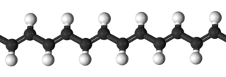
Biopolymers are natural polymers produced by the cells of living organisms. Like other polymers, biopolymers consist of monomeric units that are covalently bonded in chains to form larger molecules. There are three main classes of biopolymers, classified according to the monomers used and the structure of the biopolymer formed: polynucleotides, polypeptides, and polysaccharides. The Polynucleotides, RNA and DNA, are long polymers of nucleotides. Polypeptides include proteins and shorter polymers of amino acids; some major examples include collagen, actin, and fibrin. Polysaccharides are linear or branched chains of sugar carbohydrates; examples include starch, cellulose, and alginate. Other examples of biopolymers include natural rubbers, suberin and lignin, cutin and cutan, melanin, and polyhydroxyalkanoates (PHAs).

<span class="mw-page-title-main">Tissue engineering</span> Biomedical engineering discipline

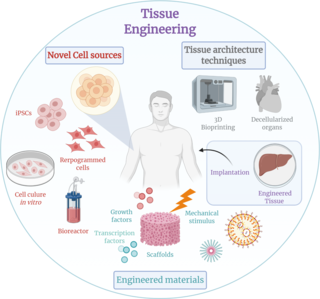
Tissue engineering is a biomedical engineering discipline that uses a combination of cells, engineering, materials methods, and suitable biochemical and physicochemical factors to restore, maintain, improve, or replace different types of biological tissues. Tissue engineering often involves the use of cells placed on tissue scaffolds in the formation of new viable tissue for a medical purpose but is not limited to applications involving cells and tissue scaffolds. While it was once categorized as a sub-field of biomaterials, having grown in scope and importance it can is considered as a field of its own.

<span class="mw-page-title-main">Hydrogel</span> Soft water-rich polymer gel

A hydrogel is a biphasic material, a mixture of porous, permeable solids and at least 10% by weight or volume of interstitial fluid composed completely or mainly by water. In hydrogels the porous permeable solid is a water insoluble three dimensional network of natural or synthetic polymers and a fluid, having absorbed a large amount of water or biological fluids. These properties underpin several applications, especially in the biomedical area. Many hydrogels are synthetic, but some are derived from nature. The term 'hydrogel' was coined in 1894.

<span class="mw-page-title-main">Chitosan</span> Chemical compound

Chitosan is a linear polysaccharide composed of randomly distributed β-(1→4)-linked D-glucosamine and N-acetyl-D-glucosamine. It is made by treating the chitin shells of shrimp and other crustaceans with an alkaline substance, such as sodium hydroxide.

<span class="mw-page-title-main">Alginic acid</span> Polysaccharide found in brown algae

Alginic acid, also called algin, is a naturally occurring, edible polysaccharide found in brown algae. It is hydrophilic and forms a viscous gum when hydrated. With metals such as sodium and calcium, its salts are known as alginates. Its colour ranges from white to yellowish-brown. It is sold in filamentous, granular, or powdered forms.
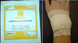
<span class="mw-page-title-main">Dressing (medical)</span> Sterile pad or compress applied to wounds

A dressing or compress is a sterile pad applied to a wound to promote healing and protect the wound from further harm. A dressing is designed to be in direct contact with the wound, as distinguished from a bandage, which is most often used to hold a dressing in place. Many modern dressings are self-adhesive.

In polymer chemistry and materials science, the term "polymer" refers to large molecules whose structure is composed of multiple repeating units. Supramolecular polymers are a new category of polymers that can potentially be used for material applications beyond the limits of conventional polymers. By definition, supramolecular polymers are polymeric arrays of monomeric units that are connected by reversible and highly directional secondary interactions–that is, non-covalent bonds. These non-covalent interactions include van der Waals interactions, hydrogen bonding, Coulomb or ionic interactions, π-π stacking, metal coordination, halogen bonding, chalcogen bonding, and host–guest interaction. The direction and strength of the interactions are precisely tuned so that the array of molecules behaves as a polymer in dilute and concentrated solution, as well as in the bulk.

Thiolated polymers – designated thiomers – are functional polymers used in biotechnology product development with the intention to prolong mucosal drug residence time and to enhance absorption of drugs. The name thiomer was coined by Andreas Bernkop-Schnürch in 2000. Thiomers have thiol bearing side chains. Sulfhydryl ligands of low molecular mass are covalently bound to a polymeric backbone consisting of mainly biodegradable polymers, such as chitosan, hyaluronic acid, cellulose derivatives, pullulan, starch, gelatin, polyacrylates, cyclodextrins, or silicones.

A nanogel is a polymer-based, crosslinked hydrogel particle on the sub-micron scale. These complex networks of polymers present a unique opportunity in the field of drug delivery at the intersection of nanoparticles and hydrogel synthesis. Nanogels can be natural, synthetic, or a combination of the two and have a high degree of tunability in terms of their size, shape, surface functionalization, and degradation mechanisms. Given these inherent characteristics in addition to their biocompatibility and capacity to encapsulate small drugs and molecules, nanogels are a promising strategy to treat disease and dysfunction by serving as delivery vehicles capable of navigating across challenging physiological barriers within the body. 

<span class="mw-page-title-main">Arginylglycylaspartic acid</span> Chemical compound

Arginylglycylaspartic acid (RGD) is the most common peptide motif responsible for cell adhesion to the extracellular matrix (ECM), found in species ranging from Drosophila to humans. Cell adhesion proteins called integrins recognize and bind to this sequence, which is found within many matrix proteins, including fibronectin, fibrinogen, vitronectin, osteopontin, and several other adhesive extracellular matrix proteins. The discovery of RGD and elucidation of how RGD binds to integrins has led to the development of a number of drugs and diagnostics, while the peptide itself is used ubiquitously in bioengineering. Depending on the application and the integrin targeted, RGD can be chemically modified or replaced by a similar peptide which promotes cell adhesion.

Smart polymers, stimuli-responsive polymers or functional polymers are high-performance polymers that change according to the environment they are in.

Polymers with the ability to kill or inhibit the growth of microorganisms such as bacteria, fungi, or viruses are classified as antimicrobial agents. This class of polymers consists of natural polymers with inherent antimicrobial activity and polymers modified to exhibit antimicrobial activity. Polymers are generally nonvolatile, chemically stable, and can be chemically and physically modified to display desired characteristics and antimicrobial activity. Antimicrobial polymers are a prime candidate for use in the food industry to prevent bacterial contamination and in water sanitation to inhibit the growth of microorganisms in drinking water.

<span class="mw-page-title-main">3D bioprinting</span> Utilization of 3D printing to fabricate biomedical parts

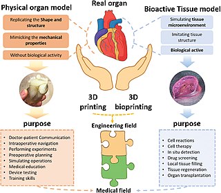
Three dimensional (3D) bioprinting is the utilization of 3D printing–like techniques to combine cells, growth factors, bio-inks, and/or biomaterials to fabricate biomedical parts that imitate natural tissue characteristics, form functional biofilms, and assist in the removal of pollutants. 3D bioprinting has uses in fields such as wastewater treatment, environmental remediation, and corrosion prevention. 3D bioprinting can produce functional biofilms which can assist in a variety of situations. The 3D bioprinted biofilms host functional microorganisms which can facilitate pollutant removal. Generally, 3D bioprinting can utilize a layer-by-layer method to deposit materials known as bio-inks to create tissue-like structures that are later used in various medical and tissue engineering fields. 3D bioprinting covers a broad range of bioprinting techniques and biomaterials. Currently, bioprinting can be used to print tissue and organ models to help research drugs and potential treatments. Nonetheless, translation of bioprinted living cellular constructs into clinical application is met with several issues due to the complexity and cell number needed to create functional organs. However, innovations span from bioprinting of extracellular matrix to mixing cells with hydrogels deposited layer by layer to produce the desired tissue. In addition, 3D bioprinting has begun to incorporate the printing of scaffolds which can be used to regenerate joints and ligaments.

Self-healing hydrogels are a specialized type of polymer hydrogel. A hydrogel is a macromolecular polymer gel constructed of a network of crosslinked polymer chains. Hydrogels are synthesized from hydrophilic monomers by either chain or step growth, along with a functional crosslinker to promote network formation. A net-like structure along with void imperfections enhance the hydrogel's ability to absorb large amounts of water via hydrogen bonding. As a result, hydrogels, self-healing alike, develop characteristic firm yet elastic mechanical properties. Self-healing refers to the spontaneous formation of new bonds when old bonds are broken within a material. The structure of the hydrogel along with electrostatic attraction forces drive new bond formation through reconstructive covalent dangling side chain or non-covalent hydrogen bonding. These flesh-like properties have motivated the research and development of self-healing hydrogels in fields such as reconstructive tissue engineering as scaffolding, as well as use in passive and preventive applications.

Nanocomposite hydrogels are nanomaterial-filled, hydrated, polymeric networks that exhibit higher elasticity and strength relative to traditionally made hydrogels. A range of natural and synthetic polymers are used to design nanocomposite network. By controlling the interactions between nanoparticles and polymer chains, a range of physical, chemical, and biological properties can be engineered. The combination of organic (polymer) and inorganic (clay) structure gives these hydrogels improved physical, chemical, electrical, biological, and swelling/de-swelling properties that cannot be achieved by either material alone. Inspired by flexible biological tissues, researchers incorporate carbon-based, polymeric, ceramic and/or metallic nanomaterials to give these hydrogels superior characteristics like optical properties and stimulus-sensitivity which can potentially be very helpful to medical and mechanical fields.

4-dimensional printing uses the same techniques of 3D printing through computer-programmed deposition of material in successive layers to create a three-dimensional object. However, in 4D printing, the resulting 3D shape is able to morph into different forms in response to environmental stimulus, with the 4th dimension being the time-dependent shape change after the printing. It is therefore a type of programmable matter, wherein after the fabrication process, the printed product reacts with parameters within the environment and changes its form accordingly.

Bio-inks are materials used to produce engineered/artificial live tissue using 3D printing. These inks are mostly composed of the cells that are being used, but are often used in tandem with additional materials that envelope the cells. The combination of cells and usually biopolymer gels are defined as a bio-ink. They must meet certain characteristics, including such as rheological, mechanical, biofunctional and biocompatibility properties, among others. Using bio-inks provides a high reproducibility and precise control over the fabricated constructs in an automated manner. These inks are considered as one of the most advanced tools for tissue engineering and regenerative medicine (TERM).

Ka Yee Christina Lee is Executive Vice President for Strategic Initiatives and the David Lee Shillinglaw Distinguished Service Professor in the Department of Chemistry University of Chicago. She works on membrane biophysics, including protein–lipid interactions, Alzheimer's disease and respiratory distress syndrome. She is a Fellow of the American Institute for Medical and Biological Engineering and American Physical Society.

Bioprinting drug delivery is a method of using the three-dimensional printing of biomaterials through an additive manufacturing technique to develop drug delivery vehicles that are biocompatible tissue-specific hydrogels or implantable devices. 3D bioprinting uses printed cells and biological molecules to manufacture tissues, organs, or biological materials in a scaffold-free manner that mimics living human tissue to provide localized and tissue-specific drug delivery, allowing for targeted disease treatments with scalable and complex geometry.

Ultrasound-triggered drug delivery using stimuli-responsive hydrogels refers to the process of using ultrasound energy for inducing drug release from hydrogels that are sensitive to acoustic stimuli. This method of approach is one of many stimuli-responsive drug delivery-based systems that has gained traction in recent years due to its demonstration of localization and specificity of disease treatment. Although recent developments in this field highlight its potential in treating certain diseases such as COVID-19, there remain many major challenges that need to be addressed and overcome before more related biomedical applications are clinically translated into standard of care.

References

  1. 1 2 3 4 5 6 7 8 9 10 11 Peppas, N. A.; Hilt, J. Z.; Khademhosseini, A.; Langer, R. (2006-06-06). "Hydrogels in Biology and Medicine: From Molecular Principles to Bionanotechnology". Advanced Materials. 18 (11): 1345–1360. Bibcode:2006AdM....18.1345P. doi:10.1002/adma.200501612. ISSN   0935-9648. S2CID   16865835.
  2. 1 2 3 4 5 6 7 8 9 Tavakoli, Shima; Klar, Agnes S. (2020-08-11). "Advanced Hydrogels as Wound Dressings". Biomolecules. 10 (8): 1169. doi: 10.3390/biom10081169 . ISSN   2218-273X. PMC   7464761 . PMID   32796593.
  3. 1 2 3 4 Ulijn, Rein V.; Bibi, Nurguse; Jayawarna, Vineetha; Thornton, Paul D.; Todd, Simon J.; Mart, Robert J.; Smith, Andrew M.; Gough, Julie E. (April 2007). "Bioresponsive hydrogels". Materials Today. 10 (4): 40–48. doi: 10.1016/S1369-7021(07)70049-4 .
  4. Percival, Steven L.; McCarty, Sara; Hunt, John A.; Woods, Emma J. (2014-02-24). "The effects of pH on wound healing, biofilms, and antimicrobial efficacy". Wound Repair and Regeneration. 22 (2): 174–186. doi: 10.1111/wrr.12125 . ISSN   1067-1927. PMID   24611980. S2CID   5393915.
  5. Xu, Cancan; Dai, Guohao; Hong, Yi (September 2019). "Recent advances in high-strength and elastic hydrogels for 3D printing in biomedical applications". Acta Biomaterialia. 95: 50–59. doi:10.1016/j.actbio.2019.05.032. PMC   6710142 . PMID   31125728.
  6. 1 2 3 4 Jones, Annie; Vaughan, David (December 2005). "Hydrogel dressings in the management of a variety of wound types: A review". Journal of Orthopaedic Nursing. 9: S1–S11. doi:10.1016/S1361-3111(05)80001-9.
  7. 1 2 Mogoşanu, George Dan; Grumezescu, Alexandru Mihai (March 2014). "Natural and synthetic polymers for wounds and burns dressing". International Journal of Pharmaceutics. 463 (2): 127–136. doi:10.1016/j.ijpharm.2013.12.015. PMID   24368109.
  8. 1 2 3 Rivas, Manuel; del Valle, Luís; Alemán, Carlos; Puiggalí, Jordi (2019-03-06). "Peptide Self-Assembly into Hydrogels for Biomedical Applications Related to Hydroxyapatite". Gels. 5 (1): 14. doi: 10.3390/gels5010014 . ISSN   2310-2861. PMC   6473879 . PMID   30845674.
  9. Varshney, Lalit (February 2007). "Role of natural polysaccharides in radiation formation of PVA–hydrogel wound dressing". Nuclear Instruments and Methods in Physics Research Section B: Beam Interactions with Materials and Atoms. 255 (2): 343–349. Bibcode:2007NIMPB.255..343V. doi:10.1016/j.nimb.2006.11.101. ISSN   0168-583X.
  10. Kasko, Andrea M. "Degradable Poly(ethylene glycol) Hydrogels for 2D and 3D Cell Culture".
  11. Hennink, W.E; van Nostrum, C.F (January 2002). "Novel crosslinking methods to design hydrogels". Advanced Drug Delivery Reviews. 54 (1): 13–36. doi:10.1016/s0169-409x(01)00240-x. ISSN   0169-409X. PMID   11755704.
  12. 1 2 Gonçalves, Catarina; Pereira, Paula; Gama, Miguel (2010-02-24). "Self-Assembled Hydrogel Nanoparticles for Drug Delivery Applications". Materials. 3 (2): 1420–1460. Bibcode:2010Mate....3.1420G. doi: 10.3390/ma3021420 . ISSN   1996-1944. PMC   5513474 .
  13. Basak, Shibaji; Nanda, Jayanta; Banerjee, Arindam (2014). "Multi-stimuli responsive self-healing metallo-hydrogels: tuning of the gel recovery property". Chem. Commun. 50 (18): 2356–2359. doi:10.1039/c3cc48896a. ISSN   1359-7345. PMID   24448590.
  14. 1 2 Wong, Vicky (2007). "Hydrogels". Catalyst : 18–21.
  15. 1 2 3 4 5 6 Salomé Veiga, Ana; Schneider, Joel P. (November 2013). "Antimicrobial hydrogels for the treatment of infection". Biopolymers. 100 (6): 637–644. doi:10.1002/bip.22412. ISSN   0006-3525. PMC   3929057 . PMID   24122459.
  16. Chan, David I.; Prenner, Elmar J.; Vogel, Hans J. (September 2006). "Tryptophan- and arginine-rich antimicrobial peptides: Structures and mechanisms of action". Biochimica et Biophysica Acta (BBA) - Biomembranes. 1758 (9): 1184–1202. doi: 10.1016/j.bbamem.2006.04.006 . ISSN   0005-2736. PMID   16756942.
  17. Park, Bae Keun; Kim, Moon-Moo (2010-12-15). "Applications of Chitin and Its Derivatives in Biological Medicine". International Journal of Molecular Sciences. 11 (12): 5152–5164. doi: 10.3390/ijms11125152 . ISSN   1422-0067. PMC   3100826 . PMID   21614199.
  18. De Giglio, E.; Cometa, S.; Ricci, M.A.; Cafagna, D.; Savino, A.M.; Sabbatini, L.; Orciani, M.; Ceci, E.; Novello, L.; Tantillo, G.M.; Mattioli-Belmonte, M. (February 2011). "Ciprofloxacin-modified electrosynthesized hydrogel coatings to prevent titanium-implant-associated infections". Acta Biomaterialia. 7 (2): 882–891. doi:10.1016/j.actbio.2010.07.030. ISSN   1742-7061. PMID   20659594.
  19. 1 2 Chang, Chiung-Hung; Lin, Yu-Hsin; Yeh, Chia-Lin; Chen, Yi-Chi; Chiou, Shu-Fen; Hsu, Yuan-Man; Chen, Yueh-Sheng; Wang, Chi-Chung (2009-11-19). "Nanoparticles Incorporated in pH-Sensitive Hydrogels as Amoxicillin Delivery for Eradication of Helicobacter pylori". Biomacromolecules. 11 (1): 133–142. doi:10.1021/bm900985h. ISSN   1525-7797. PMID   19924885.
  20. 1 2 Halpenny, Genevieve M.; Steinhardt, Rachel C.; Okialda, Krystle A.; Mascharak, Pradip K. (2009-06-24). "Characterization of pHEMA-based hydrogels that exhibit light-induced bactericidal effect via release of NO". Journal of Materials Science: Materials in Medicine. 20 (11): 2353–2360. doi:10.1007/s10856-009-3795-0. ISSN   0957-4530. PMC   2778696 . PMID   19554428.
  21. Cencer, Morgan; Liu, Yuan; Winter, Audra; Murley, Meridith; Meng, Hao; Lee, Bruce P. (2014-07-17). "Effect of pH on the Rate of Curing and Bioadhesive Properties of Dopamine Functionalized Poly(ethylene glycol) Hydrogels". Biomacromolecules. 15 (8): 2861–2869. doi:10.1021/bm500701u. ISSN   1525-7797. PMC   4130238 . PMID   25010812.
  22. 1 2 He, Xiaoyan; Liu, Liqin; Han, Huimin; Shi, Wenyu; Yang, Wu; Lu, Xiaoquan (2018-12-18). "Bioinspired and Microgel-Tackified Adhesive Hydrogel with Rapid Self-Healing and High Stretchability". Macromolecules. 52 (1): 72–80. doi:10.1021/acs.macromol.8b01678. ISSN   0024-9297. S2CID   104431319.
  23. Lei, Hai; Dong, Liang; Li, Ying; Zhang, Junsheng; Chen, Huiyan; Wu, Junhua; Zhang, Yu; Fan, Qiyang; Xue, Bin; Qin, Meng; Chen, Bin (2020-08-12). "Stretchable hydrogels with low hysteresis and anti-fatigue fracture based on polyprotein cross-linkers". Nature Communications. 11 (1): 4032. Bibcode:2020NatCo..11.4032L. doi:10.1038/s41467-020-17877-z. ISSN   2041-1723. PMC   7423981 . PMID   32788575.
  24. 1 2 Huang, Guoyou; Li, Fei; Zhao, Xin; Ma, Yufei; Li, Yuhui; Lin, Min; Jin, Guorui; Lu, Tian Jian; Genin, Guy M.; Xu, Feng (2017-10-09). "Functional and Biomimetic Materials for Engineering of the Three-Dimensional Cell Microenvironment". Chemical Reviews. 117 (20): 12764–12850. doi:10.1021/acs.chemrev.7b00094. ISSN   0009-2665. PMC   6494624 . PMID   28991456.
  25. 1 2 3 4 5 Liu, Yi; Hsu, Shan-hui (2018-10-02). "Synthesis and Biomedical Applications of Self-healing Hydrogels". Frontiers in Chemistry. 6: 449. doi: 10.3389/fchem.2018.00449 . ISSN   2296-2646. PMC   6176467 . PMID   30333970.
  26. 1 2 "Hydrogels: Sheets". Wound Source .
  27. 1 2 "Hydrogels: Amorphous". Wound Source .
  28. 1 2 "Hydrogels: Impregnated". Wound Source .
  29. 1 2 He, Jacqueline Jialu; McCarthy, Colleen; Camci-Unal, Gulden (2021-04-09). "Development of Hydrogel‐Based Sprayable Wound Dressings for Second‐ and Third‐Degree Burns". Advanced NanoBiomed Research. 1 (6): 2100004. doi: 10.1002/anbr.202100004 . ISSN   2699-9307. S2CID   233669658.
  30. Hendi, Asail; Umair Hassan, Muhammad; Elsherif, Mohamed; Alqattan, Bader; Park, Seongjun; Yetisen, Ali Kemal; Butt, Haider (June 2020). "Healthcare Applications of pH-Sensitive Hydrogel-Based Devices: A Review". International Journal of Nanomedicine. 15: 3887–3901. doi: 10.2147/ijn.s245743 . ISSN   1178-2013. PMC   7276332 . PMID   32581536.
  31. Lu, Hao; Yuan, Long; Yu, Xunzhou; Wu, Chengzhou; He, Danfeng; Deng, Jun (2018). "Recent advances of on-demand dissolution of hydrogel dressings". Burns & Trauma. 6: 35. doi: 10.1186/s41038-018-0138-8 . ISSN   2321-3876. PMC   6310937 . PMID   30619904.
  32. Witthayaprapakorn, C.; Molloy, Robert; Nalampang, K.; Tighe, B.J. (August 2008). "Design and Preparation of a Bioresponsive Hydrogel for Biomedical Application as a Wound Dressing". Advanced Materials Research. 55–57: 681–684. doi:10.4028/www.scientific.net/amr.55-57.681. ISSN   1662-8985. S2CID   136738274.
  33. Nizioł, Martyna; Paleczny, Justyna; Junka, Adam; Shavandi, Amin; Dawiec-Liśniewska, Anna; Podstawczyk, Daria (2021-06-08). "3D Printing of Thermoresponsive Hydrogel Laden with an Antimicrobial Agent towards Wound Healing Applications". Bioengineering. 8 (6): 79. doi: 10.3390/bioengineering8060079 . ISSN   2306-5354. PMC   8227034 . PMID   34201362.
  34. Mi, Luo; Xue, Hong; Li, Yuting; Jiang, Shaoyi (2011-09-07). "A Thermoresponsive Antimicrobial Wound Dressing Hydrogel Based on a Cationic Betaine Ester". Advanced Functional Materials. 21 (21): 4028–4034. doi:10.1002/adfm.201100871. ISSN   1616-301X. S2CID   96376955.
  35. Zoellner, P.; Kapp, H.; Smola, H. (March 2007). "Clinical performance of a hydrogel dressing in chronic wounds: a prospective observational study". Journal of Wound Care. 16 (3): 133–136. doi:10.12968/jowc.2007.16.3.27019. ISSN   0969-0700. PMID   17385591.
  36. Dumville, Jo C; O'Meara, Susan; Deshpande, Sohan; Speak, Katharine (2013-07-12). "Hydrogel dressings for healing diabetic foot ulcers". Cochrane Database of Systematic Reviews. 2013 (7): CD009101. doi:10.1002/14651858.cd009101.pub3. ISSN   1465-1858. PMC   6486218 . PMID   23846869.
  37. Norman, Gill; Westby, Maggie J; Rithalia, Amber D; Stubbs, Nikki; Soares, Marta O; Dumville, Jo C (2018-06-15). "Dressings and topical agents for treating venous leg ulcers". Cochrane Database of Systematic Reviews. 2018 (6): CD012583. doi:10.1002/14651858.cd012583.pub2. ISSN   1465-1858. PMC   6513558 . PMID   29906322.
  38. Li, Yuan; Jiang, Shishuang; Song, Liwan; Yao, Zhe; Zhang, Junwen; Wang, Kangning; Jiang, Liping; He, Huacheng; Lin, Cai; Wu, Jiang (2021-10-08). "Zwitterionic Hydrogel Activates Autophagy to Promote Extracellular Matrix Remodeling for Improved Pressure Ulcer Healing". Frontiers in Bioengineering and Biotechnology. 9: 740863. doi: 10.3389/fbioe.2021.740863 . ISSN   2296-4185. PMC   8531594 . PMID   34692658.
  39. Mohd Zohdi, Rozaini; Abu Bakar Zakaria, Zuki; Yusof, Norimah; Mohamed Mustapha, Noordin; Abdullah, Muhammad Nazrul Hakim (2012). "Gelam ( Melaleuca spp.) Honey-Based Hydrogel as Burn Wound Dressing". Evidence-Based Complementary and Alternative Medicine. 2012: 843025. doi: 10.1155/2012/843025 . ISSN   1741-427X. PMC   3175734 . PMID   21941590.
  40. 1 2 Nuutila, Kristo; Grolman, Josh; Yang, Lu; Broomhead, Michael; Lipsitz, Stuart; Onderdonk, Andrew; Mooney, David; Eriksson, Elof (2020-02-01). "Immediate Treatment of Burn Wounds with High Concentrations of Topical Antibiotics in an Alginate Hydrogel Using a Platform Wound Device". Advances in Wound Care. 9 (2): 48–60. doi:10.1089/wound.2019.1018. ISSN   2162-1918. PMC   6940590 . PMID   31903298.
  41. "In third-degree burn treatment, hydrogel helps grow new, scar-free skin". Science Daily . 14 December 2011.
  42. 1 2 Zhang, Lijun; Yin, Hanxiao; Lei, Xun; Lau, Johnson N. Y.; Yuan, Mingzhou; Wang, Xiaoyan; Zhang, Fangyingnan; Zhou, Fei; Qi, Shaohai; Shu, Bin; Wu, Jun (2019-11-21). "A Systematic Review and Meta-Analysis of Clinical Effectiveness and Safety of Hydrogel Dressings in the Management of Skin Wounds". Frontiers in Bioengineering and Biotechnology. 7: 342. doi: 10.3389/fbioe.2019.00342 . ISSN   2296-4185. PMC   6881259 . PMID   31824935.
  43. Jiang, X; Sun, R; Li, J (2018). "Observation on the effect of hydrogel dressing on radiation-induced skin injury". J. Pract. Clin. Nurs. 3: 91–100.
  44. Wang, J (2008). "Local treatment of canine bite wound III with silver ion dressing combined with hydrogel: randomized controlled group". Chin. J. Tissue Eng . 12: 2659–2662.
  45. Chen, L (2015). "Clinical observation of skin wound wet healing treatment". Huaxi Med . 30: 1811–1813.
  46. Huang, G (2016). "Treatment and nursing observation of 42 cases of acute skin abrasion and contusion". J. Yangtze Univ. 13: 67–68.
  47. Ong, Shin-Yeu; Wu, Jian; Moochhala, Shabbir M.; Tan, Mui-Hong; Lu, Jia (November 2008). "Development of a chitosan-based wound dressing with improved hemostatic and antimicrobial properties". Biomaterials. 29 (32): 4323–4332. doi:10.1016/j.biomaterials.2008.07.034. ISSN   0142-9612. PMID   18708251.
  48. Mattioli-Belmonte, M.; Zizzi, A.; Lucarini, G.; Giantomassi, F.; Biagini, G.; Tucci, G.; Orlando, F.; Provinciali, M.; Carezzi, F.; Morganti, P. (September 2007). "Chitin Nanofibrils Linked to Chitosan Glycolate as Spray, Gel, and Gauze Preparations for Wound Repair". Journal of Bioactive and Compatible Polymers. 22 (5): 525–538. doi:10.1177/0883911507082157. ISSN   0883-9115. S2CID   56285246.
  49. 1 2 Stubbe, Birgit; Mignon, Arn; Declercq, Heidi; Vlierberghe, Sandra; Dubruel, Peter (2019-06-25). "Development of Gelatin‐Alginate Hydrogels for Burn Wound Treatment". Macromolecular Bioscience. 19 (8): 1900123. doi:10.1002/mabi.201900123. ISSN   1616-5187. PMID   31237746. S2CID   195355185.
  50. Tavakoli, Shima; Mokhtari, Hamidreza; Kharaziha, Mahshid; Kermanpur, Ahmad; Talebi, Ardeshir; Moshtaghian, Jamal (June 2020). "A multifunctional nanocomposite spray dressing of Kappa-carrageenan-polydopamine modified ZnO/L-glutamic acid for diabetic wounds". Materials Science and Engineering: C. 111: 110837. doi:10.1016/j.msec.2020.110837. ISSN   0928-4931. PMID   32279800. S2CID   215750261.
  51. Ying, Huiyan; Zhou, Juan; Wang, Mingyu; Su, Dandan; Ma, Qiaoqiao; Lv, Guozhong; Chen, Jinghua (August 2019). "In situ formed collagen-hyaluronic acid hydrogel as biomimetic dressing for promoting spontaneous wound healing". Materials Science and Engineering: C. 101: 487–498. doi:10.1016/j.msec.2019.03.093. ISSN   0928-4931. PMID   31029343. S2CID   108904004.
  52. Kamoun, Elbadawy A.; Kenawy, El-Refaie S.; Chen, Xin (May 2017). "A review on polymeric hydrogel membranes for wound dressing applications: PVA-based hydrogel dressings". Journal of Advanced Research. 8 (3): 217–233. doi:10.1016/j.jare.2017.01.005. ISSN   2090-1232. PMC   5315442 . PMID   28239493.
  53. Mir, Mariam; Ali, Murtaza Najabat; Barakullah, Afifa; Gulzar, Ayesha; Arshad, Munam; Fatima, Shizza; Asad, Maliha (2018-02-14). "Synthetic polymeric biomaterials for wound healing: a review". Progress in Biomaterials. 7 (1): 1–21. doi:10.1007/s40204-018-0083-4. ISSN   2194-0509. PMC   5823812 . PMID   29446015.
  54. Seow, Wei Yang; Salgado, Giorgiana; Lane, E. Birgitte; Hauser, Charlotte A. E. (2016-09-07). "Transparent crosslinked ultrashort peptide hydrogel dressing with high shape-fidelity accelerates healing of full-thickness excision wounds". Scientific Reports. 6 (1): 32670. Bibcode:2016NatSR...632670S. doi:10.1038/srep32670. ISSN   2045-2322. PMC   5013444 . PMID   27600999.
  55. Cereceres, Stacy; Lan, Ziyang; Bryan, Laura; Whitely, Michael; Wilems, Thomas; Greer, Hunter; Alexander, Ellen Ruth; Taylor, Robert J.; Bernstein, Lawrence; Cohen, Noah; Whitfield-Cargile, Canaan (June 2019). "Bactericidal activity of 3D-printed hydrogel dressing loaded with gallium maltolate". APL Bioengineering. 3 (2): 026102. doi:10.1063/1.5088801. ISSN   2473-2877. PMC   6506339 . PMID   31123722.
  56. Jang, M J; Bae, S K; Jung, Y S; Kim, J C; Kim, J S; Park, S K; Suh, J S; Yi, S J; Ahn, S H; Lim, J O (2021-04-09). "Enhanced wound healing using a 3D printed VEGF-mimicking peptide incorporated hydrogel patch in a pig model". Biomedical Materials. 16 (4): 045013. doi: 10.1088/1748-605x/abf1a8 . ISSN   1748-6041. PMID   33761488. S2CID   232355932.