ILO Classification

Last updated

The ILO International Classification of Radiographs of Pneumoconioses is a system of classifying chest radiographs (X-rays) for persons with a (or, rarely, more than one) form of pneumoconiosis. The intent is to provide a standardized, uniform method of interpreting and describing abnormalities in chest x-rays that are thought to be caused by prolonged dust inhalation. In use, it provides a system for both epidemiological comparisons of many individuals exposed to dust and evaluation of an individual's potential disease relative to established standards.

Contents

History

Since 1946, the International Labour Organization [1] has been a specialized agency of the United Nations, with objectives including establishing and overseeing international labor standards and labor rights. The International Labour Office ("ILO") is the Organization’s research body and publishing house. Since 1950, the ILO has periodically published guidelines on how to classify chest X-rays for pneumoconiosis. The purpose of the Classification was to describe and codify radiographic abnormalities of the pneumoconioses in a simple, systematic, and reproducible manner, aiding international comparisons of data, epidemiology, screening and surveillance, clinical purposes, and medical research. The most recent edition of the Guidelines, [2] completed in 2011, replaced the 2000 revised edition. [3]

In 1974, after studies of surveillance programs for coal miners revealed unacceptable degrees of interreader variability, [4] the National Institute for Occupational Safety and Health (NIOSH),began the "B" reader program (so named because of the Black lung or Coal Workers' X-ray Surveillance Program), with the intent to train and certify physicians in the ILO Classification system. The "B" reader certification examination [5] went into full operation in 1978. A physician must pass the certification examination to be a "B" reader.

Basic Description

The ILO Classification system includes the printed Guidelines and sets of standard radiographs, available in both film and, as of 2011, digital forms. The reader compares the subject chest X-ray (only the appearances seen on postero-anterior, or PA, chest x-ray) with those of the standard set. The standard radiographs provide differing types ("shape and size") and severity ("profusion") of abnormalities seen in persons with pneumoconiosis, including Coal Workers’ Pneumoconiosis, silicosis, and asbestosis. The reader then classifies the subject x-ray, often recording the findings on the NIOSH Roentgenographic Interpretation form. The ILO Classification system pertains to pulmonary parenchymal abnormalities (small and large opacities), pleural changes (pleural plaques, calcification, and diffuse pleural thickening) and other features associated, or sometimes confused, with occupational lung disease.

The "Complete Set" of standard x-rays consists of 22 radiographs: two illustrating normal profusion, fifteen of differing profusion category and shape/size of small opacity (see below), three illustrating large opacity, one of "u"-sized small opacity, and one of various pleural abnormalities. The "Quad Set" consists of 14 radiographs, nine of the most commonly used standards from the Complete Set, plus five additional composite reproductions of quadrant sections from the other radiographs in the Complete Set. The film sets were new to coincide with the ILO (2000) Guidelines; the digital set is new and coincides with the 2011 Guidelines.

Methodology

In the current ILO Classification system, the reader is first asked to grade radiographic quality. There are four technical grades: (1) Good; (2) Acceptable, with no technical defect likely to impair classification; (3) Acceptable, with some technical defect but still adequate; and (4) Unacceptable. Quality defects include over- or under-exposure, underinflation, artifacts, improper positioning, and others.
Small Opacities: The reader will categorize small opacities according to shape and size. The small, rounded opacities are p (up to about 1.5 mm), q (about 1.5 mm to about 3 mm), or r (exceeding about 3mm and up to about 10 mm). Small, irregular opacities are classified by width as s, t, or u (same respective sizes as for small, rounded opacities).
Lung Zones: Each lung is mentally subdivided by the reader into 3 evenly spaced zones: upper, middle, and lower. The zones in which the small parenchymal opacities appear are recorded.
Profusion: Using the Standard X-rays, the profusion (concentration) of small opacities is classified on a 4-point major category scale (0, 1, 2, or 3), with each major category divided into three, giving 12 ordered subcategories of increasing profusion: 0/-, 0/0, 0/1, 1/0, 1/1, 1/2, 2/1, 2/2, 2/3, 3/2, 3/3, and 3/+. Category 0 refers to the absence of small opacity and category 3 represents the most profuse. The major category (first number) represents the profusion felt to best fit the subject x-ray, and the minor category (second number) represents either the profusion seriously considered as an alternative, or if none, the same profusion as the major category. For example, if the reader thinks the x-ray being read has profusion most like the standard x-ray for category 1, but serious considered category 2 as an alternative description of the profusion, then the reading is 1/2.
Close-up right lower zone 2/2 S/S Asbestosis.ILO 2-2 S-S.jpg
Close-up right lower zone 2/2 S/S
Large opacities: A large opacity is defined as any opacity greater than 1 cm in diameter. They are classified as Category A (for one or more large opacities whose combined longest dimension does not exceed about 50 mm), category B (for one or more large opacities whose combined longest dimension exceeds 50 mm but does not exceed the equivalent area of the right upper lung zone), or category C (for one or more large opacities whose combined longest dimension exceed the equivalent area of the right upper lung zone).
Pleural abnormalities are reported with respect to type (pleural plaques or diffuse pleural thickening), location (chest wall, diaphragm, or other), presence of calcification, width (only of in profile pleural thickening seen along the chest wall edge), and extent (combined distance for involved chest wall).
There are 29 "obligatory" symbols representing important features related to dust diseases of the lungs and other etiologies. These symbols are: aa atherosclerotic aorta; at significant apical pleural thickening; ax coalescence of small opacities; bu bulla(e); ca cancer; cg calcified granuloma; cn calcification of small pneumoconiotic opacities; co abnormal cardiac shape or size; cp cor pulmonale; cv cavity; di marked distortion of an intrathoracic structure; ef pleural effusion; em emphysema; es eggshell calcification of hilar lymph node; fr rib fracture(s); hi enlargement of non-calcified hilar nodes; ho honeycombing; id ill-defined diaphragm border; ih ill-defined heart border; kl septal (Kerley) lines; me mesothelioma (pleural). pa plate atelectasis; pb parenchymal bands; pi pleural thickening of an interlobar fissure; px pneumothorax; ra rounded atelectasis; rp rheumatoid pneumoconiosis; tb tuberculosis; and od other disease or significant abnormality. Finally, the reader comments on any other abnormal features of the chest radiograph or other relevant information.

Related Research Articles

Asbestosis Pneumoconiosis caused by inhalation and retention of asbestos fibers

Asbestosis is long-term inflammation and scarring of the lungs due to asbestos fibers. Symptoms may include shortness of breath, cough, wheezing, and chest tightness. Complications may include lung cancer, mesothelioma, and pulmonary heart disease.

Silicosis Pneumoconiosis caused by inhalation of silica, quartz or slate particles

Silicosis is a form of occupational lung disease caused by inhalation of crystalline silica dust. It is marked by inflammation and scarring in the form of nodular lesions in the upper lobes of the lungs. It is a type of pneumoconiosis. Silicosis is characterized by shortness of breath, cough, fever, and cyanosis. It may often be misdiagnosed as pulmonary edema, pneumonia, or tuberculosis.

Radiology (X-rays) is used in the diagnosis of tuberculosis. Abnormalities on chest radiographs may be suggestive of, but are never diagnostic of TB, but can be used to rule out pulmonary TB.

Interstitial lung disease Group of lung diseases affecting the tissue and space around the air sacs of the lungs

Interstitial lung disease (ILD), or diffuse parenchymal lung disease (DPLD), is a group of respiratory diseases affecting the interstitium (the tissue and space around the alveoli of the lungs. It concerns alveolar epithelium, pulmonary capillary endothelium, basement membrane, and perivascular and perilymphatic tissues. It may occur when an injury to the lungs triggers an abnormal healing response. Ordinarily, the body generates just the right amount of tissue to repair damage, but in interstitial lung disease, the repair process is disrupted, and the tissue around the air sacs becomes scarred and thickened. This makes it more difficult for oxygen to pass into the bloodstream. The disease presents itself with the following symptoms: shortness of breath, nonproductive coughing, fatigue, and weight loss, which tend to develop slowly, over several months. The average rate of survival for someone with this disease is between three and five years. The term ILD is used to distinguish these diseases from obstructive airways diseases.

Chest radiograph Projection X-ray of the chest

A chest radiograph, called a chest X-ray (CXR), or chest film, is a projection radiograph of the chest used to diagnose conditions affecting the chest, its contents, and nearby structures. Chest radiographs are the most common film taken in medicine.

Coalworkers pneumoconiosis


Coal workers' pneumoconiosis (CWP), also known as black lung disease or black lung, is caused by long-term exposure to coal dust. It is common in coal miners and others who work with coal. It is similar to both silicosis from inhaling silica dust and asbestosis from inhaling asbestos dust. Inhaled coal dust progressively builds up in the lungs and leads to inflammation, fibrosis, and in worse cases, necrosis.

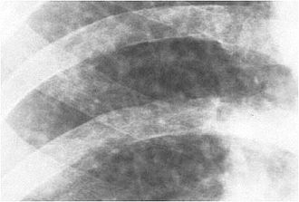
Progressive massive fibrosis (PMF), characterized by the development of large conglomerate masses of dense fibrosis, can complicate silicosis and coal worker's pneumoconiosis. Conglomerate masses may also occur in other pneumoconioses, such as talcosis, berylliosis (CBD), kaolin pneumoconiosis, and pneumoconiosis from carbon compounds, such as carbon black, graphite, and oil shale. Conglomerate masses can also develop in sarcoidosis, but usually near the hilae and with surrounding paracicatricial emphysema.

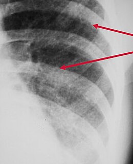
Kerley lines are a sign seen on chest radiographs with interstitial pulmonary edema. They are thin linear pulmonary opacities caused by fluid or cellular infiltration into the interstitium of the lungs. They are named after Irish neurologist and radiologist Peter Kerley.

Baritosis is a benign type of pneumoconiosis, which is caused by long-term exposure to barium dust.

Usual interstitial pneumonia

Usual interstitial pneumonia (UIP) is a form of lung disease characterized by progressive scarring of both lungs. The scarring (fibrosis) involves the supporting framework (interstitium) of the lung. UIP is thus classified as a form of interstitial lung disease.

High-resolution computed tomography

High-resolution computed tomography (HRCT) is a type of computed tomography (CT) with specific techniques to enhance image resolution. It is used in the diagnosis of various health problems, though most commonly for lung disease, by assessing the lung parenchyma.

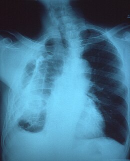
Malignant pleural effusion is a condition in which cancer causes an abnormal amount of fluid to collect between the thin layers of tissue (pleura) lining the outside of the lung and the wall of the chest cavity. Lung cancer and breast cancer account for about 50-65% of malignant pleural effusions. Other common causes include pleural mesothelioma and lymphoma.

Peribronchial cuffing

Peribronchial cuffing, also referred to as peribronchial thickening or bronchial wall thickening, is a radiologic sign which occurs when excess fluid or mucus buildup in the small airway passages of the lung causes localized patches of atelectasis. This causes the area around the bronchus to appear more prominent on an X-ray. It has also been described as donut sign, considering the edge is thicker, and the center contains air.

Pleural disease occurs in the pleural space, which is the thin fluid-filled area in between the two pulmonary pleurae in the human body. There are several disorders and complications that can occur within the pleural area, and the surrounding tissues in the lung.

Fibrothorax Medical condition involving fibrosis of the pleural space

Fibrothorax is a medical condition characterised by severe scarring (fibrosis) and fusion of the layers of the pleural space surrounding the lungs resulting in decreased movement of the lung and ribcage. The main symptom of fibrothorax is shortness of breath. There also may be recurrent fluid collections surrounding the lungs. Fibrothorax may occur as a complication of many diseases, including infection of the pleural space known as an empyema or bleeding into the pleural space known as a haemothorax.

A "B" reader is a physician certified by the National Institute for Occupational Safety and Health (NIOSH) as demonstrating proficiency in classifying radiographs of the pneumoconioses.

Ground-glass opacity Radiologic sign on radiographs and computed tomography scans

Ground-glass opacity (GGO) is a finding seen on chest x-ray (radiograph) or computed tomography (CT) imaging of the lungs. It is typically defined as an area of hazy opacification (x-ray) or increased attenuation (CT) due to air displacement by fluid, airway collapse, fibrosis, or a neoplastic process. When a substance other than air fills an area of the lung it increases that area's density. On both x-ray and CT, this appears more grey or hazy as opposed to the normally dark-appearing lungs. Although it can sometimes be seen in normal lungs, common pathologic causes include infections, interstitial lung disease, and pulmonary edema.

Tumor-like disorders of the lung pleura are a group of conditions that on initial radiological studies might be confused with malignant lesions. Radiologists must be aware of these conditions in order to avoid misdiagnosing patients. Examples of such lesions are: pleural plaques, thoracic splenosis, catamenial pneumothorax, pleural pseudotumor, diffuse pleural thickening, diffuse pulmonary lymphangiomatosis and Erdheim–Chester disease.

Asbestos-related diseases

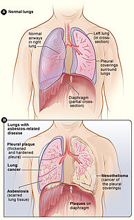
Asbestos-related diseases are disorders of the lung and pleura caused by the inhalation of asbestos fibres. Asbestos-related diseases include non-malignant disorders such as asbestosis, diffuse pleural thickening, pleural plaques, pleural effusion, rounded atelectasis and malignancies such as lung cancer and malignant mesothelioma.

Indium lung is a rare occupational lung disease caused by exposure to respirable indium in the form of indium tin oxide. It is classified as an interstitial lung disease.

References

  1. "International Labour Organization". Archived from the original on 2010-02-09. Retrieved 2010-02-12.
  2. International Labour Office. International Classification of Radiographs of Pneumoconiosis, rev ed. Occupational Safety and Health Series No. 22, Rev 2011. Geneva: ILO;2011; http://www.ilo.org/wcmsp5/groups/public/---ed_protect/---protrav/---safework/documents/publication/wcms_168260.pdf
  3. International Labour Office. International Classification of Radiographs of Pneumoconiosis, rev ed. Occupational Safety and Health Series No. 22, Rev 2000. Geneva: ILO;2000.
  4. Felson B, Morgan WKC, Bristol LJ, et al. Observations on the Results of Multiple Readings of Chest Films in Coal Miners' Pneumoconiosis. Radiol, 1973;109:19-23.
  5. Morgan RH. Proficiency Examination of Physicians for Classifying Pneumoconiosis Chest Films. Am J Radiol, 1979;132:803-808.