Armand Imbert (1850-1922) and Adolf Fick (1829-1901) both demonstrated, independently of each other, that in ocular tonometry the tension of the wall can be neutralized when the application of the tonometer produces a flat surface instead of a convex one, and the reading of the tonometer (P) then equals (T) the IOP," whence all forces cancel each other.
This principle was used by Hans Goldmann (1899–1991) who referred to it as the Imbert-Fick "law", thus giving his newly marketed tonometer (with the help of the Haag-Streit Company) a quasi-scientific basis; it is mentioned in the ophthalmic and optometric literature, but not in any books of physics. According to Goldmann,[1] "The law states that the pressure in a sphere filled with liquid and surrounded by an infinitely thin membrane is measured by the counterpressure which just flattens the membrane." "The law presupposes that the membrane is without thickness and without rigidity...practically without any extensibility."
A sphere formed from an inelastic membrane and filled with incompressible liquid cannot be indented or applanated even when the pressure inside is zero, because a sphere contains the maximum volume with the minimum surface area.[2][3] Any deformation necessarily increases surface area, which is impossible if the membrane is inelastic.
The physical basis of tonometry is Newton's third law of motion: "If you press an eyeball with an object, the object is also pressed by the eyeball."
The law is this:
Intraocular pressure = Contact force/Area of contact
The law assumes that the cornea is infinitely thin, perfectly elastic, and perfectly flexible.[4] None of these assumptions are accurate. The cornea is a membrane that has thickness and offers resistance when pressed.[5] Therefore, in Goldmann tonometry, readings are normally taken when an area of 3.06mm diameter has been flattened. At this point the opposing forces of corneal rigidity and the tear film are roughly approximate in a normal cornea and cancel each other out allowing the pressure in the eye to be inferred from the force applied.[6]
↑ Goldmann H. "Applanation Tonometry". Transactions Second Glaucoma Conference. New York, Josiah Macy, Jr Foundation. 1957.
↑ Koster W. "Beiträge zur Tonometrie und Manometrie des Auges". Graefe's Arch. Ophthalmol. 1895; 41: 113-158.
↑ Markiewitz HH. "The so-called Imbert-Fick Law". AMA Arch. Ophthalmol. 1960; 64: 189/159.
↑ Whitacre, MM, Stein, R. (1993) "Sources of error with use of Goldmann-type tonometers". Surv. Ophthalmol. 38,1-30
↑ Anders Eklund, Per Hallberg, Christina Lindén, and Olof A. Lindahl. (2003) "An Applanation Resonator Sensor for Measuring Intraocular Pressure Using Combined Continuous Force and Area Measurement". Invest. Ophthalmol. Vis. Sci. 2003 Jul;44(7):3017-24.
↑ The Glaucoma Book, Paul N. Schacknow, John R. Samples, p.79. Springer, 2010. ISBN978-0-387-76699-7.
Related Research Articles
Glaucoma is a group of eye diseases that lead to damage of the optic nerve, which transmits visual information from the eye to the brain. Glaucoma may cause vision loss if left untreated. It has been called the "silent thief of sight" because the loss of vision usually occurs slowly over a long period of time. A major risk factor for glaucoma is increased pressure within the eye, known as intraocular pressure (IOP). It is associated with old age, a family history of glaucoma, and certain medical conditions or medications. The word glaucoma comes from the Ancient Greek word γλαυκός, meaning 'gleaming, blue-green, gray'.
Eye surgery, also known as ophthalmic surgery or ocular surgery, is surgery performed on the eye or its adnexa. Eye surgery is part of ophthalmology and is performed by an ophthalmologist or eye surgeon. The eye is a fragile organ, and requires due care before, during, and after a surgical procedure to minimize or prevent further damage. An eye surgeon is responsible for selecting the appropriate surgical procedure for the patient, and for taking the necessary safety precautions. Mentions of eye surgery can be found in several ancient texts dating back as early as 1800 BC, with cataract treatment starting in the fifth century BC. It continues to be a widely practiced class of surgery, with various techniques having been developed for treating eye problems.
Radial keratotomy (RK) is a refractive surgical procedure to correct myopia (nearsightedness). It was developed in 1974 by Svyatoslav Fyodorov, a Russian ophthalmologist. It has been largely supplanted by newer, more accurate operations, such as photorefractive keratectomy, LASIK, Epi-LASIK and the phakic intraocular lens.
The corneal endothelium is a single layer of endothelial cells on the inner surface of the cornea. It faces the chamber formed between the cornea and the iris.
The ciliary body is a part of the eye that includes the ciliary muscle, which controls the shape of the lens, and the ciliary epithelium, which produces the aqueous humor. The aqueous humor is produced in the non-pigmented portion of the ciliary body. The ciliary body is part of the uvea, the layer of tissue that delivers oxygen and nutrients to the eye tissues. The ciliary body joins the ora serrata of the choroid to the root of the iris.
Intraocular pressure (IOP) is the fluid pressure inside the eye. Tonometry is the method eye care professionals use to determine this. IOP is an important aspect in the evaluation of patients at risk of glaucoma. Most tonometers are calibrated to measure pressure in millimeters of mercury (mmHg).
Physical or chemical injuries of the eye can be a serious threat to vision if not treated appropriately and in a timely fashion. The most obvious presentation of ocular (eye) injuries is redness and pain of the affected eyes. This is not, however, universally true, as tiny metallic projectiles may cause neither symptom. Tiny metallic projectiles should be suspected when a patient reports metal on metal contact, such as with hammering a metal surface. Corneal foreign bodies are one of the most common preventable occupational hazards. Intraocular foreign bodies do not cause pain because of the lack of nerve endings in the vitreous humour and retina that can transmit pain sensations. As such, general or emergency department doctors should refer cases involving the posterior segment of the eye or intraocular foreign bodies to an ophthalmologist. Ideally, ointment would not be used when referring to an ophthalmologist, since it diminishes the ability to carry out a thorough eye examination.
A red eye is an eye that appears red due to illness or injury. It is usually injection and prominence of the superficial blood vessels of the conjunctiva, which may be caused by disorders of these or adjacent structures. Conjunctivitis and subconjunctival hemorrhage are two of the less serious but more common causes.
Tonometry is the procedure that eye care professionals perform to determine the intraocular pressure (IOP), the fluid pressure inside the eye. It is an important test in the evaluation of patients at risk from glaucoma. Most tonometers are calibrated to measure pressure in millimeters of mercury (mmHg), with the normal eye pressure range between 10 and 21 mmHg (13–28 hPa).
The anterior chamber (AC) is the aqueous humor-filled space inside the eye between the iris and the cornea's innermost surface, the endothelium. Hyphema, anterior uveitis and glaucoma are three main pathologies in this area. In hyphema, blood fills the anterior chamber as a result of a hemorrhage, most commonly after a blunt eye injury. Anterior uveitis is an inflammatory process affecting the iris and ciliary body, with resulting inflammatory signs in the anterior chamber. In glaucoma, blockage of the trabecular meshwork prevents the normal outflow of aqueous humour, resulting in increased intraocular pressure, progressive damage to the optic nerve head, and eventually blindness.
A topical anesthetic is a local anesthetic that is used to numb the surface of a body part. They can be used to numb any area of the skin as well as the front of the eyeball, the inside of the nose, ear or throat, the anus and the genital area. Topical anesthetics are available in creams, ointments, aerosols, sprays, lotions, and jellies. Examples include benzocaine, butamben, dibucaine, lidocaine, oxybuprocaine, pramoxine, proxymetacaine (proparacaine), and tetracaine.
In ophthalmology, gonioscopy is a routine procedure that measures the angle between the iris and the cornea, using a goniolens together with a slit lamp or operating microscope. Its use is important in diagnosing and monitoring various eye conditions associated with glaucoma.
Glaucoma is a group of diseases affecting the optic nerve that results in vision loss and is frequently characterized by raised intraocular pressure (IOP). There are many glaucoma surgeries, and variations or combinations of those surgeries, that facilitate the escape of excess aqueous humor from the eye to lower intraocular pressure, and a few that lower IOP by decreasing the production of aqueous humor.
Ronald H. Silverman is an American ophthalmologist. He is currently Professor of Ophthalmic Science at Columbia University Medical Center. He is currently the director of the CUMC Basic Science Course in Ophthalmology, which takes place every January at the Harkness Eye Institute. He departed Weill Cornell Medical College in 2010, where he was Professor of Ophthalmology as well as a Dyson Scholar and the Research Director of the Bioacoustic Research Facility, Margaret M. Dyson Vision Research Institute at Weill Cornell.
Pseudoexfoliation syndrome, often abbreviated as PEX and sometimes as PES or PXS, is an aging-related systemic disease manifesting itself primarily in the eyes which is characterized by the accumulation of microscopic granular amyloid-like protein fibers. Its cause is unknown, although there is speculation that there may be a genetic basis. It is more prevalent in women than men, and in persons past the age of seventy. Its prevalence in different human populations varies; for example, it is prevalent in Scandinavia. The buildup of protein clumps can block normal drainage of the eye fluid called the aqueous humor and can cause, in turn, a buildup of pressure leading to glaucoma and loss of vision. As worldwide populations become older because of shifts in demography, PEX may become a matter of greater concern.
The aim of an accurate intraocular lens power calculation is to provide an intraocular lens (IOL) that fits the specific needs and desires of the individual patient. The development of better instrumentation for measuring the eye's axial length (AL) and the use of more precise mathematical formulas to perform the appropriate calculations have significantly improved the accuracy with which the surgeon determines the IOL power.
Open-globe injuries are full-thickness eye-wall wounds requiring urgent diagnosis and treatment.
Pre Descemet's endothelial keratoplasty (PDEK) is a kind of endothelial keratoplasty, where the pre descemet's layer (PDL) along with descemet's membrane (DM) and endothelium is transplanted. Conventionally in a corneal transplantation, doctors use a whole cornea or parts of the five layers of the cornea to perform correction surgeries. In May 2013, Dr Harminder Dua discovered a sixth layer between the stroma and the descemet membrane which was named after him as the Dua's layer. In the PDEK technique, doctors take the innermost two layers of the cornea, along with the Dua's layer and graft it in the patient's eye.
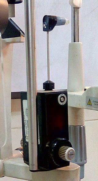
Goldmann Applanation Tonometer is an instrument that is based on Imbert-Fick law. It is considered to be the gold standard instrument for measurement of Intraocular pressure (IOP).
Manual small incision cataract surgery (MSICS) is an evolution of extracapsular cataract extraction (ECCE); the lens is removed from the eye through a self-sealing scleral tunnel wound. A well-constructed scleral tunnel is held closed by internal pressure, is watertight, and does not require suturing. The wound is relatively smaller than that in ECCE but is still markedly larger than a phacoemulsification wound. Comparative trials of MSICS against phaco in dense cataracts have found no statistically significant difference in outcomes but MSICS had shorter operating times and significantly lower costs. MSICS has become the method of choice in the developing world because it provides high-quality outcomes with less surgically induced astigmatism than ECCE, no suture-related problems, quick rehabilitation, and fewer post-operative visits. MSICS is easy and fast to learn for the surgeon, cost effective, simple, and applicable to almost all types of cataract.
This page is based on this Wikipedia article Text is available under the CC BY-SA 4.0 license; additional terms may apply. Images, videos and audio are available under their respective licenses.