Indirect DNA damage

Last updated


Indirect DNA damage: The chromophore absorbs UV-light (* denotes an excited state), and the energy of the excited state is creating singlet oxygen ( O2) or a hydroxyl radical (*OH), which then damages DNA through oxidation. Indirect DNA damage.png
Indirect DNA damage: The chromophore absorbs UV-light (* denotes an excited state), and the energy of the excited state is creating singlet oxygen ( O2) or a hydroxyl radical (•OH), which then damages DNA through oxidation.

Indirect DNA damage occurs when a UV-photon is absorbed in the human skin by a chromophore that does not have the ability to convert the energy into harmless heat very quickly. [2] Molecules that do not have this ability have a long-lived excited state. This long lifetime leads to a high probability for reactions with other molecules—so-called bimolecular reactions. [2] Melanin [ dubious ][ citation needed ] and DNA have extremely short excited state lifetimes in the range of a few femtoseconds (10−15s). [3] The excited state lifetime of compounds used in sunscreens such as menthyl anthranilate, avobenzone or padimate O is 1,000 to 1,000,000 times longer than that of melanin, [2] and therefore they may cause damage to living cells that come in contact with them. [4] [5] [6] [7]

Contents

The molecule that originally absorbs the UV-photon is called a "chromophore". Bimolecular reactions can occur either between the excited chromophore and DNA or between the excited chromophore and another species, to produce free radicals and reactive oxygen species. These reactive chemical species can reach DNA by diffusion and the bimolecular reaction damages the DNA (oxidative stress). It is important to note that, unlike direct DNA damage which causes sunburn, indirect DNA damage does not result in any warning signal or pain in the human body.

The bimolecular reactions that cause the indirect DNA damage are illustrated in the figure:

1O2 is reactive harmful singlet oxygen:

[1]

Location of the damage

Unlike direct DNA damage, which occurs in areas directly exposed to UV-B light, reactive chemical species can travel through the body and affect other areas—possibly even inner organs. [ dubious ] The traveling nature of the indirect DNA damage can be seen in the fact that the malignant melanoma can occur in places that are not directly illuminated by the sun—in contrast to basal-cell carcinoma and squamous cell carcinoma, which appear only on directly illuminated locations on the body. [ dubious ][ citation needed ]

See also

Related Research Articles

<span class="mw-page-title-main">Ultraviolet</span> Form of electromagnetic radiation

Ultraviolet (UV) is a form of electromagnetic radiation with wavelength from 10 nm to 400 nm (750 THz), shorter than that of visible light, but longer than X-rays. UV radiation is present in sunlight, and constitutes about 10% of the total electromagnetic radiation output from the Sun. It is also produced by electric arcs, Cherenkov radiation, and specialized lights, such as mercury-vapor lamps, tanning lamps, and black lights. Although long-wavelength ultraviolet is not considered an ionizing radiation because its photons lack the energy to ionize atoms, it can cause chemical reactions and causes many substances to glow or fluoresce. Many practical applications, including chemical and biological effects, derive from the way that UV radiation can interact with organic molecules. These interactions can involve absorption or adjusting energy states in molecules, but do not necessarily involve heating.

<span class="mw-page-title-main">Photochemistry</span> Sub-discipline of chemistry

Photochemistry is the branch of chemistry concerned with the chemical effects of light. Generally, this term is used to describe a chemical reaction caused by absorption of ultraviolet, visible light (400–750 nm) or infrared radiation (750–2500 nm).

<span class="mw-page-title-main">Sun tanning</span> The darkening of skin in response to ultraviolet light

Sun tanning or tanning is the process whereby skin color is darkened or tanned. It is most often a result of exposure to ultraviolet (UV) radiation from sunlight or from artificial sources, such as a tanning lamp found in indoor tanning beds. People who deliberately tan their skin by exposure to the sun engage in a passive recreational activity of sun bathing. Some people use chemical products which can produce a tanning effect without exposure to ultraviolet radiation, known as sunless tanning.

<span class="mw-page-title-main">Reactive oxygen species</span> Highly reactive molecules formed from diatomic oxygen (O2)

In chemistry, reactive oxygen species (ROS) are highly reactive chemicals formed from diatomic oxygen. Examples of ROS include peroxides, superoxide, hydroxyl radical, singlet oxygen, and alpha-oxygen.

<span class="mw-page-title-main">Erythrulose</span> Chemical compound

D-Erythrulose (also known as erythrulose) is a tetrose carbohydrate with the chemical formula C4H8O4. It has one ketone group and so is part of the ketose family. It is used in some self-tanning cosmetics, in general, combined with dihydroxyacetone (DHA).

<span class="mw-page-title-main">Sunless tanning</span> Indoor tanning lotion

Sunless tanning, also known as UV filled tanning, self tanning, spray tanning, or fake tanning, refers to the effect of a suntan without exposure to the Sun. Sunless tanning involves the use of oral agents (carotenids), or creams, lotions or sprays applied to the skin. Skin-applied products may be skin-reactive agents or temporary bronzers (colorants).

<span class="mw-page-title-main">Avobenzone</span> Oil-soluble ingredient used in sunscreen products

Avobenzone is an oil-soluble ingredient used in sunscreen products to absorb the full spectrum of UVA rays.

<span class="mw-page-title-main">Octyl methoxycinnamate</span> Organic chemical compound

Octyl methoxycinnamate or ethylhexyl methoxycinnamate (INCI) or octinoxate (USAN), trade names Eusolex 2292 and Uvinul MC80, is an organic compound that is an ingredient in some sunscreens and lip balms. It is an ester formed from methoxycinnamic acid and 2-ethylhexanol. It is a liquid that is insoluble in water.

<span class="mw-page-title-main">Photodegradation</span>

Photodegradation is the alteration of materials by light. Commonly, the term is used loosely to refer to the combined action of sunlight and air, which cause oxidation and hydrolysis. Often photodegradation is intentionally avoided, since it destroys paintings and other artifacts. It is, however, partly responsible for remineralization of biomass and is used intentionally in some disinfection technologies. Photodegradation does not apply to how materials may be aged or degraded via infrared light or heat, but does include degradation in all of the ultraviolet light wavebands.

<span class="mw-page-title-main">UV filter</span> Camera parts, features and technologies

UV filters are compounds, mixtures, or materials that block or absorb ultraviolet (UV) light. One of the major applications of UV filters is their use as sunscreens to protect skin from sunburn and other sun/UV related damage. After the invention of digital cameras changed the field of photography, UV filters have been used to coat glass discs fitted to camera lenses to protect hardware that is sensitive to UV light.

<span class="mw-page-title-main">Padimate O</span> Water-insoluble oily ingredient used in some sunscreens

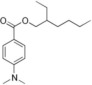
Padimate O is an organic compound related to the water-soluble compound PABA that is used as an ingredient in some sunscreens. This yellowish water-insoluble oily liquid is an ester formed by the condensation of 2-ethylhexanol with dimethylaminobenzoic acid. Other names for padimate O include 2-ethylhexyl 4-dimethylaminobenzoate, Escalol 507, octyldimethyl PABA, and OD-PABA.

Photoprotection is the biochemical process that helps organisms cope with molecular damage caused by sunlight. Plants and other oxygenic phototrophs have developed a suite of photoprotective mechanisms to prevent photoinhibition and oxidative stress caused by excess or fluctuating light conditions. Humans and other animals have also developed photoprotective mechanisms to avoid UV photodamage to the skin, prevent DNA damage, and minimize the downstream effects of oxidative stress.

UV-induced apoptosis UV-induced apoptosis is an adequate (physiological) reaction of a cell damaged by UV radiation (UVR) in a sufficiently large (lethal) dose and it prevents the disordered destruction of UV damaged cells by help necrosis. Cell elimination by apoptosis occurs when UV-induced cell damage which cannot be repaired by the intracellular repair system exceeds at it certain limit. Through apoptosis, the cells are self-disassembled into compartments with their subsequent utilization. The first time sign of the beginning of the apoptosis system is working in a UV damaged cell is the activation of restriction enzymes, which divide cell DNA into fragments convenient for utilization. But too large a dose of UVR can lead to breakdown (inactivation) of the energy-dependent mechanism of apoptosis. In this case, cell destruction occurs randomly, not orderly, and during a significantly longer time interval. UV-irradiated cells do not change their appearance for a long time [1, 6], as a result of which the researchers may make the erroneous conclusion that “revealed an unexpected response to a dose at which a higher dose of UV increased the viability of keratinocytes” [2]. The fact that UV-induced apoptosis at high doses of UVR begins to be replaced by necrosis was established in 2000 [3]. For keratinocytes, the proportion of cells that have elimination by help apoptosis, with an increase in UVR dose can reach to achieve 45%, but with a further increase in the dose of UVR, destruction of damaged cells by help necrosis and the part of cells that eliminated by apoptosis begins to decrease [4, 11]. In the dose range of UVR from “lethal” to “super-lethal”, “pro-inflammatory” apoptosis can be manifested, which was experimentally discovered in 2003 [5]. This may be the result of partial damage to the apoptosis mechanism by UV radiation [1]. If at moderate doses “pure” apoptosis does not cause an inflammatory reaction, then at sufficiently large doses, an inflammatory reaction arises due to pro-inflammatory apoptosis, which leads to the appearance of “fast” erythema for UV irradiated skin keratinocytes. Kinetic of “fast” erythema is much faster by the time of development of UV erythema caused by necrosis of UV damaged keratinocytes [6]. The most erythemogenic is UVB the spectral range of UVR, since radiation in this range is less absorbed by the outer layers of the skin, which allows UVB radiation, in contrast to UVC, to reach more deep layers skin and act on keratinocytes of the deep-lying basal layer of the epidermis of the skin. The ability to induce apoptosis for UVB and UVC radiation is due to the fact that the DNA of the nucleus [7] and / or mitochondria [8] of the cell absorbs UVR well in the UVC and UVB spectral range. Keratinocytes of the skin are in a state of programmed apoptosis, during which the keratinocytes of the basal layer are removed from it and during the transition through all layers of the epidermis within 28 days turn into flakes of the outer stratum corneum, which are subsequently desquamated. It is clear that the keratinocyte response to UV exposure will depend on what phase of programmed apoptosis the keratinocyte experienced UV exposure, and this is the main reason for the difference of the UV effect for UVC and UVB on the skin. There are also differences in the initiation of mitochondrial (internal) and caspase-dependent (external) apoptosis for the UVC and UVB spectral ranges [9]. Sunburn cells (SBS) are the keratinocytes in the process of UV-induced apoptosis. The appearance of SBC may be not associated with an inflammatory reaction, but the role of UV-induced apoptosis of skin keratinocytes in the development of UV erythema of the skin has been established, which allowed the development of a patent-protected METHOD FOR QUANTITATIVE ASSESSMENT OF APOPTOSIS SYSTEM [10], in which “the brightest lamp of skin display "(photoerythema) is used to diagnose the state of the body systems involved in the elimination of UV-induced damage. Such systems include the immune system, the intracellular repair system, the microcirculation system and not only.

<span class="mw-page-title-main">Pyrimidine dimer</span>

Pyrimidine dimers are molecular lesions formed from thymine or cytosine bases in DNA via photochemical reactions, commonly associated with direct DNA damage. Ultraviolet light induces the formation of covalent linkages between consecutive bases along the nucleotide chain in the vicinity of their carbon–carbon double bonds. The dimerization reaction can also occur among pyrimidine bases in dsRNA —uracil or cytosine. Two common UV products are cyclobutane pyrimidine dimers (CPDs) and 6–4 photoproducts. These premutagenic lesions alter the structure and possibly the base-pairing. Up to 50–100 such reactions per second might occur in a skin cell during exposure to sunlight, but are usually corrected within seconds by photolyase reactivation or nucleotide excision repair. Uncorrected lesions can inhibit polymerases, cause misreading during transcription or replication, or lead to arrest of replication. It causes sunburn and it triggers the production of melanin. Pyrimidine dimers are the primary cause of melanomas in humans.

A photocarcinogen is a substance which causes cancer when an organism is exposed to it, then illuminated. Many chemicals that are not carcinogenic can be photocarcinogenic when combined with exposure to light, especially UV. This can easily be understood from a photochemical perspective: The reactivity of a chemical substance itself might be low, but after illumination it transitions to an excited state, which is chemically much more reactive and therefore potentially harmful to biological tissue and DNA. Light can also split photocarcinogens, releasing free radicals, whose unpaired electrons cause them to be extremely reactive.

Tanning activators are chemicals that increase the effect of UV-radiation on the human skin.

<span class="mw-page-title-main">Photoaging</span> Ultraviolet light-induced biological development over time

Photoaging or photoageing is a term used for the characteristic changes to skin induced by chronic UVA and UVB exposure. Tretinoin is the best studied retinoid in the treatment of photoaging.

Mycosporine-like amino acids (MAAs) are small secondary metabolites produced by organisms that live in environments with high volumes of sunlight, usually marine environments. The exact number of compounds within this class of natural products is yet to be determined, since they have only relatively recently been discovered and novel molecular species are constantly being discovered; however, to date their number is around 30. They are commonly described as “microbial sunscreens” although their function is believed not to be limited to sun protection. MAAs represent high potential in cosmetics, and biotechnological applications. Indeed, their UV-absorbing properties would allow to create products derived from natural photoprotectors, potentially harmless to the environment and efficient against UV damage.

<span class="mw-page-title-main">Angelicin</span> Chemical compound

Angelicin is the parent compound in a family of naturally occurring organic compounds known as the angular furanocoumarins. Structurally, it can be considered as benzapyra-2-one fused with a furan moiety in the 7,8-position. Angelicin is commonly found in certain Apiaceae and Fabaceae plant species such as Bituminaria bituminosa. It has a skin permeability coefficient (LogKp) of -2.46. The maximum absorption is observed at 300 nm. The 1HNMR spectrum is available; the infrared and mass spectra of angelicin can be found in this database. The sublimation of angelicin occurs at 120 °C and the pressure of 0.13 Pa. Angelicin is a coumarine.

<span class="mw-page-title-main">Photooxygenation</span> Light-induced oxidation reaction

A photooxygenation is a light-induced oxidation reaction in which molecular oxygen is incorporated into the product(s). Initial research interest in photooxygenation reactions arose from Oscar Raab's observations in 1900 that the combination of light, oxygen and photosensitizers is highly toxic to cells. Early studies of photooxygenation focused on oxidative damage to DNA and amino acids, but recent research has led to the application of photooxygenation in organic synthesis and photodynamic therapy.

References

  1. 1 2 Ribeiro, D. T.; Madzak, C.; Sarasin, A.; Mascio, P. Di; Sies, H.; Menck, C. F. M. (January 1992). "Singlet Oxygen Induced DNA Damage and Mutagenicity in a Single-Stranded Sv40-Based Shuttle Vector". Photochemistry and Photobiology. 55 (1): 39–45. doi:10.1111/j.1751-1097.1992.tb04207.x. PMID   1318549. S2CID   9807925.
  2. 1 2 3 Cantrell, Ann; McGarvey, David J (2001). "3(Sun Protection in Man)". Comprehensive Series in Photosciences. 495: 497–519. CAN 137:43484.
  3. "Ultrafast internal conversion of DNA". Archived from the original on 2008-06-05. Retrieved 2008-02-13.
  4. Armeni, Tatiana; Damiani, Elisabetta; et al. (2004). "Lack of in vitro protection by a common sunscreen ingredient on UVA-induced cytotoxicity in keratinocytes". Toxicology. 203 (1–3): 165–178. doi:10.1016/j.tox.2004.06.008. hdl: 11566/51196 . PMID   15363592.
  5. Knowland, John; McKenzie, Edward A.; McHugh, Peter J.; Cridland, Nigel A. (1993). "Sunlight-induced mutagenicity of a common sunscreen ingredient". FEBS Letters. 324 (3): 309–313. doi:10.1016/0014-5793(93)80141-G. PMID   8405372. S2CID   23853321.
  6. Mosley, C N; Wang, L; Gilley, S; Wang, S; Yu, H (2007). "Light-Induced Cytotoxicity and Genotoxicity of a Sunscreen Agent, 2-Phenylbenzimidazol in Salmonella typhimurium TA 102 and HaCaT Keratinocytes". International Journal of Environmental Research and Public Health. 4 (2): 126–131. doi: 10.3390/ijerph2007040006 . PMC   3728577 . PMID   17617675.
  7. Xu, C.; Green, Adele; Parisi, Alfio; Parsons, Peter G (2001). "Photosensitization of the Sunscreen Octyl p-Dimethylaminobenzoate b UVA in Human Melanocytes but not in Keratinocytes". Photochemistry and Photobiology. 73 (6): 600–604. doi:10.1562/0031-8655(2001)073<0600:POTSOP>2.0.CO;2. ISSN   0031-8655. PMID   11421064. S2CID   38706861.