Related Research Articles
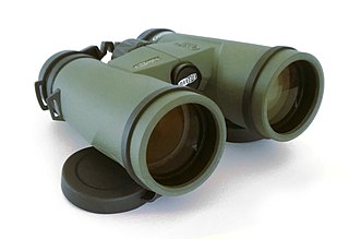
Binoculars or field glasses are two refracting telescopes mounted side-by-side and aligned to point in the same direction, allowing the viewer to use both eyes when viewing distant objects. Most binoculars are sized to be held using both hands, although sizes vary widely from opera glasses to large pedestal-mounted military models.

The optical microscope, also referred to as a light microscope, is a type of microscope that commonly uses visible light and a system of lenses to generate magnified images of small objects. Optical microscopes are the oldest design of microscope and were possibly invented in their present compound form in the 17th century. Basic optical microscopes can be very simple, although many complex designs aim to improve resolution and sample contrast.

In biology, binocular vision is a type of vision in which an animal has two eyes capable of facing the same direction to perceive a single three-dimensional image of its surroundings. Binocular vision does not typically refer to vision where an animal has eyes on opposite sides of its head and shares no field of view between them, like in some animals.

Stereoscopy is a technique for creating or enhancing the illusion of depth in an image by means of stereopsis for binocular vision. The word stereoscopy derives from Greek στερεός (stereos) 'firm, solid', and σκοπέω (skopeō) 'to look, to see'. Any stereoscopic image is called a stereogram. Originally, stereogram referred to a pair of stereo images which could be viewed using a stereoscope.

Depth perception is the ability to perceive distance to objects in the world using the visual system and visual perception. It is a major factor in perceiving the world in three dimensions. Depth perception happens primarily due to stereopsis and accommodation of the eye.

Presbyopia is physiological insufficiency of accommodation associated with the aging of the eye that results in progressively worsening ability to focus clearly on close objects. Also known as age-related farsightedness, it affects many adults over the age of 40. A common sign of presbyopia is difficulty reading small print which results in having to hold reading material farther away. Other symptoms associated can be headaches and eyestrain. Different people will have different degrees of problems. Other types of refractive errors may exist at the same time as presbyopia. This condition is similar to hypermetropia or far-sightedness which starts in childhood and exhibits similar symptoms of blur in the vision for close objects.

An autostereogram is a two-dimensional (2D) image that can create the optical illusion of a three-dimensional (3D) scene. Autostereograms use only one image to accomplish the effect while normal stereograms require two. The 3D scene in an autostereogram is often unrecognizable until it is viewed properly, unlike typical stereograms. Viewing any kind of stereogram properly may cause the viewer to experience vergence-accommodation conflict.

Refractive error is a problem with focusing light accurately on the retina due to the shape of the eye and/or cornea. The most common types of refractive error are near-sightedness, far-sightedness, astigmatism, and presbyopia. Near-sightedness results in far away objects being blurry, far-sightedness and presbyopia result in close objects being blurry, and astigmatism causes objects to appear stretched out or blurry. Other symptoms may include double vision, headaches, and eye strain.

The human eye is an organ of the sensory nervous system that reacts to visible light and allows the use of visual information for various purposes including seeing things, keeping balance, and maintaining circadian rhythm.

An eye examination is a series of tests performed to assess vision and ability to focus on and discern objects. It also includes other tests and examinations pertaining to the eyes. Eye examinations are primarily performed by an optometrist, ophthalmologist, or an orthoptist. Health care professionals often recommend that all people should have periodic and thorough eye examinations as part of routine primary care, especially since many eye diseases are asymptomatic.

Accommodation is the process by which the vertebrate eye changes optical power to maintain a clear image or focus on an object as its distance varies. In this, distances vary for individuals from the far point—the maximum distance from the eye for which a clear image of an object can be seen, to the near point—the minimum distance for a clear image. Accommodation usually acts like a reflex, including part of the accommodation-convergence reflex, but it can also be consciously controlled.
Stereopsis is the component of depth perception retrieved by means of binocular disparity through binocular vision. It is not the only contributor to depth perception, but it is a major one. Binocular vision occurs because each eye receives a different image due to their slightly different positions in one's head. These positional differences are referred to as "horizontal disparities" or, more generally, "binocular disparities". Disparities are processed in the visual cortex of the brain to yield depth perception. While binocular disparities are naturally present when viewing a real three-dimensional scene with two eyes, they can also be simulated by artificially presenting two different images separately to each eye using a method called stereoscopy. The perception of depth in such cases is also referred to as "stereoscopic depth".

A vergence is the simultaneous movement of both eyes in opposite directions to obtain or maintain single binocular vision.

Pupillary distance (PD), more correctly known as interpupillary distance (IPD) is the distance in millimeters between the centers of each pupil.
Aniseikonia is an ocular condition where there is a significant difference in the perceived size of images. It can occur as an overall difference between the two eyes, or as a difference in a particular meridian. If the ocular image size in both eyes are equal, the condition is known as iseikonia.

Emmetropia is the state of vision in which a faraway object at infinity is in sharp focus with the ciliary muscle in a relaxed state. That condition of the normal eye is achieved when the refractive power of the cornea and eye lens and the axial length of the eye balance out, which focuses rays exactly on the retina, resulting in perfectly sharp distance vision. A human eye in a state of emmetropia requires no corrective lenses for distance; the vision scores well on a visual acuity test.
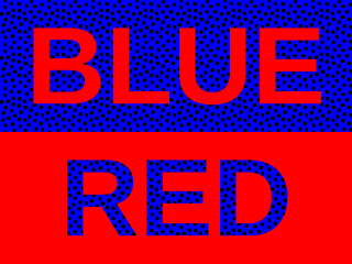
Chromostereopsis is a visual illusion whereby the impression of depth is conveyed in two-dimensional color images, usually of red–blue or red–green colors, but can also be perceived with red–grey or blue–grey images. Such illusions have been reported for over a century and have generally been attributed to some form of chromatic aberration.

The chameleon is among the most highly visually-oriented lizards, using this sense in prey capture, mating behavior, and predator avoidance. Unique features of chameleon vision include a negative lens, a positive cornea, and monocular focusing. The development of the chameleon visual system could have evolved to aid in prey capture and/or in predator avoidance.
In ophthalmology, accommodative excess occurs when an individual uses more than normal accommodation for performing certain near work. Accommodative excess has traditionally been defined as accommodation that is persistently higher than expected for the patient's age. Modern definitions simply regard it as an inability to relax accommodation readily. Excessive accommodation is seen in association with excessive convergence also.

Vergence-accommodation conflict (VAC), also known as accommodation-vergence conflict, is a visual phenomenon that occurs when the brain receives mismatching cues between vergence and accommodation of the eye. This commonly occurs in virtual reality devices, augmented reality devices, 3D movies, and other types of stereoscopic displays and autostereoscopic displays. The effect can be unpleasant and cause eye strain.
References
- 1 2 3 4 5 Richards, O. W. (1976). "Instrument myopia--microscopy". American Journal of Optometry and Physiological Optics. 53 (10): 658–663. doi:10.1097/00006324-197610000-00003. PMID 1015520. S2CID 37513722.
- ↑ Hennessy, Robert T. (1975). "Instrument myopia*". Journal of the Optical Society of America. 65 (10): 1114–1120. Bibcode:1975JOSA...65.1114H. doi:10.1364/josa.65.001114. PMID 1185295.
- ↑ Duke-Elder, S. (1973). System of ophthalmology London: Henry Kimpton.
- ↑ Fincham, E. F., & Walton, J. (1957). "The reciprocal actions of accommodation and convergence". Journal of Physiology. 137 (3): 488–508. doi:10.1113/jphysiol.1957.sp005829. PMC 1363021 . PMID 13463783.
{{cite journal}}: CS1 maint: multiple names: authors list (link) - 1 2 3 Wesner, M. F., & Miller, R. J. (1986). "Instrument myopia conceptions, misconceptions, and influencing factors". Documenta Ophthalmologica. 62 (3): 281–308. doi:10.1007/bf00212652. PMID 3698796. S2CID 20659472.
{{cite journal}}: CS1 maint: multiple names: authors list (link) - ↑ Leibowitz, H. W., & Owens, D. A. (1975). "Night myopia and the intermediate dark focus of accommodation". Journal of the Optical Society of America. 65 (10): 1121–1128. doi:10.1364/josa.65.001121. PMID 1185296.
{{cite journal}}: CS1 maint: multiple names: authors list (link) - ↑ Imbert, Henri (1899). De l'état de l'accommodation de l'oeil pendant les observations au microscope [Of the accommodation state of the eye during observations through a microscope]. Paris: Librairie J.-B. Baillière et Fils.
- ↑ Schober, H. A. W.; Dehler, H.; Kassel, R. (1970). "Accommodation during observations with optical instruments". Journal of the Optical Society of America. 60 (1): 103–107. Bibcode:1970JOSA...60..103S. doi:10.1364/josa.60.000103.