| Internal granular layer | |
|---|---|
| Details | |
| Identifiers | |
| Latin | lamina granularis interna |
| TA98 | A14.1.09.312 |
| FMA | 77811 |
| Anatomical terms of neuroanatomy | |
The internal granular layer of the cerebral cortex, also commonly referred to as the granular layer of the cortex, is the layer IV in the subdivision of the mammalian cerebral cortex into 6 layers. The adjective internal is used in opposition to the external granular layer of the cortex, the term granular refers to the granule cells found here.
This layer receives the afferent connections from the thalamus and from other cortical regions and sends connections to the other layers. The line of Gennari (occipital stripe) is also present in this layer.

The cerebral cortex, also known as the cerebral mantle, is the outer layer of neural tissue of the cerebrum of the brain in humans and other mammals. The cerebral cortex mostly consists of the six-layered neocortex, with just 10% consisting of the allocortex. It is separated into two cortices, by the longitudinal fissure that divides the cerebrum into the left and right cerebral hemispheres. The two hemispheres are joined beneath the cortex by the corpus callosum. The cerebral cortex is the largest site of neural integration in the central nervous system. It plays a key role in attention, perception, awareness, thought, memory, language, and consciousness. The cerebral cortex is part of the brain responsible for cognition.

Brodmann area 23 (BA23) is a region in the brain that lies inside the posterior cingulate cortex. It lies between Brodmann area 30 and Brodmann area 31 and is located on the medial wall of the cingulate gyrus between the callosal sulcus and the cingulate sulcus.

Brodmann area 19, or BA 19, is part of the occipital lobe cortex in the human brain. Along with area 18, it comprises the extrastriate cortex. In humans with normal sight, extrastriate cortex is a visual association area, with feature-extracting, shape recognition, attentional, and multimodal integrating functions.

Brodmann area 20, or BA20, is part of the temporal cortex in the human brain. The region encompasses most of the ventral temporal cortex, a region believed to play a part in high-level visual processing and recognition memory.

Brodmann area 21, or BA21, is part of the temporal cortex in the human brain. The region encompasses most of the lateral temporal cortex and is also known as middle temporal area 21. In the human it corresponds approximately to the middle temporal gyrus.

Brodmann area 4 refers to the primary motor cortex of the human brain. It is located in the posterior portion of the frontal lobe.
Granular layer may refer to:

Brodmann area 24 is part of the anterior cingulate in the human brain.

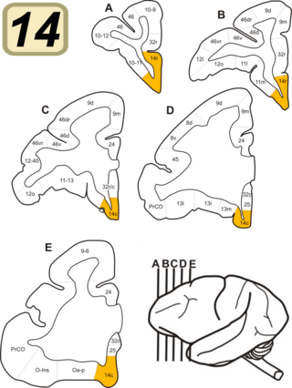
Brodmann Area 14 is one of Brodmann's subdivisions of the cerebral cortex in the brain. It was defined by Brodmann in the guenon monkey . While Brodmann, writing in 1909, argued that no equivalent structure existed in humans, later work demonstrated that area 14 has a clear homologue in the human ventromedial prefrontal cortex.

Brodmann area 12 is a subdivision of the cerebral cortex of the guenon defined on the basis of cytoarchitecture. It occupies the most rostral portion of the frontal lobe. Brodmann-1909 did not regard it as homologous, either topographically or cytoarchitecturally, to rostral area 12 of the human. Distinctive features (Brodmann-1905): a quite distinct internal granular layer (IV) separates slender pyramidal cells of the external pyramidal layer (III) and the internal pyramidal layer (V); the multiform layer (VI) is expanded, contains widely dispersed spindle cells and merges gradually with the underlying cortical white matter; all cells, including the pyramidal cells of the external and internal pyramidal layers are inordinately small; the internal pyramidal layer (V) also contains spindle cells in groups of two to five located close to its border with the internal granular layer (IV).
Brodmann Area 15 is one of Brodmann's subdivisions of the cerebral cortex in the brain.

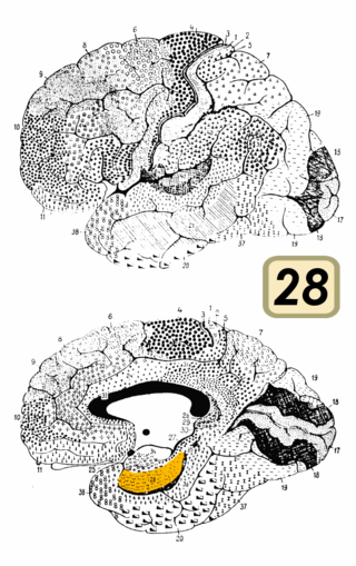
Brodmann area 28 is a subdivision of the cerebral cortex defined on the basis of cytoarchitecture. It is located on the medial aspect of the temporal lobe and is part of the entorhinal cortex (Brodmann-1909).

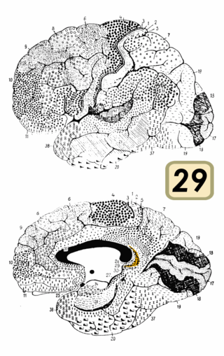
Brodmann area 29, also known as granular retrolimbic area 29 or granular retrosplenial cortex, is a cytoarchitecturally defined portion of the retrosplenial region of the cerebral cortex. In the human it is a narrow band located in the isthmus of cingulate gyrus. Cytoarchitecturally it is bounded internally by the ectosplenial area 26 and externally by the agranular retrolimbic area 30 (Brodmann-1909).
Bands of Baillarger, in neuroanatomy, are two myeloarchitectural structures, the external band of Baillarger and the internal band of Baillarger. These are myelinated fibers that course in the internal granular layer and internal pyramidal layer, respectively, of cerebral cortex. In the primary visual cortex (V1), the external band of Baillarger is enlarged and is called the line of Gennari. They are named after Jules Baillarger.

The anatomy of the cerebellum can be viewed at three levels. At the level of gross anatomy, the cerebellum consists of a tightly folded and crumpled layer of cortex, with white matter underneath, several deep nuclei embedded in the white matter, and a fluid-filled ventricle in the middle. At the intermediate level, the cerebellum and its auxiliary structures can be broken down into several hundred or thousand independently functioning modules or compartments known as microzones. At the microscopic level, each module consists of the same small set of neuronal elements, laid out with a highly stereotyped geometry.
Agranular insula is a portion of the cerebral cortex defined on the basis of internal structure in the human, the macaque, the rat, and the mouse. Classified as allocortex (periallocortex), it is in primates distinguished from adjacent neocortex (proisocortex) by absence of the external granular layer (II) and of the internal granular layer (IV). It occupies the anterior part of the insula, the posterior portion of the orbital gyri and the medial part of the temporal pole. In rodents it is located on the ventrolateral surface of the cortex rostrally, between the piriform area ventrally and the gustatory area or the visceral area dorsally.
Granular insular cortex refers to a portion of the cerebral cortex defined on the basis of internal structure in the human and macaque, the rat, and the mouse. Classified as neocortex, it is in primates distinguished from adjacent allocortex (periallocortex) by the presence of granular layers – external granular layer (II) and internal granular layer (IV) – and by differentiation of the external pyramidal layer (III) into sublayers. In primates it occupies the posterior part of the insula. In rodents it is located on the lateral surface of the cortex rostrally, dorsal to the gustatory area or, more caudally, dorsal to the agranular insula.
The external granular layer of the cerebral cortex is commonly known as layer II. It is different from the internal granular layer of the cerebral cortex.
External granular layer may refer to: