Related Research Articles
Cell biology is a branch of biology that studies the structure, function, and behavior of cells. All living organisms are made of cells. A cell is the basic unit of life that is responsible for the living and functioning of organisms. Cell biology is the study of the structural and functional units of cells. Cell biology encompasses both prokaryotic and eukaryotic cells and has many subtopics which may include the study of cell metabolism, cell communication, cell cycle, biochemistry, and cell composition. The study of cells is performed using several microscopy techniques, cell culture, and cell fractionation. These have allowed for and are currently being used for discoveries and research pertaining to how cells function, ultimately giving insight into understanding larger organisms. Knowing the components of cells and how cells work is fundamental to all biological sciences while also being essential for research in biomedical fields such as cancer, and other diseases. Research in cell biology is interconnected to other fields such as genetics, molecular genetics, molecular biology, medical microbiology, immunology, and cytochemistry.

The endoplasmic reticulum (ER) is a part of a transportation system of the eukaryotic cell, and has many other important functions such as protein folding. It is a type of organelle made up of two subunits – rough endoplasmic reticulum (RER), and smooth endoplasmic reticulum (SER). The endoplasmic reticulum is found in most eukaryotic cells and forms an interconnected network of flattened, membrane-enclosed sacs known as cisternae, and tubular structures in the SER. The membranes of the ER are continuous with the outer nuclear membrane. The endoplasmic reticulum is not found in red blood cells, or spermatozoa.

Endocytosis is a cellular process in which substances are brought into the cell. The material to be internalized is surrounded by an area of cell membrane, which then buds off inside the cell to form a vesicle containing the ingested materials. Endocytosis includes pinocytosis and phagocytosis. It is a form of active transport.
In biology, caveolae, which are a special type of lipid raft, are small invaginations of the plasma membrane in the cells of many vertebrates. They are the most abundant surface feature of many vertebrate cell types, especially endothelial cells, adipocytes and embryonic notochord cells. They were originally discovered by E. Yamada in 1955.
In molecular biology, caveolins are a family of integral membrane proteins that are the principal components of caveolae membranes and involved in receptor-independent endocytosis. Caveolins may act as scaffolding proteins within caveolar membranes by compartmentalizing and concentrating signaling molecules. They also induce positive (inward) membrane curvature by way of oligomerization, and hairpin insertion. Various classes of signaling molecules, including G-protein subunits, receptor and non-receptor tyrosine kinases, endothelial nitric oxide synthase (eNOS), and small GTPases, bind Cav-1 through its 'caveolin-scaffolding domain'.

Nucleoside-diphosphate kinases are enzymes that catalyze the exchange of terminal phosphate between different nucleoside diphosphates (NDP) and triphosphates (NTP) in a reversible manner to produce nucleotide triphosphates. Many NDP serve as acceptor while NTP are donors of phosphate group. The general reaction via ping-pong mechanism is as follows: XDP + YTP ←→ XTP + YDP. NDPK activities maintain an equilibrium between the concentrations of different nucleoside triphosphates such as, for example, when guanosine triphosphate (GTP) produced in the citric acid (Krebs) cycle is converted to adenosine triphosphate (ATP). Other activities include cell proliferation, differentiation and development, signal transduction, G protein-coupled receptor, endocytosis, and gene expression.

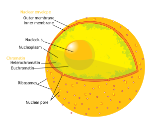
The nuclear envelope, also known as the nuclear membrane, is made up of two lipid bilayer membranes that in eukaryotic cells surround the nucleus, which encloses the genetic material.

Autocrine motility factor receptor, isoform 2 is a protein that in humans is encoded by the AMFR gene.

Caveolin-2 is a protein that in humans is encoded by the CAV2 gene.
Mitophagy is the selective degradation of mitochondria by autophagy. It often occurs to defective mitochondria following damage or stress. The process of mitophagy was first described in 1915 by Margaret Reed Lewis and Warren Harmon Lewis. Ashford and Porter used electron microscopy to observe mitochondrial fragments in liver lysosomes by 1962, and a 1977 report suggested that "mitochondria develop functional alterations which would activate autophagy." The term "mitophagy" was in use by 1998.
Membrane contact sites (MCS) are close appositions between two organelles. Ultrastructural studies typically reveal an intermembrane distance in the order of the size of a single protein, as small as 10 nm or wider, with no clear upper limit. These zones of apposition are highly conserved in evolution. These sites are thought to be important to facilitate signalling, and they promote the passage of small molecules, including ions, lipids and reactive oxygen species. MCS are important in the function of the endoplasmic reticulum (ER), since this is the major site of lipid synthesis within cells. The ER makes close contact with many organelles, including mitochondria, Golgi, endosomes, lysosomes, peroxisomes, chloroplasts and the plasma membrane. Both mitochondria and sorting endosomes undergo major rearrangements leading to fission where they contact the ER. Sites of close apposition can also form between most of these organelles most pairwise combinations. First mentions of these contact sites can be found in papers published in the late 1950s mainly visualized using electron microscopy (EM) techniques. Copeland and Dalton described them as “highly specialized tubular form of endoplasmic reticulum in association with the mitochondria and apparently in turn, with the vascular border of the cell”.

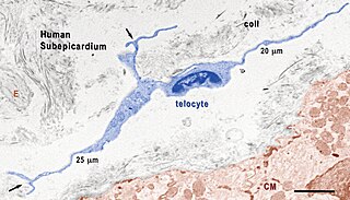
Telocytes are a type of interstitial (stromal) cells with very long and very thin prolongations called telopodes.

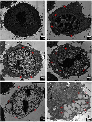
Paraptosis is a type of programmed cell death, morphologically distinct from apoptosis and necrosis. The defining features of paraptosis are cytoplasmic vacuolation, independent of caspase activation and inhibition, and lack of apoptotic morphology. Paraptosis lacks several of the hallmark characteristics of apoptosis, such as membrane blebbing, chromatin condensation, and nuclear fragmentation. Like apoptosis and other types of programmed cell death, the cell is involved in causing its own death, and gene expression is required. This is in contrast to necrosis, which is non-programmed cell death that results from injury to the cell.
Reductive stress (RS) is defined as an abnormal accumulation of reducing equivalents despite being in the presence of intact oxidation and reduction systems. A redox reaction involves the transfer of electrons from reducing agents (reductants) to oxidizing agents (oxidants) and redox couples are accountable for the majority of the cellular electron flow. RS is a state where there are more reducing equivalents compared to reductive oxygen species (ROS) in the form of known biological redox couples such as GSH/GSSG, NADP+/NADPH, and NAD+/NADH. Reductive stress is the counterpart to oxidative stress, where electron acceptors are expected to be mostly reduced. Reductive stress is likely derived from intrinsic signals that allow for the cellular defense against pro-oxidative conditions. There is a feedback regulation balance between reductive and oxidative stress where chronic RS induce oxidative species (OS), resulting in an increase in production of RS, again.

HCT116 is a human colon cancer cell line used in therapeutic research and drug screenings.

Jean Vance is a British-Canadian biochemist. She is known for her pioneering work on subcellular organelles and for her discovery of a connection between the endoplasmic reticulum and the mitochondrial membrane. She is a Professor of Medicine at the University of Alberta, Canada and a Fellow of the Royal Society of Canada.

Suliana Manley is an American biophysicist. Her research focuses on the development of high-resolution optical instruments, and their application in studying the organization and dynamics of proteins. She is a professor at École Polytechnique Fédérale de Lausanne and heads the Laboratory of Experimental Biophysics.

Robert Clarke is a Northern Irish cancer researcher and academic administrator. He is the executive director of The Hormel Institute, a professor of biochemistry, Molecular Biology and Biophysics at the University of Minnesota, and an adjunct professor of oncology at Georgetown University.
Ribosome-associated vesicles, also known as RAVs, are novel sub-compartments of the rough endoplasmic reticulum (ER), a membranous cellular network that is important for the synthesis and transport of proteins. RAVs have been observed via multiple imaging techniques and appear as discrete spherical vesicles that are associated with actively translated ribosomes. It is hypothesized that RAVs may arise from structural and/or functional changes in local membrane curvature along the rough endoplasmic reticulum's tubular membrane network.

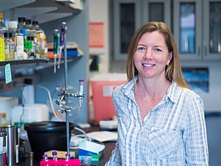
Gia Voeltz is an American cell biologist. She is a professor of Molecular, Cellular and Developmental Biology at the University of Colorado Boulder and a Howard Hughes Medical Institute Investigator. She is known for her research identifying the factors and unraveling the mechanisms that determine the structure and dynamics of the largest organelle in the cell: the endoplasmic reticulum. Her lab has produced paradigm shifting studies on organelle membrane contact sites that have revealed that most cytoplasmic organelles are not isolated entities but are instead physically tethered to an interconnected ER membrane network.
References
- 1 2 "Ivan Robert Nabi". University of British Columbia .
- ↑ "AI-Driven Breakthroughs in Cells Study: SFU-UBC Collaboration Introduces "MCS-DETECT" for Advancements in Super-Resolution Microscopy". Simon Fraser University .
- ↑ Nabi, I. R. (June 2020). "Biography-Ivan Robert Nabi". Cancer and Metastasis Reviews . 39 (2): 333. doi:10.1007/s10555-020-09893-8. PMID 32577925.
- ↑ "Ivan Robert Nabi - Vision Research". University of British Columbia .
- ↑ "Ivan Robert Nabi". breastcancerinyoungwomen.org.
- ↑ "Ivan Robert Nabi". Loop.
- ↑ "Ivan R. Nabi". Institute of Electrical and Electronics Engineers .
- ↑ Nabi, Ivan R. (May 2022). "A TMPRSS2 inhibitor acts as a pan-SARS-CoV-2 prophylactic and therapeutic". Nature. 605 (7909): 340–348. Bibcode:2022Natur.605..340S. doi:10.1038/s41586-022-04661-w. PMC 9095466 . PMID 35344983.
- ↑ Nabi, Ivan Robert (8 July 2019). "Super-resolution modularity analysis shows polyhedral caveolin-1 oligomers combine to form scaffolds and caveolae". Scientific Reports . 9: 9888. Bibcode:2019NatSR...9.9888K. doi:10.1038/s41598-019-46174-z. PMC 6614455 . PMID 31285524.
- ↑ "SFU and UBC researchers collaborate to understand the role of caveolin-1 in cancer". Simon Fraser University .