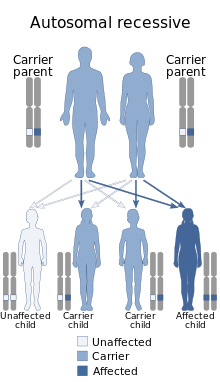
A genetic disorder is a health problem caused by one or more abnormalities in the genome. It can be caused by a mutation in a single gene (monogenic) or multiple genes (polygenic) or by a chromosome abnormality. Although polygenic disorders are the most common, the term is mostly used when discussing disorders with a single genetic cause, either in a gene or chromosome. The mutation responsible can occur spontaneously before embryonic development, or it can be inherited from two parents who are carriers of a faulty gene or from a parent with the disorder. When the genetic disorder is inherited from one or both parents, it is also classified as a hereditary disease. Some disorders are caused by a mutation on the X chromosome and have X-linked inheritance. Very few disorders are inherited on the Y chromosome or mitochondrial DNA.

Joubert syndrome is a rare autosomal recessive genetic disorder that affects the cerebellum, an area of the brain that controls balance and coordination.

Osteogenesis imperfecta, colloquially known as brittle bone disease, is a group of genetic disorders that all result in bones that break easily. The range of symptoms—on the skeleton as well as on the body's other organs—may be mild to severe. Symptoms found in various types of OI include whites of the eye (sclerae) that are blue instead, short stature, loose joints, hearing loss, breathing problems and problems with the teeth. Potentially life-threatening complications, all of which become more common in more severe OI, include: tearing (dissection) of the major arteries, such as the aorta; pulmonary valve insufficiency secondary to distortion of the ribcage; and basilar invagination.

Enamelin is an enamel matrix protein (EMPs), that in humans is encoded by the ENAM gene. It is part of the non-amelogenins, which comprise 10% of the total enamel matrix proteins. It is one of the key proteins thought to be involved in amelogenesis. The formation of enamel's intricate architecture is thought to be rigorously controlled in ameloblasts through interactions of various organic matrix protein molecules that include: enamelin, amelogenin, ameloblastin, tuftelin, dentine sialophosphoprotein, and a variety of enzymes. Enamelin is the largest protein (~168kDa) in the enamel matrix of developing teeth and is the least abundant of total enamel matrix proteins. It is present predominantly at the growing enamel surface.

Laurence–Moon syndrome (LMS) is a rare autosomal recessive genetic disorder associated with retinitis pigmentosa, spastic paraplegia, and mental disabilities.

Cyclic nucleotide-gated cation channel alpha-3 is a protein that in humans is encoded by the CNGA3 gene.

Cyclic nucleotide gated channel beta 3, also known as CNGB3, is a human gene encoding an ion channel protein.

WD repeat-containing protein 72 is a protein that in humans is encoded by the WDR72 gene. WDR72 contains 7 WD40 repeats, which are predicted to form the blades of a 7 beta propeller structure.

Amelogenesis imperfecta (AI) is a congenital disorder which presents with a rare abnormal formation of the enamel or external layer of the crown of teeth, unrelated to any systemic or generalized conditions. Enamel is composed mostly of mineral, that is formed and regulated by the proteins in it. Amelogenesis imperfecta is due to the malfunction of the proteins in the enamel as a result of abnormal enamel formation via amelogenesis.

FAM20A is a protein that in humans is encoded by the FAM20A gene.

Kohlschütter–Tönz syndrome (KTS), also called amelo-cerebro-hypohidrotic syndrome, is a rare inherited syndrome characterized by epilepsy, psychomotor delay or regression, intellectual disability, and yellow teeth caused by amelogenesis imperfecta. It is a type A ectodermal dysplasia.

Oculoauricular syndrome is a rare genetic condition affecting the eyes and ears. It is due to mutations in the H6 family homeobox 1 (HMX1) gene. It is also known as the Schorderet-Munier-Franceschetti syndrome.

Enamel-renal syndrome is a rare autosomal recessive condition. This condition is also known as idiopathic multicentric osteolysis with nephropathy. It is characterised by dental abnormalities and nephrocalcinosis.

Waardenburg anophthalmia syndrome is a rare autosomal recessive genetic disorder which is characterized by either microphthalmia or anophthalmia, osseous synostosis, ectrodactylism, polydactylism, and syndactylism. So far, 29 cases from families in Brazil, Italy, Turkey, and Lebanon have been reported worldwide. This condition is caused by homozygous mutations in the SMOC1 gene, in chromosome 14.

Retinal cone dystrophy 3B is a very rare genetic disorder which is characterized by ocular anomalies. Approximately 34 cases from 20 families across the world have been described in medical literature (OMIM). This disorder is associated with autosomal recessive mutations in the KCNV2 and PDE6H genes.

Boucher-Neuhäuser syndrome is a very rare genetic disorder which is characterized by a triad consisting of cerebellar ataxia, chorioretinal dystrophy, and hypogonadism.

Amaurosis congenita, cone-rod type, with congenital hypertrichosis is a very rare genetic disorder which is characterized by ocular anomalies and trichomegaly. It is inherited in an autosomal recessive manner. Only 2 cases have been described in medical literature.

Rhizomelic dysplasia, scoliosis, and retinitis pigmentosa is a very rare genetic disorder which is characterized by ocular/visual, dental and osseous anomalies. Only 2 cases have been described in medical literature.
Posterior column ataxia-retinitis pigmentosa syndrome (PCARP) is an autosomal recessive genetic disorder of the human eye, attributed to mutation of a gene originally dubbed AXPC1 which was identified as a mutation in the FLCVR1 gene. Generally rare, a Pennsylvania Mennonite variant has been estimated to have a population allele prevalence close to 1% due to founder effects.


