Related Research Articles

The large intestine,also known as the large bowel,is the last part of the gastrointestinal tract and of the digestive system in vertebrates. Water is absorbed here and the remaining waste material is stored as feces before being removed by defecation.

Colorectal cancer (CRC),also known as bowel cancer,colon cancer,or rectal cancer,is the development of cancer from the colon or rectum. Signs and symptoms may include blood in the stool,a change in bowel movements,weight loss,and fatigue.

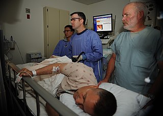
Colonoscopy or coloscopy is the endoscopic examination of the large bowel and the distal part of the small bowel with a CCD camera or a fiber optic camera on a flexible tube passed through the anus. It can provide a visual diagnosis and grants the opportunity for biopsy or removal of suspected colorectal cancer lesions.

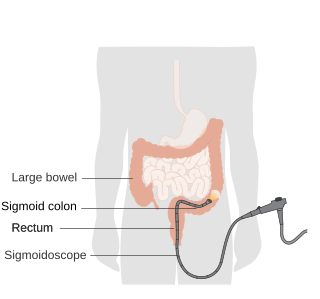
Sigmoidoscopy is the minimally invasive medical examination of the large intestine from the rectum through the nearest part of the colon,the sigmoid colon. There are two types of sigmoidoscopy:flexible sigmoidoscopy,which uses a flexible endoscope,and rigid sigmoidoscopy,which uses a rigid device. Flexible sigmoidoscopy is generally the preferred procedure. A sigmoidoscopy is similar to,but not the same as,a colonoscopy. A sigmoidoscopy only examines up to the sigmoid,the most distal part of the colon,while colonoscopy examines the whole large bowel.

In anatomy,a polyp is an abnormal growth of tissue projecting from a mucous membrane. If it is attached to the surface by a narrow elongated stalk,it is said to be pedunculated;if it is attached without a stalk,it is said to be sessile. Polyps are commonly found in the colon,stomach,nose,ear,sinus(es),urinary bladder,and uterus. They may also occur elsewhere in the body where there are mucous membranes,including the cervix,vocal folds,and small intestine. Some polyps are tumors (neoplasms) and others are non-neoplastic,for example hyperplastic or dysplastic,which are benign. The neoplastic ones are usually benign,although some can be pre-malignant,or concurrent with a malignancy.

A lower gastrointestinal series is a medical procedure used to examine and diagnose problems with the human colon. Radiographs are taken while barium sulfate,a radiocontrast agent,fills the colon via an enema through the rectum.

Digital rectal examination is an internal examination of the rectum,performed by a healthcare provider.

Fecal occult blood (FOB) refers to blood in the feces that is not visibly apparent. A fecal occult blood test (FOBT) checks for hidden (occult) blood in the stool (feces).

A stool test involves the collection and analysis of fecal matter to diagnose the presence or absence of a medical condition.

Virtual colonoscopy is the use of CT scanning or magnetic resonance imaging (MRI) to produce two- and three-dimensional images of the colon,from the lowest part,the rectum,to the lower end of the small intestine,and to display the images on an electronic display device. The procedure is used to screen for colon cancer and polyps,and may detect diverticulosis. A virtual colonoscopy can provide 3D reconstructed endoluminal views of the bowel. VC provides a secondary benefit of revealing diseases or abnormalities outside the colon.

Blood in stool looks different depending on how early it enters the digestive tract—and thus how much digestive action it has been exposed to—and how much there is. The term can refer either to melena,with a black appearance,typically originating from upper gastrointestinal bleeding;or to hematochezia,with a red color,typically originating from lower gastrointestinal bleeding. Evaluation of the blood found in stool depends on its characteristics,in terms of color,quantity and other features,which can point to its source,however,more serious conditions can present with a mixed picture,or with the form of bleeding that is found in another section of the tract. The term "blood in stool" is usually only used to describe visible blood,and not fecal occult blood,which is found only after physical examination and chemical laboratory testing.

Lower gastrointestinal bleeding,commonly abbreviated LGIB,is any form of gastrointestinal bleeding in the lower gastrointestinal tract. LGIB is a common reason for seeking medical attention at a hospital's emergency department. LGIB accounts for 30–40% of all gastrointestinal bleeding and is less common than upper gastrointestinal bleeding (UGIB). It is estimated that UGIB accounts for 100–200 per 100,000 cases versus 20–27 per 100,000 cases for LGIB. Approximately 85% of lower gastrointestinal bleeding involves the colon,10% are from bleeds that are actually upper gastrointestinal bleeds,and 3–5% involve the small intestines.

The stool guaiac test or guaiac fecal occult blood test (gFOBT) is one of several methods that detects the presence of fecal occult blood. The test involves placing a fecal sample on guaiac paper and applying hydrogen peroxide which,in the presence of blood,yields a blue reaction product within seconds.

A colorectal polyp is a polyp occurring on the lining of the colon or rectum. Untreated colorectal polyps can develop into colorectal cancer.
Wendy Sheila Atkin was Professor of Gastrointestinal Epidemiology at Imperial College London.
Ronald Marc Summers is an American radiologist and senior investigator at the Diagnostic Radiology Department at the NIH Clinical Center in Bethesda,Maryland. He is chief of the Clinical Image Processing Service and directs the Imaging Biomarkers and Computer-Aided Diagnosis (CAD) Laboratory. A researcher in the field of radiology and computer-aided diagnosis,he has co-authored over 500 journal articles and conference proceedings papers and is a coinventor on 12 patents. His lab has conducted research applying artificial intelligence and deep learning to radiology.
Charles Daniel Johnson is an American radiologist.
Serrated polyposis syndrome (SPS),previously known as hyperplastic polyposis syndrome,is a disorder characterized by the appearance of serrated polyps in the colon. While serrated polyposis syndrome does not cause symptoms,the condition is associated with a higher risk of colorectal cancer (CRC). The lifelong risk of CRC is between 25 and 40%. SPS is the most common polyposis syndrome affecting the colon,but is under recognized due to a lack of systemic long term monitoring. Diagnosis requires colonoscopy,and is defined by the presence of either of two criteria:≥5 serrated lesions/polyps proximal to the rectum,or >20 serrated lesions/polyps of any size distributed throughout the colon with 5 proximal to the rectum.
Postpolypectomy Coagulation Syndrome is a condition that occurs following colonoscopy with electrocautery polypectomy,which results in a burn injury to the wall of the gastrointestinal tract. The condition results in abdominal pain,fever,elevated white blood cell count and elevated serum C-reactive protein.
Mark Pochapin is a gastroenterologist and educator whose work is focused on the prevention,early detection,and treatment of gastrointestinal cancers.
References
- ↑ Rubin, Geoff (October 17, 2018). "Serving Vulnerable Populations From Coast to Coast" (PDF). acr.org. Retrieved September 8, 2020.
- 1 2 3 4 "Judy Yee, M.D." einstein.yu.edu. Retrieved September 8, 2020.
- ↑ "Judy Yee". c250.columbia.edu. Retrieved September 8, 2020.
- ↑ Boyd, Kevin (May 30, 2001). "Virtual colonoscopy as effective at colon cancer screening as standard invasive colonoscopy, SFVAMC". ucsf.edu. Retrieved September 8, 2020.
- ↑ "Highlights Around the Medical Center" (PDF). sanfrancisco.va.gov. 2007. p. 6. Retrieved September 8, 2020.
- 1 2 "Honorary Fellow" (PDF). cdn.ymaws.com. 2019. Retrieved September 8, 2020.
- ↑ Tokar, Steve (September 18, 2008). "Virtual colonoscopy as good as other colon cancer screening methods, study says". ucsf.edu. Retrieved September 8, 2020.
- ↑ "Latest study is step forward for virtual colonoscopy". research.va.gov. 2012. Retrieved September 8, 2020.
- ↑ "Judy Yee, MD, FACR, Becomes President of the Society of Abdominal Radiology". radiology.ucsf.edu. May 1, 2015. Retrieved September 8, 2020.
- ↑ "3-D Virtual Reality Colonoscopy: Pursuing a Better Path to Colorectal Cancer Prevention". ucsf.edu. July 5, 2016. Retrieved September 8, 2020.
- ↑ "Montefiore Health System and Albert Einstein College of Medicine Announce New Chair of Radiology". einstein.yu.edu. September 28, 2017. Retrieved September 8, 2020.