
Positron emission tomography (PET) is a functional imaging technique that uses radioactive substances known as radiotracers to visualize and measure changes in metabolic processes, and in other physiological activities including blood flow, regional chemical composition, and absorption. Different tracers are used for various imaging purposes, depending on the target process within the body.

A computed tomography scan is a medical imaging technique used to obtain detailed internal images of the body. The personnel that perform CT scans are called radiographers or radiology technologists.

Radiology is the medical specialty that uses medical imaging to diagnose diseases and guide their treatment, within the bodies of humans and other animals. It began with radiography, but today it includes all imaging modalities, including those that use no ionizing electromagnetic radiation, as well as others that do, such as computed tomography (CT), fluoroscopy, and nuclear medicine including positron emission tomography (PET). Interventional radiology is the performance of usually minimally invasive medical procedures with the guidance of imaging technologies such as those mentioned above.

Medical imaging is the technique and process of imaging the interior of a body for clinical analysis and medical intervention, as well as visual representation of the function of some organs or tissues (physiology). Medical imaging seeks to reveal internal structures hidden by the skin and bones, as well as to diagnose and treat disease. Medical imaging also establishes a database of normal anatomy and physiology to make it possible to identify abnormalities. Although imaging of removed organs and tissues can be performed for medical reasons, such procedures are usually considered part of pathology instead of medical imaging.

Optical coherence tomography (OCT) is an imaging technique that uses interferometry with short-coherence-length light to obtain micrometer-level depth resolution and uses transverse scanning of the light beam to form two- and three-dimensional images from light reflected from within biological tissue or other scattering media. Short-coherence-length light can be obtained using a superluminescent diode (SLD) with a broad spectral bandwidth or a broadly tunable laser with narrow linewidth. The first demonstration of OCT imaging was published by a team from MIT and Harvard Medical School in a 1991 article in the journal Science. The article introduced the term "OCT" to credit its derivation from optical coherence-domain reflectometry, in which the axial resolution is based on temporal coherence. The first demonstrations of in vivo OCT imaging quickly followed.
Iterative reconstruction refers to iterative algorithms used to reconstruct 2D and 3D images in certain imaging techniques. For example, in computed tomography an image must be reconstructed from projections of an object. Here, iterative reconstruction techniques are usually a better, but computationally more expensive alternative to the common filtered back projection (FBP) method, which directly calculates the image in a single reconstruction step. In recent research works, scientists have shown that extremely fast computations and massive parallelism is possible for iterative reconstruction, which makes iterative reconstruction practical for commercialization.
Albany Medical College (AMC) is a private medical school in Albany, New York. It was founded in 1839 by Alden March and James H. Armsby and is one of the oldest medical schools in the nation. The college is part of the Albany Medical Center, which includes the Albany Medical Center Hospital.

Neuroimaging is the use of quantitative (computational) techniques to study the structure and function of the central nervous system, developed as an objective way of scientifically studying the healthy human brain in a non-invasive manner. Increasingly it is also being used for quantitative research studies of brain disease and psychiatric illness. Neuroimaging is highly multidisciplinary involving neuroscience, computer science, psychology and statistics, and is not a medical specialty. Neuroimaging is sometimes confused with neuroradiology.

Optical tomography is a form of computed tomography that creates a digital volumetric model of an object by reconstructing images made from light transmitted and scattered through an object. Optical tomography is used mostly in medical imaging research. Optical tomography in industry is used as a sensor of thickness and internal structure of semiconductors.

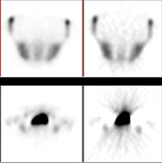
In scientific visualization, a maximum intensity projection (MIP) is a method for 3D data that projects in the visualization plane the voxels with maximum intensity that fall in the way of parallel rays traced from the viewpoint to the plane of projection. This implies that two MIP renderings from opposite viewpoints are symmetrical images if they are rendered using orthographic projection.
The American College of Radiology (ACR), founded in 1923, is a professional medical society representing nearly 40,000 diagnostic radiologists, radiation oncologists, interventional radiologists, nuclear medicine physicians and medical physicists.

Quantitative computed tomography (QCT) is a medical technique that measures bone mineral density (BMD) using a standard X-ray computed tomography (CT) scanner with a calibration standard to convert Hounsfield units (HU) of the CT image to bone mineral density values. Quantitative CT scans are primarily used to evaluate bone mineral density at the lumbar spine and hip.
Abass Alavi is an Iranian-American physician-scientist specializing in the field of molecular imaging, most notably in the imaging modality of positron emission tomography (PET). In August 1976, he was part of the team that performed the first human PET studies of the brain and whole body using the radiotracer [18F]Fluorodeoxyglucose (FDG). Alavi holds the position of Professor of Radiology and Neurology, as well as Director of Research Education in the Department of Radiology at the University of Pennsylvania. Over a career spanning five decades, he has amassed over 2,300 publications and 60,000 citations, earning an h-index of 125 and placing his publication record in the top percentile of scientists.
The partial volume effect can be defined as the loss of apparent activity in small objects or regions because of the limited resolution of the imaging system. It occurs in medical imaging and more generally in biological imaging such as positron emission tomography (PET) and single-photon emission computed tomography (SPECT). If the object or region to be imaged is less than twice the full width at half maximum (FWHM) resolution in x-, y- and z-dimension of the imaging system, the resultant activity in the object or region is underestimated. A higher resolution decreases this effect, as it better resolves the tissue.

David Edmund Kuhl was an American scientist specializing in nuclear medicine. He was well known for his pioneering work in positron emission tomography. Dr. Kuhl served as the Chief of the Division of Nuclear Medicine at the University of Michigan for 20 years and retired in June 2011.
Burton Drayer, MD, FACR, FANN, is an American radiologist and nationally recognized authority on the use of computed tomography and magnetic resonance imaging for diagnosing neurological disorders. From 2003 to 2008, he served as president, The Mount Sinai Hospital. As of 2020, he is the Charles M. and Marilyn Newman Professor and System Chair, Radiology, for The Mount Sinai Health System and Icahn School of Medicine at Mount Sinai Hospital in New York City.

Cone beam computed tomography is a medical imaging technique consisting of X-ray computed tomography where the X-rays are divergent, forming a cone.
A medical procedure is defined as non-invasive when no break in the skin is created and there is no contact with the mucosa, or skin break, or internal body cavity beyond a natural or artificial body orifice. For example, deep palpation and percussion are non-invasive but a rectal examination is invasive. Likewise, examination of the ear-drum or inside the nose or a wound dressing change all fall outside the definition of non-invasive procedure. There are many non-invasive procedures, ranging from simple observation, to specialised forms of surgery, such as radiosurgery. Extracorporeal shock wave lithotripsy is a non-invasive treatment of stones in the kidney, gallbladder or liver, using an acoustic pulse. For centuries, physicians have employed many simple non-invasive methods based on physical parameters in order to assess body function in health and disease, such as pulse-taking, the auscultation of heart sounds and lung sounds, temperature examination, respiratory examination, peripheral vascular examination, oral examination, abdominal examination, external percussion and palpation, blood pressure measurement, change in body volumes, audiometry, eye examination, and many others.
Medical Image Analysis (MedIA) is a peer-reviewed academic journal which focuses on medical and biological image analysis. The journal publishes papers which contribute to the basic science of analyzing and processing biomedical images acquired through means such as magnetic resonance imaging, ultrasound, computed tomography, nuclear medicine, x-ray, optical and confocal microscopy, among others. Common topics covered in the journal include feature extraction, image segmentation, image registration, and other image processing methods with applications to diagnosis, prognosis, and computer-assisted interventions.
Richard Frederick Spaide is an American ophthalmologist and retinal specialist known for his work in retinal diseases and advancements in ocular imaging techniques.