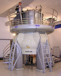
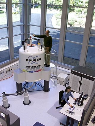
The nuclear Overhauser effect (NOE) is the transfer of nuclear spin polarization from one population of spin-active nuclei to another via cross-relaxation. A phenomenological definition of the NOE in nuclear magnetic resonance spectroscopy (NMR) is the change in the integrated intensity of one NMR resonance that occurs when another is saturated by irradiation with an RF field. The change in resonance intensity of a nucleus is a consequence of the nucleus being close in space to those directly affected by the RF perturbation.
In nuclear magnetic resonance (NMR) spectroscopy, the chemical shift is the resonant frequency of an atomic nucleus relative to a standard in a magnetic field. Often the position and number of chemical shifts are diagnostic of the structure of a molecule. Chemical shifts are also used to describe signals in other forms of spectroscopy such as photoemission spectroscopy.

In chemistry, conformational isomerism is a form of stereoisomerism in which the isomers can be interconverted just by rotations about formally single bonds. While any two arrangements of atoms in a molecule that differ by rotation about single bonds can be referred to as different conformations, conformations that correspond to local minima on the potential energy surface are specifically called conformational isomers or conformers. Conformations that correspond to local maxima on the energy surface are the transition states between the local-minimum conformational isomers. Rotations about single bonds involve overcoming a rotational energy barrier to interconvert one conformer to another. If the energy barrier is low, there is free rotation and a sample of the compound exists as a rapidly equilibrating mixture of multiple conformers; if the energy barrier is high enough then there is restricted rotation, a molecule may exist for a relatively long time period as a stable rotational isomer or rotamer. When the time scale for interconversion is long enough for isolation of individual rotamers, the isomers are termed atropisomers. The ring-flip of substituted cyclohexanes constitutes another common form of conformational isomerism.
In chemistry the descriptor vicinal, abbreviated vic, is a descriptor that identifies two functional groups as bonded to two adjacent carbon atoms. It may arise from vicinal difunctionalization.

Nuclear magnetic resonance spectroscopy, most commonly known as NMR spectroscopy or magnetic resonance spectroscopy (MRS), is a spectroscopic technique based on re-orientation of atomic nuclei with non-zero nuclear spins in an external magnetic field. This re-orientation occurs with absorption of electromagnetic radiation in the radio frequency region from roughly 4 to 900 MHz, which depends on the isotopic nature of the nucleus and increased proportionally to the strength of the external magnetic field. Notably, the resonance frequency of each NMR-active nucleus depends on its chemical environment. As a result, NMR spectra provide information about individual functional groups present in the sample, as well as about connections between nearby nuclei in the same molecule. As the NMR spectra are unique or highly characteristic to individual compounds and functional groups, NMR spectroscopy is one of the most important methods to identify molecular structures, particularly of organic compounds.

Solid-state nuclear magnetic resonance (ssNMR) is a spectroscopy technique used to characterize atomic-level structure and dynamics in solid materials. ssNMR spectra are broader due to nuclear spin interactions which can be categorized as dipolar coupling, chemical shielding, quadrupolar interactions, and j-coupling. These interactions directly affect the lines shapes of experimental ssNMR spectra which can be seen in powder and dipolar patterns. There are many essential solid-state techniques alongside advanced ssNMR techniques that may be applied to elucidate the fundamental aspects of solid materials. ssNMR is often combined with magic angle spinning (MAS) to remove anisotropic interactions and improve the sensitivity of the technique. The applications of ssNMR further extend to biology and medicine.
Carbon-13 (C13) nuclear magnetic resonance is the application of nuclear magnetic resonance (NMR) spectroscopy to carbon. It is analogous to proton NMR and allows the identification of carbon atoms in an organic molecule just as proton NMR identifies hydrogen atoms. 13C NMR detects only the 13
C
isotope. The main carbon isotope, 12
C
does not produce an NMR signal. Although ca. 1 mln. times less sensitive than 1H NMR spectroscopy, 13C NMR spectroscopy is widely used for characterizing organic and organometallic compounds, primarily because 1H-decoupled 13C-NMR spectra are more simple, have a greater sensitivity to differences in the chemical structure, and, thus, are better suited for identifying molecules in complex mixtures. At the same time, such spectra lack quantitative information about the atomic ratios of different types of carbon nuclei, because nuclear Overhauser effect used in 1H-decoupled 13C-NMR spectroscopy enhances the signals from carbon atoms with a larger number of hydrogen atoms attached to them more than from carbon atoms with a smaller number of H's, and because full relaxation of 13C nuclei is usually not attained, and the nuclei with shorter relaxation times produce more intense signals.

Proton nuclear magnetic resonance is the application of nuclear magnetic resonance in NMR spectroscopy with respect to hydrogen-1 nuclei within the molecules of a substance, in order to determine the structure of its molecules. In samples where natural hydrogen (H) is used, practically all the hydrogen consists of the isotope 1H.
Nuclear magnetic resonance spectroscopy of proteins is a field of structural biology in which NMR spectroscopy is used to obtain information about the structure and dynamics of proteins, and also nucleic acids, and their complexes. The field was pioneered by Richard R. Ernst and Kurt Wüthrich at the ETH, and by Ad Bax, Marius Clore, Angela Gronenborn at the NIH, and Gerhard Wagner at Harvard University, among others. Structure determination by NMR spectroscopy usually consists of several phases, each using a separate set of highly specialized techniques. The sample is prepared, measurements are made, interpretive approaches are applied, and a structure is calculated and validated.

Two-Dimensional Nuclear Magnetic Resonance is an advanced spectroscopic technique that builds upon the capabilities of one-dimensional (1D) NMR by incorporating an additional frequency dimension. This extension allows for a more comprehensive analysis of molecular structures. In 2D NMR, signals are distributed across two frequency axes, providing improved resolution and separation of overlapping peaks, particularly beneficial for studying complex molecules. This technique identifies correlations between different nuclei within a molecule, facilitating the determination of connectivity, spatial proximity, and dynamic interactions.
In nuclear chemistry and nuclear physics, J-couplings are mediated through chemical bonds connecting two spins. It is an indirect interaction between two nuclear spins that arises from hyperfine interactions between the nuclei and local electrons. In NMR spectroscopy, J-coupling contains information about relative bond distances and angles. Most importantly, J-coupling provides information on the connectivity of chemical bonds. It is responsible for the often complex splitting of resonance lines in the NMR spectra of fairly simple molecules.

The residual dipolar coupling between two spins in a molecule occurs if the molecules in solution exhibit a partial alignment leading to an incomplete averaging of spatially anisotropic dipolar couplings.
Carbon satellites in physics and spectroscopy, are small peaks that can be seen shouldering the main peaks in the nuclear magnetic resonance (NMR) spectrum. These peaks can occur in the NMR spectrum of any NMR active atom where those atoms adjoin a carbon atom. However, Carbon satellites are most often encountered in proton NMR.

Fluorine-19 nuclear magnetic resonance spectroscopy is an analytical technique used to detect and identify fluorine-containing compounds. 19F is an important nucleus for NMR spectroscopy because of its receptivity and large chemical shift dispersion, which is greater than that for proton nuclear magnetic resonance spectroscopy.
Carbohydrate NMR spectroscopy is the application of nuclear magnetic resonance (NMR) spectroscopy to structural and conformational analysis of carbohydrates. This method allows the scientists to elucidate structure of monosaccharides, oligosaccharides, polysaccharides, glycoconjugates and other carbohydrate derivatives from synthetic and natural sources. Among structural properties that could be determined by NMR are primary structure, saccharide conformation, stoichiometry of substituents, and ratio of individual saccharides in a mixture. Modern high field NMR instruments used for carbohydrate samples, typically 500 MHz or higher, are able to run a suite of 1D, 2D, and 3D experiments to determine a structure of carbohydrate compounds.
Nuclear magnetic resonance decoupling is a special method used in nuclear magnetic resonance (NMR) spectroscopy where a sample to be analyzed is irradiated at a certain frequency or frequency range to eliminate or partially the effect of coupling between certain nuclei. NMR coupling refers to the effect of nuclei on each other in atoms within a couple of bonds distance of each other in molecules. This effect causes NMR signals in a spectrum to be split into multiple peaks. Decoupling fully or partially eliminates splitting of the signal between the nuclei irradiated and other nuclei such as the nuclei being analyzed in a certain spectrum. NMR spectroscopy and sometimes decoupling can help determine structures of chemical compounds.

Nuclear magnetic resonance (NMR) is a physical phenomenon in which nuclei in a strong constant magnetic field are disturbed by a weak oscillating magnetic field and respond by producing an electromagnetic signal with a frequency characteristic of the magnetic field at the nucleus. This process occurs near resonance, when the oscillation frequency matches the intrinsic frequency of the nuclei, which depends on the strength of the static magnetic field, the chemical environment, and the magnetic properties of the isotope involved; in practical applications with static magnetic fields up to ca. 20 tesla, the frequency is similar to VHF and UHF television broadcasts (60–1000 MHz). NMR results from specific magnetic properties of certain atomic nuclei. High-resolution nuclear magnetic resonance spectroscopy is widely used to determine the structure of organic molecules in solution and study molecular physics and crystals as well as non-crystalline materials. NMR is also routinely used in advanced medical imaging techniques, such as in magnetic resonance imaging (MRI). The original application of NMR to condensed matter physics is nowadays mostly devoted to strongly correlated electron systems. It reveals large many-body couplings by fast broadband detection and should not be confused with solid state NMR, which aims at removing the effect of the same couplings by Magic Angle Spinning techniques.
Nucleic acid NMR is the use of nuclear magnetic resonance spectroscopy to obtain information about the structure and dynamics of nucleic acid molecules, such as DNA or RNA. It is useful for molecules of up to 100 nucleotides, and as of 2003, nearly half of all known RNA structures had been determined by NMR spectroscopy.
In the context of nuclear magnetic resonance (NMR), the term magnetic inequivalence refers to the distinction between magnetically active nuclear spins by their NMR signals, owing to a difference in either chemical shift or spin–spin coupling (J-coupling). Since chemically inequivalent spins are expected to also be magnetically distinct, and since an observed difference in chemical shift makes their inequivalence clear, the term magnetic inequivalence most commonly refers solely to the latter type, i.e. to situations of chemically equivalent spins differing in their coupling relationships.
PREDITOR is a freely available web-server for the prediction of protein torsion angles from chemical shifts. For many years it has been known that protein chemical shifts are sensitive to protein secondary structure, which in turn, is sensitive to backbone torsion angles. torsion angles are internal coordinates that can be used to describe the conformation of a polypeptide chain. They can also be used as constraints to help determine or refine protein structures via NMR spectroscopy. In proteins there are four major torsion angles of interest: phi, psi, omega and chi-1. Traditionally protein NMR spectroscopists have used vicinal J-coupling information and the Karplus relation to determine approximate backbone torsion angle constraints for phi and chi-1 angles. However, several studies in the early 1990s pointed out the strong relationship between 1H and 13C chemical shifts and torsion angles, especially with backbone phi and psi angles. Later a number of other papers pointed out additional chemical shift relationships with chi-1 and even omega angles. PREDITOR was designed to exploit these experimental observations and to help NMR spectroscopists easily predict protein torsion angles from chemical shift assignments. Specifically, PREDITOR accepts protein sequence and/or chemical shift data as input and generates torsion angle predictions for phi, psi, omega and chi-1 angles. The algorithm that PREDITOR uses combines sequence alignment, chemical shift alignment and a number of related chemical shift analysis techniques to predict torsion angles. PREDITOR is unusually fast and exhibits a very high level of accuracy. In a series of tests 88% of PREDITOR’s phi/psi predictions were within 30 degrees of the correct values, 84% of chi-1 predictions were correct and 99.97% of PREDITOR’s predicted omega angles were correct. PREDITOR also estimates the torsion angle errors so that its torsion angle constraints can be used with standard protein structure refinement software, such as CYANA, CNS, XPLOR and AMBER. PREDITOR also supports automated protein chemical shift re-referencing and the prediction of proline cis/trans states. PREDITOR is not the only torsion angle prediction software available. Several other computer programs including TALOS, TALOS+ and DANGLE have also been developed to predict backbone torsion angles from protein chemical shifts. These stand-alone programs exhibit similar prediction performance to PREDITOR but are substantially slower.


