Labral reconstruction

Last updated
Labral reconstruction
Reconlabrum.jpg
Arthroscopic view of the superior acetabular labrum status-post circumferential labral reconstruction with ITB allograft. Note restoration of the labral suction-seal. Surgery performed by Dr. Andrew B Wolff of Washington Orthopaedics and Sports Medicine.
Specialty orthopedic

Labral reconstruction is a type of hip arthroscopy in which the patient's native labrum is partially or completely removed and reconstructed using either autograft or allograft tissue. Originally described in 2009 [1] using the ligamentum teres capitis, arthroscopic labral reconstruction using a variety of graft tissue has demonstrated promising short and mid-term clinical outcomes. [2] [3] [4] Most importantly, labral reconstruction has demonstrated utility when the patient's native labral tissue is far too damaged for debridement or repair. [5]

Contents

Anatomy

The acetabular labrum is a fibrocartilagenous structure similar in composition to the meniscus. It is a ring of tissue that surrounds the acetabulum of the pelvis, and allows the head of the femur to articulate smoothly and efficiently with the acetabulum. The labrum plays an important role in maintaining the biomechanical stability of the hip joint. Studies [6] have shown that damage to the labral tissue can result in disruption of the labral suction-seal, a fluid force paramount in maintaining hip joint integrity. An intact labrum also helps to buttress the hip joint to distraction forces. [7] The labrum, when damaged, is also a pain generator, due to a large concentration of type II pain-associated free nerve endings found throughout the tissue, most pronounced at the labral base. [8]

Hip joint, front view. The capsular ligament has been largely removed. Gray342.png
Hip joint, front view. The capsular ligament has been largely removed.

History

Labral reconstruction was first described in 2009 by Sierra et al. [1] The procedure described in their article described reconstructing a patient's native labrum with a ligamentum teres capitis graft. This was done in the setting of an open surgical hip dislocation. Prior to the introduction of labral reconstruction, complex labral tears were often treated with removal of damaged tissue (debridement) or focal repair. The applicability of these methods to severe or widespread labral damage is less than ideal. Since then, surgeons have reported on a variety of graft choices and surgical techniques, and an arthroscopic approach has usurped open dislocation, due to fewer complications, a lower need for revision surgery and quicker recovery time. [9] [10]

Indications

Labral reconstruction, while still a relatively novel technique, has demonstrated utility and efficacy in treating labral tears in patient's whose native labral tissue is far too damaged for arthroscopic debridement or repair. [5] It is most often utilized in order to surgically correct the damage resulting from femoroacetabular impingement (FAI), a condition in which the femoral head articulates imperfectly with the acetabular cup. Labral damage resulting from FAI or other conditions exists on a spectrum, with varying degrees of labral damage necessitating different surgical management. The most mild degrees of labral damage can be managed with arthroscopic debridement, a procedure in which the damaged tissue is excised with an arthroscopic shaver or electrocautery device. More moderate damage responds better to arthroscopic labral repair, a procedure in which surgical anchors are drilled into the bony acetabular rim and sutures are used to reapproximate the damaged labral tissue. The most severe degrees of labral pathology is often unresponsive to labral repair, with damage far too diffuse for focal debridement. In these cases, labral reconstruction is the best option for not only restoring the biomechanics of the acetabular labrum, but for treatment of the patient's pain.

Diagram illustrating the two subtypes of FAI. Femoral acetabular impingement FAI de.svg
Diagram illustrating the two subtypes of FAI.

A recent multicenter epidemiological study found that the majority of patients undergoing labral reconstruction are middle-aged females whose pain is localized around the groin. [11] Patient pain is ofter exacerbated by sitting and athletic activities. Many patients undergoing labral reconstruction have failed conservative therapy, which typically includes intra-articular injections and physical therapy. A majority of patients have abnormal acetabular or femoral bony morphology typical of femoroacetabular impingement (FAI).[ citation needed ]

Technique

Graft selection

A variety of graft options for labral reconstruction have been proposed. Although the literature for ACL reconstruction has demonstrated more favorable outcomes with autograft tissue versus allograft, no such relationship has been found to exist for labral reconstruction. Drs. Brian White and Andrew Wolff, two sports medicine trained surgeons specializing in hip arthroscopy, both prefer the utilization of allograft tissue. [12] [13] The surgeons who advocate for the use of allograft tissue feel that the control over graft thickness, consistency and size, in addition to the absence of donor site morbidity make it the preferred graft choice for this procedure. Other graft options include iliotibial band autograft, hamstrings autograft or quadriceps tendon autograft. [5] Currently, there is a lack of sufficient data to claim one graft choice superior to the others.

Segmental vs. circumferential/front-to-back

Traditionally, only the damaged labral tissue was resected, and the graft was attached to both the acetabulum and the native labral tissue. This method demonstrated superiority over straight debridement in the treatment of irreparable labral tears. [5] There was concern by some surgeons, however, that the junction points between the native labrum and graft were inherently weak, and thus prone to failure. [12] There was also concern that despite resection of the visibly damaged tissue there existed the possibility for underresection, which could lead to persistent pain despite restoration of the labral biomechanics.[ citation needed ]

Recently, surgeons have begun experimenting with circumferential (front-to-back) reconstruction in which the entirety of the native labral tissue is debrided, and the labrum is completely reconstructed. [12] This technique has shown promising outcomes when utilized in patients whose native labral tissue is far too damaged for repair or debridement. A recent study comparing primary labral reconstruction versus primary labral repair demonstrated higher failure rates in the repair cohort versus the reconstruction cohort. [14]

Outcomes

Arthroscopic labral reconstruction has demonstrated favorable outcomes, which have become more pronounced as techniques and technology continue to develop. Arthroscopic labral reconstruction has shown comparable outcomes to labral repair, despite the fact that those patients who undergo reconstruction typically have far more severe labral damage. [3] Recent studies have shown not only equivalent outcomes between labral reconstruction and labral repair, but improved outcomes for labral reconstruction for patients with more moderate degrees of labral damage. Labral reconstruction also has proven utility in the hips of elite athletes and other high-demand patients. [15]

Complications

The complications encountered during and after labral reconstruction are similar to arthroscopic procedures involving the hip. [16]

Anesthetic complications

Anesthetic complications are rare, but include urinary retention, gastrointestinal upset, cardiac complications and even death.[ citation needed ]

Operative complications

As with all surgery, arthroscopic labral reconstruction has a small risk of infection. Damage to the surrounding neurovasculature is possible, but this risk is minimized through meticulous surgical technique. The most commonly injured nerve is the lateral femoral cutaneous nerve, although this risk is very low with proper arthroscopic portal placement. A new postless table designed by Stryker has nearly eliminated the risk of postoperative saddle parasthesia, which was previously a common complication. Post-operative deep-vein thrombosis is also possible, but the rate of this complication can be minimized through the use of blood thinning medications and early ambulation.[ citation needed ]

Related Research Articles

Arthroscopy

Arthroscopy is a minimally invasive surgical procedure on a joint in which an examination and sometimes treatment of damage is performed using an arthroscope, an endoscope that is inserted into the joint through a small incision. Arthroscopic procedures can be performed during ACL reconstruction.

Anterior cruciate ligament Type of cruciate ligament in the human knee

The anterior cruciate ligament (ACL) is one of a pair of cruciate ligaments in the human knee. The two ligaments are also called "cruciform" ligaments, as they are arranged in a crossed formation. In the quadruped stifle joint, based on its anatomical position, it is also referred to as the cranial cruciate ligament. The term cruciate translates to cross. This name is fitting because the ACL crosses the posterior cruciate ligament to form an “X”. It is composed of strong, fibrous material and assists in controlling excessive motion. This is done by limiting mobility of the joint. The anterior cruciate ligament is one of the four main ligaments of the knee, providing 85% of the restraining force to anterior tibial displacement at 30 and 90° of knee flexion. The ACL is the most injured ligament of the four located in the knee.

Anterior cruciate ligament reconstruction Surgical process

Anterior cruciate ligament reconstruction is a surgical tissue graft replacement of the anterior cruciate ligament, located in the knee, to restore its function after an injury. The torn ligament can either be removed from the knee, or preserved before reconstruction an arthroscopic procedure. ACL repair is also a surgical option. This involves repairing the ACL by re-attaching it, instead of performing a reconstruction. Theoretical advantages of repair include faster recovery and a lack of donor site morbidity, but randomised controlled trials and long-term data regarding re-rupture rates using contemporary surgical techniques are lacking.

Autotransplantation is the transplantation of organs, tissues, or even particular proteins from one part of the body to another in the same person.

A meniscus transplant or meniscal transplant is a transplant of the meniscus of the knee, which separates the thigh bone (femur) from the lower leg bone (tibia). The worn or damaged meniscus is removed and is replaced with a new one from a donor. The meniscus to be transplanted is taken from a cadaver, and, as such, is known as an allograft. Meniscal transplantation is technically difficult, as it must be sized accurately for each person, positioned properly and secured to the tibial plateau. As of 2012, only a few surgeons have significant volume of experience in meniscus transplantation worldwide.

SLAP tear Medical condition

A SLAP tear or SLAP lesion is an injury to the glenoid labrum. SLAP is an acronym for "superior labral tear from anterior to posterior".

A bursectomy is the removal of a bursa, which is a small sac filled with synovial fluid that cushions adjacent bone structures and reduces friction in joint movement. This procedure is usually carried out to relieve chronic inflammation (bursitis) or infection, when conservative management has failed to improve patient outcomes.

Cranioplasty

Cranioplasty is a surgical operation on the repairing of cranial defects caused by previous injuries or operations, such as decompressive craniectomy. It is performed by filling the defective area with a range of materials, usually a bone piece from the patient or a synthetic material. Cranioplasty is carried out by incision and reflection of the scalp after applying anaesthetics and antibiotics to the patient. The temporalis muscle is reflected, and all surrounding soft tissues are removed, thus completely exposing the cranial defect. The cranioplasty flap is placed and secured on the cranial defect. The wound is then sealed.

Acetabular labrum Ring of cartilage that surrounds the acetabulum of the hip

The acetabular labrum is a ring of cartilage that surrounds the acetabulum of the hip. The anterior portion is most vulnerable when the labrum tears.

Dislocated shoulder Injury

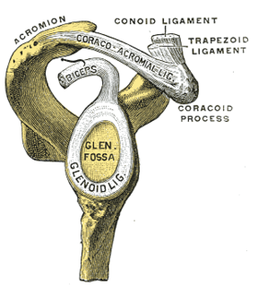
A dislocated shoulder is a condition in which the head of the humerus is detached from the shoulder joint. Symptoms include shoulder pain and instability. Complications may include a Bankart lesion, Hill-Sachs lesion, rotator cuff tear, or injury to the axillary nerve.

Shoulder surgery is a means of treating injured shoulders. Many surgeries have been developed to repair the muscles, connective tissue, or damaged joints that can arise from traumatic or overuse injuries to the shoulder.

Bankart lesion Medical condition

A Bankart lesion is an injury of the anterior (inferior) glenoid labrum of the shoulder. When this happens, a pocket at the front of the glenoid forms that allows the humeral head to dislocate into it. It is an indication for surgery and often accompanied by a Hill-Sachs lesion, damage to the posterior humeral head.

The aim of an articular cartilage repair treatment is to restore the surface of an articular joint's hyaline cartilage. Over the last decades, surgeons and researchers have made progress in elaborating surgical cartilage repair interventions. Though these solutions do not perfectly restore articular cartilage, some of the latest technologies start to bring very promising results in repairing cartilage from traumatic injuries or chondropathies. These treatments are especially targeted by patients who suffer from articular cartilage damage. They provide pain relief while at the same time slowing down the progression of damage or considerably delaying joint replacement surgery. Articular cartilage repair treatments help patients to return to their original lifestyle; regaining mobility, going back to work and even practicing sports again.

Femoroacetabular impingement Medical condition

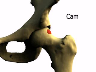
Femoroacetabular impingement (FAI) is a condition involving one or more anatomical abnormalities of the hip joint, which is a ball and socket joint. It is a common cause of hip pain and discomfort in young and middle-aged adults. It occurs when the ball shaped femoral head contacts the acetabulum abnormally or does not permit a normal range of motion in the acetabular socket. Damage can occur to the articular cartilage, or labral cartilage, or both. The condition may be symptomatic or asymptomatic. It may cause osteoarthritis of the hip. Treatment options range from conservative management to surgery.

Hip arthroscopy

Hip arthroscopy refers to the viewing of the interior of the acetabulofemoral (hip) joint through an arthroscope and the treatment of hip pathology through a minimally invasive approach. This technique is sometimes used to help in the treatment of various joint disorders and has gained popularity because of the small incisions used and shorter recovery times when compared with conventional surgical techniques. Hip arthroscopy was not feasible until recently, new technology in both the tools used and the ability to distract the hip joint has led to a recent surge in the ability to do hip arthroscopy and the popularity of it.

Posterolateral corner injuries of the knee are injuries to a complex area formed by the interaction of multiple structures. Injuries to the posterolateral corner can be debilitating to the person and require recognition and treatment to avoid long term consequences. Injuries to the PLC often occur in combination with other ligamentous injuries to the knee; most commonly the anterior cruciate ligament (ACL) and posterior cruciate ligament (PCL). As with any injury, an understanding of the anatomy and functional interactions of the posterolateral corner is important to diagnosing and treating the injury.

Cartilage repair techniques are the current focus of large amounts of research. Many different strategies have been proposed as solutions for cartilage defects. Surgical techniques currently being studied include:

Nerve allotransplantation is the transplantation of a nerve to a receiver from a donor of the same species. For example, nerve tissue is transplanted from one person to another. Allotransplantation is a commonly used type of transplantation of which nerve repair is one specific aspect.

Wrist arthroscopy

Wrist arthroscopy can be used to look inside the joint of the wrist. It is a minimally invasive technique which can be utilized for diagnostic purposes as well as for therapeutic interventions. Wrist arthroscopy has been used for diagnostic purposes since it was first introduced in 1979. However, it only became accepted as diagnostic tool around the mid-1980s. At that time, arthroscopy of the wrist was an innovative technique to determine whether a problem could be found in the wrist. A few years later, wrist arthroscopy could also be used as a therapeutic tool.

Gingival grafting

Gingival grafting, also called gum grafting or periodontal plastic surgery, is a generic term for the performance of any of a number of periodontal surgical procedures in which the gum tissue is grafted. The aim may be to cover exposed root surfaces or merely to augment the band of keratinized tissue.

References

  1. 1 2 Sierra, Rafael J.; Trousdale, Robert T. (2009-03-01). "Labral Reconstruction Using the Ligamentum Teres Capitis: Report of a New Technique". Clinical Orthopaedics and Related Research. 467 (3): 753–759. doi:10.1007/s11999-008-0633-5. ISSN   0009-921X. PMC   2635467 . PMID   19048354.
  2. Philippon, Marc J.; Briggs, Karen K.; Hay, Connor J.; Kuppersmith, David A.; Dewing, Christopher B.; Huang, Michael J. (2010). "Arthroscopic Labral Reconstruction in the Hip Using Iliotibial Band Autograft: Technique and Early Outcomes". Arthroscopy: The Journal of Arthroscopic and Related Surgery. 26 (6): 750–756. doi:10.1016/j.arthro.2009.10.016. PMID   20511032.
  3. 1 2 Matsuda, Dean K.; Burchette, Raoul J. (May 2013). "Arthroscopic hip labral reconstruction with a gracilis autograft versus labral refixation: 2-year minimum outcomes". The American Journal of Sports Medicine. 41 (5): 980–987. doi:10.1177/0363546513482884. ISSN   1552-3365. PMID   23548806. S2CID   27181307.
  4. Philippon, Marc J.; Briggs, Karen K.; Hay, Connor J.; Kuppersmith, David A.; Dewing, Christopher B.; Huang, Michael J. (June 2010). "Arthroscopic labral reconstruction in the hip using iliotibial band autograft: technique and early outcomes". Arthroscopy: The Journal of Arthroscopic and Related Surgery. 26 (6): 750–756. doi:10.1016/j.arthro.2009.10.016. ISSN   1526-3231. PMID   20511032.
  5. 1 2 3 4 Domb, Benjamin G.; El Bitar, Youssef F.; Stake, Christine E.; Trenga, Anthony P.; Jackson, Timothy J.; Lindner, Dror (January 2014). "Arthroscopic labral reconstruction is superior to segmental resection for irreparable labral tears in the hip: a matched-pair controlled study with minimum 2-year follow-up". The American Journal of Sports Medicine. 42 (1): 122–130. doi:10.1177/0363546513508256. ISSN   1552-3365. PMID   24186974. S2CID   206528292.
  6. Philippon, Marc J.; Nepple, Jeffrey J.; Campbell, Kevin J.; Dornan, Grant J.; Jansson, Kyle S.; LaPrade, Robert F.; Wijdicks, Coen A. (2014-04-01). "The hip fluid seal—Part I: the effect of an acetabular labral tear, repair, resection, and reconstruction on hip fluid pressurization". Knee Surgery, Sports Traumatology, Arthroscopy. 22 (4): 722–729. doi:10.1007/s00167-014-2874-z. ISSN   0942-2056. PMID   24519614. S2CID   8109881.
  7. Nepple, Jeffrey J.; Philippon, Marc J.; Campbell, Kevin J.; Dornan, Grant J.; Jansson, Kyle S.; LaPrade, Robert F.; Wijdicks, Coen A. (2014-04-01). "The hip fluid seal—Part II: The effect of an acetabular labral tear, repair, resection, and reconstruction on hip stability to distraction". Knee Surgery, Sports Traumatology, Arthroscopy. 22 (4): 730–736. doi:10.1007/s00167-014-2875-y. ISSN   0942-2056. PMID   24509878. S2CID   9258547.
  8. Haversath, M.; Hanke, J.; Landgraeber, S.; Herten, M.; Zilkens, C.; Krauspe, R.; Jäger, M. (June 2013). "The distribution of nociceptive innervation in the painful hip: a histological investigation". The Bone & Joint Journal. 95-B (6): 770–776. doi:10.1302/0301-620X.95B6.30262. ISSN   2049-4408. PMID   23723270.
  9. Botser, Itamar B.; Smith, Thomas W.; Nasser, Rima; Domb, Benjamin G. (2011). "Open Surgical Dislocation Versus Arthroscopy for Femoroacetabular Impingement: A Comparison of Clinical Outcomes". Arthroscopy: The Journal of Arthroscopic and Related Surgery. 27 (2): 270–278. doi:10.1016/j.arthro.2010.11.008. PMID   21266277.
  10. Ng, Vincent Y.; Arora, Naveen; Best, Thomas M.; Pan, Xueliang; Ellis, Thomas J. (November 2010). "Efficacy of surgery for femoroacetabular impingement: a systematic review". The American Journal of Sports Medicine. 38 (11): 2337–2345. doi:10.1177/0363546510365530. ISSN   1552-3365. PMID   20489213. S2CID   41692112.
  11. Kivlan, Benjamin R.; Nho, Shane J.; Christoforetti, John J.; Ellis, Thomas J.; Matsuda, Dean K.; Salvo, John P.; Wolff, Andrew B.; Van Thiel, Geoffrey S.; Stubbs, Allston J. (January–February 2017). "Multicenter Outcomes After Hip Arthroscopy: Epidemiology (MASH Study Group). What Are We Seeing in the Office, and Who Are We Choosing to Treat?". American Journal of Orthopedics (Belle Mead, N.J.). 46 (1): 35–41. ISSN   1934-3418. PMID   28235111.
  12. 1 2 3 Wolff, Andrew B.; Grossman, Jamie (July 2016). "Management of the Acetabular Labrum". Clinics in Sports Medicine. 35 (3): 345–360. doi:10.1016/j.csm.2016.02.004. ISSN   1556-228X. PMID   27343389.
  13. White, Brian J.; Herzog, Mackenzie M. (February 2016). "Arthroscopic Labral Reconstruction of the Hip Using Iliotibial Band Allograft and Front-to-Back Fixation Technique". Arthroscopy Techniques. 5 (1): e89–97. doi:10.1016/j.eats.2015.08.009. ISSN   2212-6287. PMC   4809662 . PMID   27073784.
  14. White, Brian J.; Patterson, Julie; Herzog, Mackenzie M. (February 2018). "Bilateral Hip Arthroscopy: Direct Comparison of Primary Acetabular Labral Repair and Primary Acetabular Labral Reconstruction". Arthroscopy: The Journal of Arthroscopic and Related Surgery. 34 (2): 433–440. doi:10.1016/j.arthro.2017.08.240. ISSN   1526-3231. PMID   29100774.
  15. Boykin, Robert E.; Patterson, Diana; Briggs, Karen K.; Dee, Ashley; Philippon, Marc J. (October 2013). "Results of arthroscopic labral reconstruction of the hip in elite athletes". The American Journal of Sports Medicine. 41 (10): 2296–2301. doi:10.1177/0363546513498058. ISSN   1552-3365. PMID   23928321. S2CID   7843955.
  16. Ilizaliturri, Victor M. (March 2009). "Complications of arthroscopic femoroacetabular impingement treatment: a review". Clinical Orthopaedics and Related Research. 467 (3): 760–768. doi:10.1007/s11999-008-0618-4. ISSN   1528-1132. PMC   2635434 . PMID   19018604.