Articles related to anatomy include:

The brainstem is the posterior stalk-like part of the brain that connects the cerebrum with the spinal cord. In the human brain the brainstem is composed of the midbrain, the pons, and the medulla oblongata. The midbrain is continuous with the thalamus of the diencephalon through the tentorial notch, and sometimes the diencephalon is included in the brainstem.
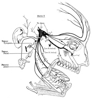
The trigeminal nerve (the fifth cranial nerve, or simply CN V) is a nerve responsible for sensation in the face and motor functions such as biting and chewing; it is the most complex of the cranial nerves. Its name ("trigeminal" = tri-, or three, and - geminus, or twin: thrice-twinned) derives from each of the two nerves (one on each side of the pons) having three major branches: the ophthalmic nerve (V1), the maxillary nerve (V2), and the mandibular nerve (V3). The ophthalmic and maxillary nerves are purely sensory, whereas the mandibular nerve supplies motor as well as sensory (or "cutaneous") functions. Adding to the complexity of this nerve is that autonomic nerve fibers as well as special sensory fibers (taste) are contained within it.

The midbrain or mesencephalon is the forward-most portion of the brainstem and is associated with vision, hearing, motor control, sleep and wakefulness, arousal (alertness), and temperature regulation. The name comes from the Greek mesos, "middle", and enkephalos, "brain".

The spinothalamic tract is a sensory pathway to the thalamus. From the ventral posterolateral nucleus in the thalamus, sensory information is relayed upward to the somatosensory cortex of the postcentral gyrus.

Medial medullary syndrome, also known as inferior alternating syndrome, hypoglossal alternating hemiplegia, lower alternating hemiplegia, or Dejerine syndrome, is a type of alternating hemiplegia characterized by a set of clinical features resulting from occlusion of the anterior spinal artery. This results in the infarction of medial part of the medulla oblongata.

The marginal nucleus of spinal cord, or posteromarginal nucleus, Rexed lamina I, is located at the most dorsal aspect of the dorsal horn of the spinal cord. The neurons located here receive input primarily from Lissauer's tract and relay information related to pain and temperature sensation. Pain sensation relayed here cannot be modulated, e.g. pain from burning the skin The axons of neurons contribute to the lateral spinothalamic tract.
The vestibulospinal tract is a neural tract in the central nervous system. Specifically, it is a component of the extrapyramidal system and is classified as a component of the medial pathway. Like other descending motor pathways, the vestibulospinal fibers of the tract relay information from nuclei to motor neurons. The vestibular nuclei receive information through the vestibulocochlear nerve about changes in the orientation of the head. The nuclei relay motor commands through the vestibulospinal tract. The function of these motor commands is to alter muscle tone, extend, and change the position of the limbs and head with the goal of supporting posture and maintaining balance of the body and head.

The nucleus raphe magnus, is located directly rostral to the nucleus raphe obscurus, and receives input from the spinal cord and cerebellum.
Dissociated sensory loss is a pattern of neurological damage caused by a lesion to a single tract in the spinal cord which involves preservation of fine touch and proprioception withselective loss of pain and temperature

The posterolateral tract is a small strand situated in relation to the tip of the posterior column close to the entrance of the posterior nerve roots. It is present throughout the spinal cord, and is most developed in the upper cervical regions.
The spinotectal tract arises in the spinothalamic tract and terminates in the inferior and superior colliculi.

The ventral posterior nucleus is the somato-sensory relay nucleus in thalamus of the brain.
The ventral posterolateral nucleus (VPL) is a nucleus of the thalamus. Together with the ventral posteromedial nucleus (VPM), ventral posterior inferior nucleus (VPI) and ventromedial posterior nucleus (VMpo), it constitutes the ventral posterior nucleus. There is uncertainty in the location of VMpo, as determined by spinothalamic tract (STT) terminations and staining for calcium-binding proteins, and several authorities do not consider its existence as being proved.

Brown-Séquard syndrome is caused by damage to one half of the spinal cord, i.e. hemisection of the spinal cord resulting in paralysis and loss of proprioception on the same side as the injury or lesion, and loss of pain and temperature sensation on the opposite side as the lesion. It is named after physiologist Charles-Édouard Brown-Séquard, who first described the condition in 1850.
The spinoreticular tract is an ascending pathway in the white matter of the spinal cord, positioned closely to the lateral spinothalamic tract. The tract is from spinal cord—to reticular formation— to thalamus.

The medial vestibular nucleus is one of the vestibular nuclei. It is located in the medulla oblongata.

The spinal cord is a long, thin, tubular structure made up of nervous tissue, which extends from the medulla oblongata in the brainstem to the lumbar region of the vertebral column. It encloses the central canal of the spinal cord, which contains cerebrospinal fluid. The brain and spinal cord together make up the central nervous system (CNS). In humans, the spinal cord begins at the occipital bone, passing through the foramen magnum and entering the spinal canal at the beginning of the cervical vertebrae. The spinal cord extends down to between the first and second lumbar vertebrae, where it ends. The enclosing bony vertebral column protects the relatively shorter spinal cord. It is around 45 cm (18 in) in men and around 43 cm (17 in) long in women. The diameter of the spinal cord ranges from 13 mm in the cervical and lumbar regions to 6.4 mm in the thoracic area.
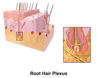
A hair plexus or root hair plexus is a special group of nerve fiber endings and serves as a very sensitive mechanoreceptor for touch sensation. Each hair plexus forms a network around a hair follicle and is a receptor, which means it sends and receives nerve impulses to and from the brain when the hair moves.