
The levator ani is a broad, thin muscle group, situated on either side of the pelvis. It is formed from three muscle components: the pubococcygeus, the iliococcygeus, and the puborectalis.

The levator scapulae is a slender skeletal muscle situated at the back and side of the neck. It originates from the transverse processes of the four uppermost cervical vertebrae; it inserts onto the upper portion of the medial border of the scapula. It is innervated by the cervical nerves C3-C4, and frequently also by the dorsal scapular nerve. As the Latin name suggests, its main function is to lift the scapula.

The cervical plexus is a nerve plexus of the anterior rami of the first four cervical spinal nerves C1-C4. The cervical plexus provides motor innervation to some muscles of the neck, and the diaphragm; it provides sensory innervation to parts of the head, neck, and chest.
The coccygeus muscle or ischiococcygeus is a muscle of the pelvic floor located posterior to levator ani and anterior to the sacrospinous ligament.

The levator palpebrae superioris is the muscle in the orbit that elevates the upper eyelid.

The zygomaticus minor muscle is a muscle of facial expression. It originates from the zygomatic bone, lateral to the rest of the levator labii superioris muscle, and inserts into the outer part of the upper lip. It draws the upper lip backward, upward, and outward and is used in smiling. It is innervated by the facial nerve (VII).

The levator labii superioris is a muscle of the human body used in facial expression. It is a broad sheet, the origin of which extends from the side of the nose to the zygomatic bone.

The sternothyroid muscle is an infrahyoid muscle of the neck. It acts to depress the hyoid bone.

The thyrohyoid muscle is a small skeletal muscle of the neck. Above, it attaches onto the greater cornu of the hyoid bone; below, it attaches onto the oblique line of the thyroid cartilage. It is innervated by fibres derived from the cervical spinal nerve 1 that run with the hypoglossal nerve to reach this muscle. The thyrohyoid muscle depresses the hyoid bone and elevates the larynx during swallowing. By controlling the position and shape of the larynx, it aids in making sound.
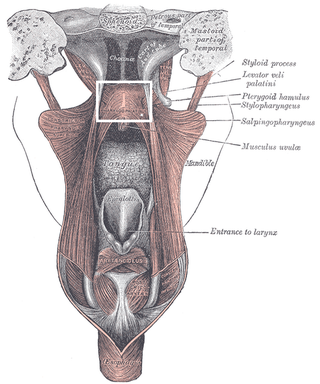
The palatopharyngeusmuscle is a small muscle in the roof of the mouth.
Terminologia Anatomica is the international standard for human anatomical terminology. It is developed by the Federative International Programme on Anatomical Terminology, a program of the International Federation of Associations of Anatomists (IFAA).

The palatine aponeurosis a thin, firm, fibrous lamella which gives strength and support to soft palate. It serves as the insertion for the tensor veli palatini and levator veli palatini, and the origin for the musculus uvulae, palatopharyngeus, and palatoglossus.

The oblique arytenoid is bilaterally paired intrinsic muscle of the larynx. It is superficial to the transverse arytenoid; the oblique and transverse arytenoids are often considered two parts of a single muscle - the interarytenoid muscle.

The inferior carotid triangle, is bounded, in front, by the median line of the neck from the hyoid bone to the sternum; behind, by the anterior margin of the sternocleidomastoid; above, by the superior belly of the omohyoid.

The pretracheal fascia is a layer of the deep cervical fascia at the front of the neck. It attaches to the hyoid bone above, and - extending down into the thorax - blends with the fibrous pericardium below. It encloses the thyroid gland and parathyroid glands, trachea, and esophagus. It extends medially in front of the carotid vessels. It assists in forming the carotid sheath.

The deep branch of the perineal nerve is a nerve of the perineum. It is a branch of the perineal nerve, from the pudendal nerve. It supplies the superficial transverse perineal muscle, bulbospongiosus muscle, ischiocavernosus muscle, the bulb of penis, levator ani, and the external anal sphincter.
Levator muscle can refer to:

The pharynx is the part of the throat behind the mouth and nasal cavity, and above the esophagus and trachea. It is found in vertebrates and invertebrates, though its structure varies across species. The pharynx carries food to the esophagus and air to the larynx. The flap of cartilage called the epiglottis stops food from entering the larynx.

The vaginal support structures are those muscles, bones, ligaments, tendons, membranes and fascia, of the pelvic floor that maintain the position of the vagina within the pelvic cavity and allow the normal functioning of the vagina and other reproductive structures in the female. Defects or injuries to these support structures in the pelvic floor leads to pelvic organ prolapse. Anatomical and congenital variations of vaginal support structures can predispose a woman to further dysfunction and prolapse later in life. The urethra is part of the anterior wall of the vagina and damage to the support structures there can lead to incontinence and urinary retention.
The pubovaginal muscle is a pelvic floor muscle that attaches to the muscles of lateral walls of the midsection of the vagina and the pubis. It is relatively short compared to the other levator ani muscles and extends between the pubic bones and the vagina. Other muscles that are part of the levator ani are: the pubococcygeus muscle which is made up of the puboperineal, pubovaginal, and puboanal muscles; the puborectal muscle; and the iliococcygeal muscle. The pubovaginal muscle was identified by anatomists as early as 1912.
