Lipotoxicity

Last updated
Two mice; the mouse on the left has more fat stores than the mouse on the right. Fatmouse.jpg
Two mice; the mouse on the left has more fat stores than the mouse on the right.

Lipotoxicity is a metabolic syndrome that results from the accumulation of lipid intermediates in non-adipose tissue, leading to cellular dysfunction and death. The tissues normally affected include the kidneys, liver, heart and skeletal muscle. Lipotoxicity is believed to have a role in heart failure, obesity, and diabetes, and is estimated to affect approximately 25% of the adult American population. [1]

Contents

Cause

In normal cellular operations, there is a balance between the production of lipids, and their oxidation or transport. In lipotoxic cells, there is an imbalance between the amount of lipids produced and the amount used. Upon entrance of the cell, fatty acids can be converted to different types of lipids for storage. Triacylglycerol consists of three fatty acids bound to a glycerol molecule and is considered the most neutral and harmless type of intracellular lipid storage. Alternatively, fatty acids can be converted to lipid intermediates like diacylglycerol, ceramides and fatty acyl-CoAs. These lipid intermediates can impair cellular function, which is referred to as lipotoxicity. [2]

Adipocytes, the cells that normally function as lipid store of the body, are well equipped to handle the excess lipids. Yet, too great of an excess will overburden these cells and cause a spillover into non-adipose cells, which do not have the necessary storage space. When the storage capacity of non-adipose cells is exceeded, cellular dysfunction and/or death result. The mechanism by which lipotoxicity causes death and dysfunction is not well understood. The cause of apoptosis and extent of cellular dysfunction is related to the type of cell affected, as well as the type and quantity of excess lipids. [3] A theory has been put forward by Cambridge researchers relating the development of lipotoxicity to the perturbation of membrane glycerophospholipid/sphingolipid homeostasis and their associated signalling events. [4]

Currently, there is no universally accepted theory for why certain individuals are afflicted with lipotoxicity. Research is ongoing into a genetic cause, but no individual gene has been named as the causative agent. The causative role of obesity in lipotoxicity is controversial. Some researchers claim that obesity has protective effects against lipotoxicity as it results in extra adipose tissue in which excess lipids can be stored. Others claim obesity is a risk factor for lipotoxicity. Both sides accept that high fat diets put patients at increased risk for lipotoxic cells. Individuals with high numbers of lipotoxic cells usually experience both leptin and insulin resistance. However, no causative mechanism has been found for this correlation. [5]

Effects in different organs

Kidneys

Renal lipotoxicity occurs when excess long-chain nonesterified fatty acids are stored in the kidney and proximal tubule cells. It is believed that these fatty acids are delivered to the kidneys via serum albumin. This condition leads to tubulointerstitial inflammation and fibrosis in mild cases, and to kidney failure and death in severe cases. The current accepted treatments for lipotoxicity in renal cells are fibrate therapy and intensive insulin therapy. [6]

Liver

An excess of free fatty acids in liver cells plays a role in Nonalcoholic Fatty Liver Disease (NAFLD). In the liver, it is the type of fatty acid, not the quantity, that determines the extent of the lipotoxic effects. In hepatocytes, the ratio of monounsaturated fatty acids and saturated fatty acids leads to apoptosis and liver damage. There are several potential mechanisms by which the excess fatty acids can cause cell death and damage. They may activate death receptors, stimulate apoptotic pathways, or initiate cellular stress response in the endoplasmic reticulum. These lipotoxic effects have been shown to be prevented by the presence of excess triglycerides within the hepatocytes. [7]

Heart

Lipotoxicity in cardiac tissue is attributed to excess saturated fatty acids. The apoptosis that follows is believed to be caused by unfolded protein response in the endoplasmic reticulum. Researchers are working on treatments that will increase the oxidation of these fatty acids within the heart in order to prevent the lipotoxic effects. [8]

Pancreas

Lipotoxicity affects the pancreas when excess free fatty acids are found in beta cells, causing their dysfunction and death. The effects of the lipotoxicity is treated with leptin therapy and insulin sensitizers. [9]

Skeletal muscle

The skeletal muscle accounts for more than 80 percent of the postprandial whole body glucose uptake and therefore plays an important role in glucose homeostasis. Skeletal muscle lipid levels – [2] Intramyocellular lipids are mainly stored in lipid droplets, the organelles for fat storage. Recent research indicates that creating intramyocellular neutral lipid storage capacity for example by increasing the abundance of lipid droplet coat proteins [2] [10] protects against obesity-associated insulin resistance in skeletal muscle.

Prevention and treatment

The methods to prevent and treat lipotoxicity are divided into three main groups.

The first strategy focuses on decreasing the lipid content of non-adipose tissues. This can be accomplished by either increasing the oxidation of the lipids, or increasing their secretion and transport. Current treatments involve extreme weight loss and leptin treatment. [11]

Another strategy is focusing on diverting excess lipids away from non-adipose tissues, and towards adipose tissues. This is accomplished with thiazolidinediones, a group of medications that activate nuclear receptor proteins responsible for lipid metabolism. [12]

The final strategy focuses on inhibiting the apoptotic pathways and signaling cascades. This is accomplished by using drugs that inhibit production of specific chemicals required for the pathways to be functional. While this may prove to the most effective protection against cell death, it will also require the most research and development due to the specificity required of the medications. [3]

See also

Related Research Articles

<span class="mw-page-title-main">Metabolic syndrome</span> Medical condition

Metabolic syndrome is a clustering of at least three of the following five medical conditions: abdominal obesity, high blood pressure, high blood sugar, high serum triglycerides, and low serum high-density lipoprotein (HDL).

Insulin resistance (IR) is a pathological condition in which cells either fail to respond normally to the hormone insulin or downregulate insulin receptors in response to hyperinsulinemia.

<span class="mw-page-title-main">Lipolysis</span> Metabolism involving breakdown of lipids

Lipolysis is the metabolic pathway through which lipid triglycerides are hydrolyzed into a glycerol and free fatty acids. It is used to mobilize stored energy during fasting or exercise, and usually occurs in fat adipocytes. The most important regulatory hormone in lipolysis is insulin; lipolysis can only occur when insulin action falls to low levels, as occurs during fasting. Other hormones that affect lipolysis include glucagon, epinephrine, norepinephrine, growth hormone, atrial natriuretic peptide, brain natriuretic peptide, and cortisol.

Carbohydrate metabolism is the whole of the biochemical processes responsible for the metabolic formation, breakdown, and interconversion of carbohydrates in living organisms.

<span class="mw-page-title-main">Adipose tissue</span> Loose connective tissue composed mostly by adipocytes

Adipose tissue (also known as body fat, or simply fat) is a loose connective tissue composed mostly of adipocytes. In addition to adipocytes, adipose tissue contains the stromal vascular fraction(SVF) of cells including preadipocytes, fibroblasts, vascular endothelial cells and a variety of immune cells such as adipose tissue macrophages. Adipose tissue is derived from preadipocytes. Its main role is to store energy in the form of lipids, although it also cushions and insulates the body. Far from being hormonally inert, adipose tissue has, in recent years, been recognized as a major endocrine organ, as it produces hormones such as leptin, estrogen, resistin, and cytokines (especially TNFα). In obesity, adipose tissue is also implicated in the chronic release of pro-inflammatory markers known as adipokines, which are responsible for the development of metabolic syndrome, a constellation of diseases, including type 2 diabetes, cardiovascular disease and atherosclerosis. The two types of adipose tissue are white adipose tissue (WAT), which stores energy, and brown adipose tissue (BAT), which generates body heat. The formation of adipose tissue appears to be controlled in part by the adipose gene. Adipose tissue – more specifically brown adipose tissue – was first identified by the Swiss naturalist Conrad Gessner in 1551.

<span class="mw-page-title-main">Adipocyte</span> Cells that primarily compose adipose tissue, specialized in storing energy as fat

Adipocytes, also known as lipocytes and fat cells, are the cells that primarily compose adipose tissue, specialized in storing energy as fat. Adipocytes are derived from mesenchymal stem cells which give rise to adipocytes through adipogenesis. In cell culture, adipocyte progenitors can also form osteoblasts, myocytes and other cell types.

<span class="mw-page-title-main">Adiponectin</span> Mammalian protein found in Homo sapiens

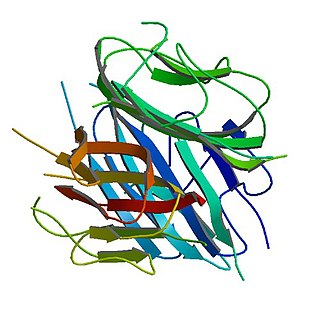
Adiponectin is a protein hormone and adipokine, which is involved in regulating glucose levels and fatty acid breakdown. In humans, it is encoded by the ADIPOQ gene and is produced primarily in adipose tissue, but also in muscle and even in the brain.

<span class="mw-page-title-main">Ceramide</span> Family of waxy lipid molecules

Ceramides are a family of waxy lipid molecules. A ceramide is composed of sphingosine and a fatty acid joined by an amide bond. Ceramides are found in high concentrations within the cell membrane of eukaryotic cells, since they are component lipids that make up sphingomyelin, one of the major lipids in the lipid bilayer. Contrary to previous assumptions that ceramides and other sphingolipids found in cell membrane were purely supporting structural elements, ceramide can participate in a variety of cellular signaling: examples include regulating differentiation, proliferation, and programmed cell death (PCD) of cells.

In biochemistry, lipogenesis is the conversion of fatty acids and glycerol into fats, or a metabolic process through which acetyl-CoA is converted to triglyceride for storage in fat. Lipogenesis encompasses both fatty acid and triglyceride synthesis, with the latter being the process by which fatty acids are esterified to glycerol before being packaged into very-low-density lipoprotein (VLDL). Fatty acids are produced in the cytoplasm of cells by repeatedly adding two-carbon units to acetyl-CoA. Triacylglycerol synthesis, on the other hand, occurs in the endoplasmic reticulum membrane of cells by bonding three fatty acid molecules to a glycerol molecule. Both processes take place mainly in liver and adipose tissue. Nevertheless, it also occurs to some extent in other tissues such as the gut and kidney. A review on lipogenesis in the brain was published in 2008 by Lopez and Vidal-Puig. After being packaged into VLDL in the liver, the resulting lipoprotein is then secreted directly into the blood for delivery to peripheral tissues.

Glucose transporter type 4 (GLUT4), also known as solute carrier family 2, facilitated glucose transporter member 4, is a protein encoded, in humans, by the SLC2A4 gene. GLUT4 is the insulin-regulated glucose transporter found primarily in adipose tissues and striated muscle. The first evidence for this distinct glucose transport protein was provided by David James in 1988. The gene that encodes GLUT4 was cloned and mapped in 1989.

Starvation response in animals is a set of adaptive biochemical and physiological changes, triggered by lack of food or extreme weight loss, in which the body seeks to conserve energy by reducing the amount of food energy it consumes.

<span class="mw-page-title-main">Acyl-CoA</span> Group of coenzymes that metabolize fatty acids

Acyl-CoA is a group of coenzymes that metabolize fatty acids. Acyl-CoA's are susceptible to beta oxidation, forming, ultimately, acetyl-CoA. The acetyl-CoA enters the citric acid cycle, eventually forming several equivalents of ATP. In this way, fats are converted to ATP, the universal biochemical energy carrier.

<span class="mw-page-title-main">White adipose tissue</span> Fatty tissue composed of white adipocytes

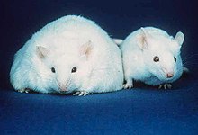
White adipose tissue or white fat is one of the two types of adipose tissue found in mammals. The other kind is brown adipose tissue. White adipose tissue is composed of monolocular adipocytes.

<span class="mw-page-title-main">Fatty acid-binding protein</span>

The fatty-acid-binding proteins (FABPs) are a family of transport proteins for fatty acids and other lipophilic substances such as eicosanoids and retinoids. These proteins are thought to facilitate the transfer of fatty acids between extra- and intracellular membranes. Some family members are also believed to transport lipophilic molecules from outer cell membrane to certain intracellular receptors such as PPAR. The FABPs are intracellular carriers that “solubilize” the endocannabinoid anandamide (AEA), transporting AEA to the breakdown by FAAH, and compounds that bind to FABPs block AEA breakdown, raising its level. The cannabinoids are also discovered to bind human FABPs that function as intracellular carriers, as THC and CBD inhibit the cellular uptake and catabolism of AEA by targeting FABPs. Competition for FABPs may in part or wholly explain the increased circulating levels of endocannabinoids reported after consumption of cannabinoids. Levels of fatty-acid-binding protein have been shown to decline with ageing in the mouse brain, possibly contributing to age-associated decline in synaptic activity.

<span class="mw-page-title-main">Stearoyl-CoA 9-desaturase</span> Class of enzymes

Stearoyl-CoA desaturase (Δ-9-desaturase) is an endoplasmic reticulum enzyme that catalyzes the rate-limiting step in the formation of monounsaturated fatty acids (MUFAs), specifically oleate and palmitoleate from stearoyl-CoA and palmitoyl-CoA. Oleate and palmitoleate are major components of membrane phospholipids, cholesterol esters and alkyl-diacylglycerol. In humans, the enzyme is encoded by the SCD gene.

<span class="mw-page-title-main">Neutral lipid storage disease</span> Congenital autosomal recessive disorder

Neutral lipid storage disease is a congenital autosomal recessive disorder characterized by accumulation of triglycerides in the cytoplasm of leukocytes, muscle, liver, fibroblasts, and other tissues. It commonly occurs as one of two subtypes, cardiomyopathic neutral lipid storage disease (NLSD-M), or ichthyotic neutral lipid storage disease (NLSD-I) which is also known as Chanarin–Dorfman syndrome), which are characterized primarily by myopathy and ichthyosis, respectively. Normally, the ichthyosis that is present is typically non-bullous congenital ichthyosiform erythroderma which appears as white scaling.

Lipid droplets, also referred to as lipid bodies, oil bodies or adiposomes, are lipid-rich cellular organelles that regulate the storage and hydrolysis of neutral lipids and are found largely in the adipose tissue. They also serve as a reservoir for cholesterol and acyl-glycerols for membrane formation and maintenance. Lipid droplets are found in all eukaryotic organisms and store a large portion of lipids in mammalian adipocytes. Initially, these lipid droplets were considered to merely serve as fat depots, but since the discovery in the 1990s of proteins in the lipid droplet coat that regulate lipid droplet dynamics and lipid metabolism, lipid droplets are seen as highly dynamic organelles that play a very important role in the regulation of intracellular lipid storage and lipid metabolism. The role of lipid droplets outside of lipid and cholesterol storage has recently begun to be elucidated and includes a close association to inflammatory responses through the synthesis and metabolism of eicosanoids and to metabolic disorders such as obesity, cancer, and atherosclerosis. In non-adipocytes, lipid droplets are known to play a role in protection from lipotoxicity by storage of fatty acids in the form of neutral triacylglycerol, which consists of three fatty acids bound to glycerol. Alternatively, fatty acids can be converted to lipid intermediates like diacylglycerol (DAG), ceramides and fatty acyl-CoAs. These lipid intermediates can impair insulin signaling, which is referred to as lipid-induced insulin resistance and lipotoxicity. Lipid droplets also serve as platforms for protein binding and degradation. Finally, lipid droplets are known to be exploited by pathogens such as the hepatitis C virus, the dengue virus and Chlamydia trachomatis among others.

Intramyocellular lipids are fats stored in droplets in muscle cells. They provide an important energy source for working muscle. During exercise, a large amount of circulating free fatty acids are directed into muscle cells for energy; during rest, incoming fatty acids are instead stored in the muscle cell as triglycerides for later burning. However, an increase in muscle insulin resistance, caused by obesity, diabetes mellitus type 2, and metabolic syndrome, will result in an excess accumulation of intramyocellular lipids.

<span class="mw-page-title-main">Perilipin-5</span> Mammalian protein found in Homo sapiens

Perilipin 5, also known as Oxpatperilipin 5 or PLIN5, is a protein that belongs to perilipin family. This protein group has been shown to be responsible for lipid droplet's biogenesis, structure and degradation. In particular, Perilipin 5 is a lipid droplet-associated protein whose function is to keep the balance between lipolysis and lipogenesis, as well as maintaining lipid droplet homeostasis. For example, in oxidative tissues, muscular tissues and cardiac tissues, PLIN5 promotes association between lipid droplets and mitochondria.

<span class="mw-page-title-main">Antonio Vidal-Puig</span> Spanish medical doctor and scientist

Antonio Vidal-Puig is a Spanish medical doctor and scientist who works as a Professor of Molecular Nutrition and Metabolism at the University of Cambridge (UK), best known for advancing the concept that pharmacological targeting of brown fat may serve to treat overweight and obesity in affected individuals, as well as for introducing the concept of adipose tissue "expandability" as an important factor in the pathogenesis of insulin resistance in the context of positive energy balance. His published work focuses on areas such as adipose tissue metabolism and lipotoxicity, regulation of insulin secretion, and the pathophysiology of metabolic syndrome, obesity, and type 2 diabetes.

References

  1. Garbarino, Jeanne; Stephen L. Sturley (2009). "Saturated with fat: new perspectives on lipotoxicity". Current Opinion in Clinical Nutrition and Metabolic Care. 12 (2): 110–116. doi:10.1097/mco.0b013e32832182ee. PMID   19202381. S2CID   7169311.
  2. 1 2 3 Bosma M, Kersten S, Hesselink MKC, and Schrauwen P. Re-evaluating lipotoxic triggers in skeletal muscle: Relating intramyocellular lipid metabolism to insulin sensitivity. Prog Lipid Res 2012; 51: 36-49|doi=10.1016/j.plipres.2011.11.003
  3. 1 2 Schaffer, Jean (June 2003). "Lipotoxicity: when tissues overeat". Current Opinion in Lipidology. 14 (3): 281–287. doi:10.1097/00041433-200306000-00008. PMID   12840659. S2CID   23895380.
  4. Rodriguez-Cuenca, S.; Pellegrinelli, V.; Campbell, M.; Oresic, M.; Vidal-Puig, A. (2017). "Sphingolipids and glycerophospholipids - The "ying and yang" of lipotoxicity in metabolic diseases". Progress in Lipid Research. 66: 14–29. doi:10.1016/j.plipres.2017.01.002. ISSN   1873-2194. PMID   28104532.
  5. Unger, Roger (June 2010). "Gluttony, Sloth and the Metabolic Syndrome: A Roadmap to Lipotoxicity". Trends in Endocrinology & Metabolism. 21 (6): 345–352. doi:10.1016/j.tem.2010.01.009. PMC   2880185 . PMID   20223680.
  6. Weinberg, J.M (2006). "Lipotoxicity". Kidney International. 70 (9): 1560–1566. doi: 10.1038/sj.ki.5001834 . PMID   16955100.
  7. Alkhouri, Naim; Dixon and Feldstein (August 2009). "Lipotoxicity in Nonalcoholic Fatty Liver Disease: Not All Lipids Are Created Equal". Expert Review of Gastroenterology & Hepatology . 3 (4): 445–451. doi:10.1586/egh.09.32. PMC   2775708 . PMID   19673631.
  8. Wende, Adam (March 2010). "Lipotoxicity in the Heart". Biochimica et Biophysica Acta (BBA) - Molecular and Cell Biology of Lipids. 1801 (3): 311–319. doi:10.1016/j.bbalip.2009.09.023. PMC   2823976 . PMID   19818871.
  9. Leitão, Cristiane (March 2010). "Lipotoxicity and Decreased Islet Graft Survival". Diabetes Care. 33 (3): 658–660. doi:10.2337/dc09-1387. PMC   2827526 . PMID   20009097.
  10. Bosma, M.; Sparks, L. M.; Hooiveld, G.; Jorgensen, J.; Houten, S. M.; Schrauwen, P.; Hesselink, M. K. C. (2013). "Overexpression of PLIN5 in skeletal muscle promotes oxidative gene expression and intramyocellular lipid content without compromising insulin sensitivity" (PDF). Biochimica et Biophysica Acta (BBA) - Molecular and Cell Biology of Lipids. 1831 (4): 844–52. doi:10.1016/j.bbalip.2013.01.007. PMID   23353597.
  11. Unger, Roger (January 2005). "Longevity, lipotoxicity and leptin: the adipocyte defense against feasting and famine". Biochimie. 87 (1): 57–64. doi:10.1016/j.biochi.2004.11.014. PMID   15733738.
  12. Smith, U; Hammarstedt (March 2010). "Antagonistic effects of thiazolidinediones and cytokines in lipotoxicity". Biochimica et Biophysica Acta (BBA) - Molecular and Cell Biology of Lipids. 1801 (3): 377–380. doi:10.1016/j.bbalip.2009.11.006. PMID   19941972.