Lumbar provocative discography

Last updated
Lumbar provocative discography
Discograph degenerative disc disease L5-S1.jpg
Lumbar provocative discography showing a degenerated disc, L5-S1 with normal disc L4-L5
Other codes CPT 62290 (lumbar/sacral)
CPT 62291 (Cervical/Thoracic)
CPT 72295 radiological supervision (lumbar/sacral)
CPT 72285 radiological supervision (cervical/thoracic)

Lumbar provocative discography (also referred to as "discography" or discogram) is an invasive diagnostic procedure for evaluation for intervertebral disc pathology. It is usually reserved for persons with persistent, severe low back pain (LBP) who have abnormal spaces between vertebrae on magnetic resonance imaging (MRI), where other diagnostic tests have failed to reveal clear confirmation of a suspected disc as the source of pain, and surgical intervention is being considered.

Contents

Uses

Spinal MRI is the imaging of choice to investigate the spine and intervertebral discs. [1] Meanwhile, lumbar discography is used to demonstrate degeneration and herniation of lumbar intervertebral discs by injecting a dye into the nucleus pulposus of the discs. The procedure is also used to reproduce pain back pain for those who has such symptoms. Lumbar discography is also used to access the response of a subject to anaesthetic injection. Other uses includes: suspected discogenic pain without radicular sign (pain travelling to lower limbs along a dermatome), and confirmation of normal intervertebral discs above and below a site before spinal fusion. [1] However, evidence supporting the usefulness of lumbar discography in recognising intervertebral disc problems are limited. [1] There is no role for cervical or thoracic discography in diagnosing disc pathologies at the cervical or thoracic regions. [1] [2]

For those with local or distant sepsis, lumbar discography can add to the risk of infective discitis. Those with co-morbities that caused them not to be suitable candidate for surgery is also relatively contraindicated for this procedure. [1]

Procedure

Non-ionic contrast media such as iopamidol and iohexol are used. [1] Needles are inserted through the back into the disc near the suspect area, guided by fluoroscopic imaging. Fluid is then injected to pressurize the disc, and any pain responses are recorded.
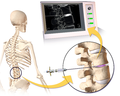
Lumbar Discography. Discography.png
Lumbar Discography.

This is repeated in random order for the various discs, without the patient knowing which disc is pressurized. This can be used to detect patients who may be exaggerating their symptoms, or to assess their pain response and hence their suitability for recovery from possible surgery (often a Discectomy).

A contrast agent is introduced, and after the procedure, a CT scan identifies leakage from the discs to identify any spinal disc herniation.

Possible complications include nerve damage, discitis, an infection of the disc. Strong antibiotics are usually administered as a precaution.

Its use in predicting surgical outcomes is sometimes controversial.

History

Lindgren from Scandinavia first performed a puncture of normal intervertebral disc for diagnostic purposes in 1941. [3]

Related Research Articles

<span class="mw-page-title-main">Intervertebral disc</span> Cartilage of spine

An intervertebral disc lies between adjacent vertebrae in the vertebral column. Each disc forms a fibrocartilaginous joint, to allow slight movement of the vertebrae, to act as a ligament to hold the vertebrae together, and to function as a shock absorber for the spine.

<span class="mw-page-title-main">Sciatica</span> Lower back pain that extends down leg

Sciatica is pain going down the leg from the lower back. This pain may go down the back, outside, or front of the leg. Onset is often sudden following activities like heavy lifting, though gradual onset may also occur. The pain is often described as shooting. Typically, symptoms are only on one side of the body. Certain causes, however, may result in pain on both sides. Lower back pain is sometimes present. Weakness or numbness may occur in various parts of the affected leg and foot.

<span class="mw-page-title-main">Discectomy</span> Surgical removal of an intervertebral disc

A discectomy is the surgical removal of abnormal disc material that presses on a nerve root or the spinal cord. The procedure involves removing a portion of an intervertebral disc, which causes pain, weakness or numbness by stressing the spinal cord or radiating nerves. The traditional open discectomy, or Love's technique, was published by Ross and Love in 1971. Advances have produced visualization improvements to traditional discectomy procedures, or endoscopic discectomy. In conjunction with the traditional discectomy or microdiscectomy, a laminotomy is often involved to permit access to the intervertebral disc. Laminotomy means a significant amount of typically normal bone is removed from the vertebra, allowing the surgeon to better see and access the area of disc herniation.

<span class="mw-page-title-main">Schmorl's nodes</span> Medical condition

Schmorl's nodes are protrusions of the nucleus pulposus of the intervertebral disc through the vertebral body endplate and into the adjacent vertebra.

<span class="mw-page-title-main">Lumbar spinal stenosis</span> Medical condition of the spine

Lumbar spinal stenosis (LSS) is a medical condition in which the spinal canal narrows and compresses the nerves and blood vessels at the level of the lumbar vertebrae. Spinal stenosis may also affect the cervical or thoracic region, in which case it is known as cervical spinal stenosis or thoracic spinal stenosis. Lumbar spinal stenosis can cause pain in the low back or buttocks, abnormal sensations, and the absence of sensation (numbness) in the legs, thighs, feet, or buttocks, or loss of bladder and bowel control.

<span class="mw-page-title-main">Back injury</span> Damage or wear to bones, muscles or other tissues of the back

Back injuries result from damage, wear, or trauma to the bones, muscles, or other tissues of the back. Common back injuries include sprains and strains, herniated discs, and fractured vertebrae. The lumbar spine is often the site of back pain. The area is susceptible because of its flexibility and the amount of body weight it regularly bears. It is estimated that low-back pain may affect as much as 80 to 90 percent of the general population in the United States.

<span class="mw-page-title-main">Spondylosis</span> Degeneration of the vertebral column

Spondylosis is the degeneration of the vertebral column from any cause. In the more narrow sense it refers to spinal osteoarthritis, the age-related degeneration of the spinal column, which is the most common cause of spondylosis. The degenerative process in osteoarthritis chiefly affects the vertebral bodies, the neural foramina and the facet joints. If severe, it may cause pressure on the spinal cord or nerve roots with subsequent sensory or motor disturbances, such as pain, paresthesia, imbalance, and muscle weakness in the limbs.

Myelopathy describes any neurologic deficit related to the spinal cord. The most common form of myelopathy in humans, cervical spondylotic myelopathy (CSM), also called degenerative cervical myelopathy, results from narrowing of the spinal canal ultimately causing compression of the spinal cord. When due to trauma, myelopathy is known as (acute) spinal cord injury. When inflammatory, it is known as myelitis. Disease that is vascular in nature is known as vascular myelopathy.

<span class="mw-page-title-main">Degenerative disc disease</span> Medical condition

Degenerative disc disease (DDD) is a medical condition typically brought on by the normal aging process in which there are anatomic changes and possibly a loss of function of one or more intervertebral discs of the spine. DDD can take place with or without symptoms, but is typically identified once symptoms arise. The root cause is thought to be loss of soluble proteins within the fluid contained in the disc with resultant reduction of the oncotic pressure, which in turn causes loss of fluid volume. Normal downward forces cause the affected disc to lose height, and the distance between vertebrae is reduced. The anulus fibrosus, the tough outer layers of a disc, also weakens. This loss of height causes laxity of the longitudinal ligaments, which may allow anterior, posterior, or lateral shifting of the vertebral bodies, causing facet joint malalignment and arthritis; scoliosis; cervical hyperlordosis; thoracic hyperkyphosis; lumbar hyperlordosis; narrowing of the space available for the spinal tract within the vertebra ; or narrowing of the space through which a spinal nerve exits with resultant inflammation and impingement of a spinal nerve, causing a radiculopathy.

<span class="mw-page-title-main">Spinal fusion</span> Immobilization or ankylosis of two or more vertebrae by fusion of the vertebral bodies

Spinal fusion, also called spondylodesis or spondylosyndesis, is a neurosurgical or orthopedic surgical technique that joins two or more vertebrae. This procedure can be performed at any level in the spine and prevents any movement between the fused vertebrae. There are many types of spinal fusion and each technique involves using bone grafting—either from the patient (autograft), donor (allograft), or artificial bone substitutes—to help the bones heal together. Additional hardware is often used to hold the bones in place while the graft fuses the two vertebrae together. The placement of hardware can be guided by fluoroscopy, navigation systems, or robotics.

Middle back pain, also known as thoracic back pain, is back pain that is felt in the region of the thoracic vertebrae, which are between the bottom of the neck and top of the lumbar spine. It has a number of potential causes, ranging from muscle strain to collapse of a vertebra or rare serious diseases. The upper spine is very strong and stable to support the weight of the upper body, as well as to anchor the rib cage which provides a cavity to allow the heart and lungs to function and protect them.

Failed back syndrome or post-laminectomy syndrome is a condition characterized by chronic pain following back surgeries. Many factors can contribute to the onset or development of FBS, including residual or recurrent spinal disc herniation, persistent post-operative pressure on a spinal nerve, altered joint mobility, joint hypermobility with instability, scar tissue (fibrosis), depression, anxiety, sleeplessness, spinal muscular deconditioning and even Cutibacterium acnes infection. An individual may be predisposed to the development of FBS due to systemic disorders such as diabetes, autoimmune disease and peripheral blood vessels (vascular) disease.

<span class="mw-page-title-main">Spinal disc herniation</span> Injury to the connective tissue between spinal vertebrae

A spinal disc herniation is an injury to the cushioning and connective tissue between vertebrae, usually caused by excessive strain or trauma to the spine. It may result in back pain, pain or sensation in different parts of the body, and physical disability. The most conclusive diagnostic tool for disc herniation is MRI, and treatment may range from painkillers to surgery. Protection from disc herniation is best provided by core strength and an awareness of body mechanics including posture.

<span class="mw-page-title-main">Radiculopathy</span> Medical condition

Radiculopathy, also commonly referred to as pinched nerve, refers to a set of conditions in which one or more nerves are affected and do not work properly. Radiculopathy can result in pain, weakness, altered sensation (paresthesia) or difficulty controlling specific muscles. Pinched nerves arise when surrounding bone or tissue, such as cartilage, muscles or tendons, put pressure on the nerve and disrupt its function.

<span class="mw-page-title-main">Laminotomy</span> Surgical procedure

A laminotomy is an orthopaedic neurosurgical procedure that removes part of the lamina of a vertebral arch in order to relieve pressure in the vertebral canal. A laminotomy is less invasive than conventional vertebral column surgery techniques, such as laminectomy because it leaves more ligaments and muscles attached to the spinous process intact and it requires removing less bone from the vertebra. As a result, laminotomies typically have a faster recovery time and result in fewer postoperative complications. Nevertheless, possible risks can occur during or after the procedure like infection, hematomas, and dural tears. Laminotomies are commonly performed as treatment for lumbar spinal stenosis and herniated disks. MRI and CT scans are often used pre- and post surgery to determine if the procedure was successful.

<span class="mw-page-title-main">Laminoplasty</span>

Laminoplasty is an orthopaedic/neurosurgical surgical procedure for treating spinal stenosis by relieving pressure on the spinal cord. The main purpose of this procedure is to provide relief to patients who may have symptoms of numbness, pain, or weakness in arm movement. The procedure involves cutting the lamina on both sides of the affected vertebrae and then "swinging" the freed flap of bone open thus relieving the pressure on the spinal cord. The spinous process may be removed to allow the lamina bone flap to be swung open. The bone flap is then propped open using small wedges or pieces of bone such that the enlarged spinal canal will remain in place.

<span class="mw-page-title-main">Spinal stenosis</span> Disease of the bony spine that results in narrowing of the spinal canal

Spinal stenosis is an abnormal narrowing of the spinal canal or neural foramen that results in pressure on the spinal cord or nerve roots. Symptoms may include pain, numbness, or weakness in the arms or legs. Symptoms are typically gradual in onset and improve with leaning forward. Severe symptoms may include loss of bladder control, loss of bowel control, or sexual dysfunction.

The TESSYS method is a minimally-invasive, endoscopic spinal procedure for the treatment of a herniated disc. It was a further development of the YESS method by the Dutch Dr Thomas Hoogland in the Alpha Klinik in Munich in 1989 and was first called THESSYS. The procedure involves performing a small foramenotomy and removal of soft tissue compressing the nerve root.

Vertebral osteomyelitis is a type of osteomyelitis that affects the vertebrae. It is a rare bone infection concentrated in the vertebral column. Cases of vertebral osteomyelitis are so rare that they constitute only 2%-4% of all bone infections. The infection can be classified as acute or chronic depending on the severity of the onset of the case, where acute patients often experience better outcomes than those living with the chronic symptoms that are characteristic of the disease. Although vertebral osteomyelitis is found in patients across a wide range of ages, the infection is commonly reported in young children and older adults. Vertebral osteomyelitis often attacks two vertebrae and the corresponding intervertebral disk, causing narrowing of the disc space between the vertebrae. The prognosis for the disease is dependent on where the infection is concentrated in the spine, the time between initial onset and treatment, and what approach is used to treat the disease.

Minimally invasive spine surgery, also known as MISS, has no specific meaning or definition. It implies a lack of severe surgical invasion. The older style of open-spine surgery for a relatively small disc problem used to require a 5-6 inch incision and a month in the hospital. MISS techniques utilize more modern technology, advanced imaging techniques and special medical equipment to reduce tissue trauma, bleeding, radiation exposure, infection risk, and decreased hospital stays by minimizing the size of the incision. Modern endoscopic procedures can be done through a 2 to 5 mm skin opening. By contrast, procedures done with a microscope require skin openings of approximately one inch, or more.

References

  1. 1 2 3 4 5 6 Watson N, Jones H (2018). Chapman and Nakielny's Guide to Radiological Procedures. Elsevier. pp. 337–339. ISBN   9780702071669.
  2. "Discography". Archived from the original on 30 May 2022. Retrieved 30 May 2022.
  3. Tomecek, FJ; Anthony, CS; Boxell, C; Warren, J. "Discography Interpretation and Techniques in the Lumbar Spine". Medscape. Archived from the original on 3 April 2017. Retrieved 30 May 2022.