
The femur, or thigh bone, is the only bone in the thigh — the region of the lower limb between the hip and the knee. In many four-legged animals the femur is the upper bone of the hindleg.

The acetabulum, also called the cotyloid cavity, is a concave surface of the pelvis. The head of the femur meets with the pelvis at the acetabulum, forming the hip joint.

Hip replacement is a surgical procedure in which the hip joint is replaced by a prosthetic implant, that is, a hip prosthesis. Hip replacement surgery can be performed as a total replacement or a hemi/semi(half) replacement. Such joint replacement orthopaedic surgery is generally conducted to relieve arthritis pain or in some hip fractures. A total hip replacement consists of replacing both the acetabulum and the femoral head while hemiarthroplasty generally only replaces the femoral head. Hip replacement is one of the most common orthopaedic operations, though patient satisfaction varies widely between different techniques and implants. Approximately 58% of total hip replacements are estimated to last 25 years. The average cost of a total hip replacement in 2012 was $40,364 in the United States, and about $7,700 to $12,000 in most European countries.

In vertebrate anatomy, the hip, or coxa in medical terminology, refers to either an anatomical region or a joint on the outer (lateral) side of the pelvis.
Osteochondrosis is a family of orthopedic diseases of the joint that occur in children, adolescents and rapidly growing animals, particularly pigs, horses, dogs, and broiler chickens. They are characterized by interruption of the blood supply of a bone, in particular to the epiphysis, followed by localized bony necrosis, and later, regrowth of the bone. This disorder is defined as a focal disturbance of endochondral ossification and is regarded as having a multifactorial cause, so no one thing accounts for all aspects of this disease.

The ball-and-socket joint is a type of synovial joint in which the ball-shaped surface of one rounded bone fits into the cup-like depression of another bone. The distal bone is capable of motion around an indefinite number of axes, which have one common center. This enables the joint to move in many directions.
In vertebrates, the pubis or pubic bone forms the lower and anterior part of each side of the hip bone. The pubis is the most forward-facing of the three bones that make up the hip bone. The left and right pubic bones are each made up of three sections; a superior ramus, an inferior ramus, and a body.

The acetabular fossa is the non-articular depressed region at the centre of the floor of the acetabulum. It is surrounded by the articular lunate surface. The floor of the fossa is formed mostly by the ischium; it is rough and thin. The space of the fossa is continuous inferiorly with the acetabular notch.

The obturator artery is a branch of the internal iliac artery that passes antero-inferiorly on the lateral wall of the pelvis, to the upper part of the obturator foramen, and, escaping from the pelvic cavity through the obturator canal, it divides into an anterior branch and a posterior branch.
The acetabular notch is a deep notch in the inferior portion of the rim of the acetabulum. It is bridged by the transverse acetabular ligament, converting it into a foramen. It is continuous with space of the acetabular fossa. The lunate surface of acetabulum is discontinued opposite the notch.

The acetabular labrum is a fibrocartilaginous ring which surrounds the circumference of the acetabulum of the hip, deepening the acetabulum. The labrum is attached onto the bony rim and transverse acetabular ligament. It is triangular in cross-section.

The femoral head is the highest part of the thigh bone (femur). It is supported by the femoral neck.

The acetabular branch is an artery in the hip that arises from the medial circumflex femoral artery opposite the acetabular notch and enters the hip-joint beneath the transverse ligament in company with an articular branch from the obturator artery. It supplies the fat in the bottom of the acetabulum, and is continued along the ligament to the head of the femur.
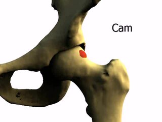
Femoroacetabular impingement (FAI) is a condition involving one or more anatomical abnormalities of the hip joint, which is a ball and socket joint. It is a common cause of hip pain and discomfort in young and middle-aged adults. It occurs when the ball shaped femoral head contacts the acetabulum abnormally or does not permit a normal range of motion in the acetabular socket. Damage can occur to the articular cartilage, or labral cartilage, or both. The condition may be symptomatic or asymptomatic. It may cause osteoarthritis of the hip. Treatment options range from conservative management to surgery.
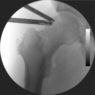
Hip arthroscopy refers to the viewing of the interior of the acetabulofemoral (hip) joint through an arthroscope and the treatment of hip pathology through a minimally invasive approach. This technique is sometimes used to help in the treatment of various joint disorders and has gained popularity because of the small incisions used and shorter recovery times when compared with conventional surgical techniques. Hip arthroscopy was not feasible until recently, new technology in both the tools used and the ability to distract the hip joint has led to a recent surge in the ability to do hip arthroscopy and the popularity of it.
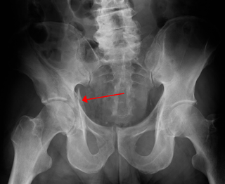
Fractures of the acetabulum occur when the head of the femur is driven into the pelvis. This injury is caused by a blow to either the side or front of the knee and often occurs as a dashboard injury accompanied by a fracture of the femur.

Pain in the hip is the experience of pain in the muscles or joints in the hip/ pelvic region, a condition commonly arising from any of a number of factors. Sometimes it is closely associated with lower back pain.
X-rays of hip dysplasia are one of the two main methods of medical imaging to diagnose hip dysplasia, the other one being medical ultrasonography. Ultrasound imaging yields better results defining the anatomy until the cartilage is ossified. When the infant is around 3 months old a clear roentgenographic image can be achieved. Unfortunately the time the joint gives a good x-ray image is also the point at which nonsurgical treatment methods cease to give good results.

Ultrasound-guided hip joint injection is a joint injection in the hip, assisted by medical ultrasound. Hip and groin pain often presents a diagnostic and therapeutic challenge. The differential diagnosis is extensive, comprising intra-articular and extra-articular pathology and referred pain from lumbar spine, knee and elsewhere in the pelvis. Various ultrasound-guided techniques have been described in the hip and groin region for diagnostic and therapeutic purposes. Ultrasound has many advantages over other imaging modalities, including portability, lack of ionizing radiation and real-time visualization of soft tissues and neurovascular structures. Many studies have demonstrated the safety, accuracy and efficacy of ultrasound-guided techniques, although there is lack of standardization regarding the injectates used and long-term benefit remains uncertain.

An acetabular labrum tear or hip labrum tear is a common injury of the acetabular labrum resulting from a number of causes including running, hip dislocation, and deterioration with ageing. Most are thought to result from a gradual tear due to repetitive microtrauma.
