Macular hypoplasia

Last updated
Human retina cross-section Human eye cross-sectional view grayscale.png
Human retina cross-section
Macular hypoplasia
Other namesFoveal hypoplasia
Autosomal dominant - en.svg
This condition is inherited via autosomal dominant manner.

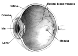
Macular hypoplasia (or foveal hypoplasia) is a rare medical condition involving the underdevelopment of the macula, [1] a small area on the retina (the eye's internal surface) responsible for seeing in detail and sensing light. [2] Macular hypoplasia is often associated with albinism. [1]

Contents

When the foveal area of the eye is compromised, visual clarity and color perception are reduced. [2] Diagnosing is done by an ophthalmologist. [2] The foveal area of the eye is located in the back of the eyeball. It is placed in front of the optic nerve and is responsible for light sensory and visual perceptiveness. [3] [4]

Other diseases with foveal hypoplasia besides albinism include aniridia, retinopathy of prematurity, and Alport syndrome. [5] [6]

Causes

Macular hypoplasia occurs the most in people that have a diagnosis of albinism. [7] There are four gene mutations that occur in albinism and are linked to macular hypoplasia. [7] The four mutations can occur on the phenotypes of FH, PAX6, SLC38A8, and AHR. [7] The most common gene mutation is the FH phenotype and has a 67.5% correlation rate to macular hypoplasia. [7]

The disorder can occur through two distinct genetic abnormalities. [8] The difference among mutated genes results in a difference in phenotypic display of macular hypoplasia. [8] In phenotype FVH1, there is a mutation of the PAX6 gene. [8] FVH1 occurs through autosomal dominant inheritance. [8] The mutation is passed down to the recipient from the mother or father. [8] FVH1 type of macular hypoplasia coincides with cataracts in the eyes. [8] In phenotype, FVH1 is caused by a SLC38A8 gene mutation. FVH2 occurs by autosomal recessive inheritance. [8] Both parents pass the mutated gene to the child. [8] Macular hypoplasia prevails due to improper placement of the optic nerve. [8]

Diagnosis

A lack of foveal pigmentation or circumfoveal light reflex is a common finding of macular hypoplasia; however, diagnosis is challenging for those that have a darker pigmentation of the skin, hair, and iris. [9] Originally, findings of nystagmus, or involuntary movement, and lack of blood flow to the retina using fluorescein angiography (FA) were used to detect macular hypoplasia. [9] [10] FA uses light to look at the retina and blood vessel development in the eye using a dye. [9]

Today, a newer technology, optical coherence tomography (OCT) is used to detect foveal hypoplasia and does not require a dye. [9] OCT allows professionals to see the structures in the eye, usually the thickness of the retina and optic nerve. [11] This is a noninvasive procedure where patients rest their chin and focus on a green light within the machine. [11] Eye dryness and fatigue are the limited risks associated with this scan. [11]

Other diseases that can be diagnosed using OCT are glaucoma, macular degeneration, and diabetes-related retinopathy. [11]

Treatment

Currently, there is no specific pharmacotherapy that prevents or reserves macular hypoplasia; however, reading glasses or other vision devices can be used to enhance the quality of life for individuals. [12]

Related Research Articles

Retinitis pigmentosa Gradual retinal degeneration leading to progressive sight loss

Retinitis pigmentosa (RP) is a genetic disorder of the eyes that causes loss of vision. Symptoms include trouble seeing at night and decreasing peripheral vision. As peripheral vision worsens, people may experience "tunnel vision". Complete blindness is uncommon. Onset of symptoms is generally gradual and often begins in childhood.

Retinoschisis Eye disease involving splitting of the retina

Retinoschisis is an eye disease characterized by the abnormal splitting of the retina's neurosensory layers, usually in the outer plexiform layer. Retinoschisis can be divided into degenerative forms which are very common and almost exclusively involve the peripheral retina and hereditary forms which are rare and involve the central retina as sometimes the peripheral retina. The degenerative forms are asymptomatic and involve the peripheral retina only and do not affect the visual acuity. Some rarer forms result in a loss of vision in the corresponding visual field.

Alport syndrome Medical condition

Alport syndrome is a genetic disorder affecting around 1 in 5,000-10,000 children, characterized by glomerulonephritis, end-stage kidney disease, and hearing loss. Alport syndrome can also affect the eyes, though the changes do not usually affect vision, except when changes to the lens occur in later life. Blood in urine is universal. Proteinuria is a feature as kidney disease progresses.

Aniridia Absence of the iris, usually involving both eyes

Aniridia is the absence of the iris, a muscular structure that opens and closes the pupil to allow light into the eye. It is also responsible for eye color. Without it the central eye appears all black. It can be congenital, in which both eyes are usually involved, or caused by a penetrant injury. Isolated aniridia is a congenital disorder which is not limited to a defect in iris development, but is a panocular condition with macular and optic nerve hypoplasia, cataract, and corneal changes. Vision may be severely compromised and the disorder is frequently associated with a number of ocular complications: nystagmus, amblyopia, buphthalmos, and cataract. Aniridia in some individuals occurs as part of a syndrome, such as WAGR syndrome, or Gillespie syndrome.

Coloboma Hole in one of the structures of the eye

A coloboma is a hole in one of the structures of the eye, such as the iris, retina, choroid, or optic disc. The hole is present from birth and can be caused when a gap called the choroid fissure, which is present during early stages of prenatal development, fails to close up completely before a child is born. Ocular coloboma is relatively uncommon, affecting less than one in every 10,000 births.

Collie eye anomaly (CEA) is a congenital, inherited, bilateral eye disease of dogs, which affects the retina, choroid, and sclera. It can be a mild disease or cause blindness. CEA is caused by a simple autosomal recessive gene defect. There is no treatment.

Vitelliform macular dystrophy Medical condition

Vitelliform macular dystrophy is an irregular autosomal dominant eye disorder which can cause progressive vision loss. This disorder affects the retina, specifically cells in a small area near the center of the retina called the macula. The macula is responsible for sharp central vision, which is needed for detailed tasks such as reading, driving, and recognizing faces. The condition is characterized by yellow, slightly elevated, round structures similar to the yolk of an egg.

Stargardt disease is the most common inherited single-gene retinal disease. In terms of the first description of the disease, it follows an autosomal recessive inheritance pattern, which has been later linked to bi-allelic ABCA4 gene variants (STGD1). However, there are Stargardt-like diseases with mimicking phenotypes that are referred to as STGD3 and STGD4, and have a autosomal dominant inheritance due to defects with ELOVL4 or PROM1 genes, respectively. It is characterized by macular degeneration that begins in childhood, adolescence or adulthood, resulting in progressive loss of vision.

Papillorenal syndrome Medical condition

Papillorenal syndrome is an autosomal dominant genetic disorder marked by underdevelopment (hypoplasia) of the kidney and colobomas of the optic nerve.

Optic pit Medical condition

Optic pit, optic nerve pit, or optic disc pit (ODP) is rare a congenital excavation of the optic disc, resulting from a malformation during development of the eye. The incidence of ODP is 1 in 10,000 people with no predilection for either gender. There is currently no known risk factors for their development. Optic pits are important because they are associated with posterior vitreous detachments (PVD) and even serous retinal detachments.

Ocular albinism is a form of albinism which, in contrast to oculocutaneous albinism, presents primarily in the eyes. There are multiple forms of ocular albinism, which are clinically similar.

Oculocutaneous albinism type I or type 1A is an autosomal recessive skin disease. This subtype of oculocutaneous albinism is caused when the gene for tyrosinase does not function properly.

Ocular albinism type 1 Most common type of ocular albinism

Ocular albinism type 1(OA1) is the most common type of ocular albinism, with a prevalence rate of 1:50,000. It is an inheritable classical Mendelian type X-linked recessive disorder wherein the retinal pigment epithelium lacks pigment while hair and skin appear normal. Since it is usually an X-linked disorder, it occurs mostly in males, while females are carriers unless they are homozygous. About 60 missense and nonsense mutations, insertions, and deletions have been identified in Oa1. Mutations in OA1 have been linked to defective glycosylation and thus improper intracellular transportation.

Gillespie syndrome Medical condition

Gillespie syndrome, also called aniridia, cerebellar ataxia and mental deficiency, is a rare genetic disorder. The disorder is characterized by partial aniridia, ataxia, and, in most cases, intellectual disability. It is heterogeneous, inherited in either an autosomal dominant or autosomal recessive manner. Gillespie syndrome was first described by American ophthalmologist Fredrick Gillespie in 1965.

Berlin's edema a common condition caused by blunt injury to the eye. It is characterized by decreased vision in the injured eye a few hours after the injury. Under examination the retina appears opaque and white in colour in the periphery but the blood vessels are normally seen along with "cherry red spot" in the foveal region. This whitening is indicative of cell damage, which occurs in the retinal pigment epithelium and outer segment layer of photoreceptors. Damage to the outer segment often results in photoreceptor death through uncertain mechanisms. Usually there is no leakage of fluid and therefore it is not considered a true edema. The choroidal fluorescence in fluorescent angiography is absent. Visual acuity ranges from 20/20 to 20/400.

Hypotrichosis with juvenile macular dystrophy Medical condition

Hypotrichosis with juvenile macular dystrophy is an extremely rare congenital disease characterized by sparse hair growth (hypotrichosis) from birth and progressive macular corneal dystrophy.

Occult macular dystrophy (OMD) is a rare inherited degradation of the retina, characterized by progressive loss of function in the most sensitive part of the central retina (macula), the location of the highest concentration of light-sensitive cells (photoreceptors) but presenting no visible abnormality. "Occult" refers to the degradation in the fundus being difficult to discern. The disorder is called "dystrophy" instead of "degradation" to distinguish its genetic origin from other causes, such as age. OMD was first reported by Y. Miyake et al. in 1989.

Sickle cell retinopathy can be defined as retinal changes due to blood vessel damage in the eye of a person with a background of sickle cell disease. It can likely progress to loss of vision in late stages due to vitreous hemorrhage or retinal detachment. Sickle cell disease is a structural red blood cell disorder leading to consequences in multiple systems. It is characterized by chronic red blood cell destruction, vascular injury, and tissue ischemia causing damage to the brain, eyes, heart, lungs, kidneys, spleen, and musculoskeletal system.

Ocular albinism late onset sensorineural deafness (OASD) is a rare, X-linked recessive disease characterized by intense visual impairments, reduced retinal pigments, translucent pale-blue irises and moderately severe hearing loss from adolescence to middle-age. It is a subtype of Ocular Albinism (OA) that is linked to Ocular albinism type I (OA1). OA1 is the most common form of ocular albinism, affecting at least 1/60,000 males.

Goldmann-Favre syndrome Medical condition

Goldmann-Favre syndrome is a rare genetic disorder characterized by early-onset nyctalopia, decreased visual acuity, and abnormal findings of the fundus. It is a type of progressive vitreotapetoretinal degeneration.

References

  1. 1 2 "OMIM Entry - # 136520 - FOVEAL HYPOPLASIA 1; FVH1". omim.org. Retrieved 2017-07-28.
  2. 1 2 3 "Foveal Hypoplasia 1 | Hereditary Ocular Diseases". disorders.eyes.arizona.edu. Retrieved 2022-07-13.
  3. Rehman, Ibraheem; Mahabadi, Navid; Motlagh, Mahsaw; Ali, Tayyeba (2022), "Anatomy, Head and Neck, Eye Fovea", StatPearls, Treasure Island (FL): StatPearls Publishing, PMID   29493926 , retrieved 2022-07-28
  4. "Foveal Hypoplasia 1 | Hereditary Ocular Diseases". disorders.eyes.arizona.edu. Retrieved 2022-07-28.
  5. Thomas MG, Papageorgiou E, Kuht HJ, Gottlob I (November 2020). "Normal and abnormal foveal development". Br J Ophthalmol. doi:10.1136/bjophthalmol-2020-316348. PMID   33148537.
  6. Hess K, Pfau M, Wintergerst MW, Loeffler KU, Holz FG, Herrmann P (February 2020). "Phenotypic Spectrum of the Foveal Configuration and Foveal Avascular Zone in Patients With Alport Syndrome". Invest Ophthalmol Vis Sci. 61 (2): 5. doi:10.1167/iovs.61.2.5. PMC   7324255 . PMID   32031577.
  7. 1 2 3 4 Kuht, Helen J.; Maconachie, Gail D. E.; Han, Jinu; Kessel, Line; Genderen, Maria M. van; McLean, Rebecca J.; Hisaund, Michael; Tu, Zhanhan; Hertle, Richard W.; Gronskov, Karen; Bai, Dayong (2022-06-01). "Genotypic and Phenotypic Spectrum of Foveal Hypoplasia: A Multicenter Study". Ophthalmology. 129 (6): 708–718. doi:10.1016/j.ophtha.2022.02.010. ISSN   0161-6420. PMID   35157951.
  8. 1 2 3 4 5 6 7 8 9 Matsushita, Itsuka; Morita, Hirofumi; Kondo, Hiroyuki (2020-11-01). "Autosomal dominant foveal hypoplasia without visible macular abnormalities and PAX6 mutations". Japanese Journal of Ophthalmology. 64 (6): 635–641. doi:10.1007/s10384-020-00766-9. ISSN   1613-2246.
  9. 1 2 3 4 Recchia, Franco M.; Carvalho-Recchia, Cynthia A.; Trese, Michael T. (2002-11-01). "Optical Coherence Tomography in the Diagnosis of Foveal Hypoplasia". Archives of Ophthalmology. 120 (11): 1587–1588. ISSN   0003-9950.
  10. "Nystagmus". www.hopkinsmedicine.org. 2021-12-13. Retrieved 2022-07-25.
  11. 1 2 3 4 "Optical Coherence Tomography: What It Is & How It's Used". Cleveland Clinic. Retrieved 2022-07-25.
  12. "Foveal Hypoplasia 2 | Hereditary Ocular Diseases". disorders.eyes.arizona.edu. Retrieved 2022-07-25.