Malignant chondroid syringoma

Last updated • 9 min readFrom Wikipedia, The Free Encyclopedia
Malignant chondroid syringoma
Other namesMCS, Malignant mixed tumor
Malignant Chondroid Syringoma.jpg
Histopathological examination of malignant chondroid syringoma
CausesPrior physical injury, excessive mucoid matrix, chondroid syringoma less than three centimetres in size
Diagnostic method Clinical diagnosis needed
TreatmentExcision, chemotherapy or radiotherapy is occasionally recommended

A malignant chondroid syringoma (also known as a "malignant mixed tumour" or "MCS" ) is a very uncommon cutaneous (skin) condition characterised by an adnexal eccrine tumour. [1] :667

Contents

It is commonly reported to present on the trunk and extremities and behave in an aggressive manner. [1] [2] MCS is a cancerous subtype of its benign counterpart, chondroid syringoma, and is the least common variation that has an approximated prevalence of less than 0.005%. [3] These tumours mainly arise "from sebaceous glands, sweat glands, and ectopic salivary glands" (Tural, Selçukbiricik, Günver, Karışmaz, and Serdengecti, 2013) and are rarely encountered in radiopathological and clinical practice. [4] [5] The tumours commonly appear with an asymptomatic "slow-growing, painless, solid subcutaneous or intradermal nodules with a normal margin" (Obaidat, Alsaad, and Ghazarian, 2007) and make up for less than one percent of all primary skin tumours. [6] Commonly appearing in the limbs and body, these asymmetrical tumours range from two millimetres to more than three centimetres. [2] [7] MCS is one of the rarest subtypes of tumours and usually requires aggressive surgery to terminate. [2] Despite accounting for only a small number of tumours recorded each year, malignant mixed tumours are easily confused with other skin conditions (such as epidermal cyst, pilar cyst, calcifying epithelioma, or a solitary trichoepithelioma (Tural, Selçukbiricik, Günver, Karışmaz, and Serdengecti, 2013)) and have high potential for recurrence after surgical excision. [8] [9] The aggressiveness of malignant chondroid syringoma varies, as 49% of cases have had local recurrence whereas some demonstrate regional lymph node or osseous metastasis. The rare neoplasms generally do not follow a determinate development path and are often difficult to diagnose. [10]

Histologically, these malignant mixed tumours have epithelial and mesenchymal components and are very large, nodular, circumscribed, and nonulcerated. [11] They are morphologically identical to pleomorphic adenoma and have a female predilection. [4] They range from developing deep dermal to subcutaneous nodules and metastasise at a very high rate. [4] [8] Malignant mixed tumours can emerge "de nova or more rarely develop from a cartilaginous syringoma."(IADVL, 2004) [11]

Causes

Currently malignant mixed tumours do not have a definitive predisposing factor; however, prior physical injury has been proposed as a potential risk for the emergence of these tumours. [4] Chondroid syringoma less than three centimetres are shown to have a heightened risk of malignancy. Other critical indicators include excessive mucoid matrix, – a connective tissue tumour with a myxoid background that is composed of "clear, mucoid substance – numerous mitoses, and poorly differentiated chondroid components." (Metzler, Schaumburg-Lever, Hornstein, and Rassner, 1996) [12]

Diagnosis

Diagnosing malignant mixed tumours is difficult as there many types of tumours and their variants. [9] Malignant chondroid syringoma is particularly challenging to distinguish from other skin tumours as it is prone to the absence of definitive symptoms and an ambiguous clinical presentation. [3] Some signs that help determine diagnosis of malignancy include: "mitosis, nuclear atypia, pleomorphism, lymphatic invasion, and local recurrence" (IADVL, 2004). [11] Though it is usually found within women in the forties, the youngest-recorded case was fourteen years old and the oldest was eighty-six. [12] The tumour is described as invasive [12] and is clinically characterised by faster growth than the benign chondroid syringoma. They can generally be diagnosed when presenting "an epithelial component, eccrine or apocrine differentiation and myoepithelial component of fluctuating prominence" (IADVL, 2004). [11] Diagnosis is recommended to follow microscopic analysis to ensure correct examination. [8]

Treatment

There currently is no established guideline for the standardised treatment of malignant chondroid syringomas, due to their rarity. However, the efficacy of treatment for malignant mixed tumours depends on the time of diagnosis. [11] An immediate and broad surgical excision "with clear margins" (Tural, Selçukbiricik, Günver, Karışmaz, and Serdengecti, 2013) is considered curative and to be the most successful treatment. [5] [8] [11] [13] This treatment aims for the most efficacious disease control. [5] It is largely agreed that chemotherapy and radiotherapy are ineffective yet are sometimes still recommended, [2] [5] [11] yet radiotherapy reacts positively to skeletal metastasis. [8] Combination chemotherapy for MCS is reported as unbeneficial. [8] MCS is reported to have a high and continual risk of recurrence of metastasis, some patients experiencing metastasis nearly twenty years after the primary examination. [14] Thus this necessitates consistent clinical follow-up in order to maintain a benign state of the tumour and achieve disease control. [9]

Prognosis

Malignant mixed tumours have a poor prognosis [11] that is deemed unpredictable due to its spread being lymphatic or blood-borne. [4] As there have only been a limited number of cases that have been reported, prognostic points are challenging to confirm, however mainly "include size, histological type, lymph node involvement, and distant metastasis" (Garcia, Atun, and Fernando, 2016). [3] The outcome of the prognosis is dependent on early diagnosis and complete resection. [4] The standard duration between diagnosis and reappearance was "23 months, 50 months, and 66 months for local recurrence, nodal metastasis, and distant metastasis, [respectively]" (Watarai, Amoh, Aki, Takasu, Katsuoka, 2011). [10] In terms of prognosis regarding recorded patients, "27% of patients died from their disease."(Tural, Selçukbiricik, Günver, Karışmaz, and Serdengecti, 2013) [4] As early as "nine weeks"( Malik, Saxena, and Kamath, 2013) succeeding clinical surgeries, had reports of patients' deaths been recorded. [2] It has also been recorded that "one patient survived twelve years after their diagnosis."(Ka, Gnangnon, and Diouf, 2016) [9] The tumour has a "60% rate of metastasis and mortality in the order of 25%" (Ka, Gnangnon, and Diouf, 2016) with the highest recordings of metastases being found in "lymph nodes and lung and less commonly, bone and brain." (Ka, Gnangnon, and Diouf, 2016) [9]

Recorded Cases

Facial localisation of a malignant chondroid syringoma

A 34-year-old female was admitted with a slowly growing nodular lesion at the left nasolabial region for three years. (Tural, Selcukbiricik, Gunver, Karismaz, and Serdengecti, 2013) 34 year old woman with malignant chondroid syringoma.jpg
A 34-year-old female was admitted with a slowly growing nodular lesion at the left nasolabial region for three years. (Tural, Selçukbiricik, Günver, Karışmaz, and Serdengecti, 2013)

Written by Tural Selçukbiricik, Günver, Karışmaz, and Serdengecti, this report investigates the case of a slow growing bump on the face of a thirty-four-year-old woman. After undergoing an excisional biopsy, it was revealed that the nodule had developed into neurofibroma. [8] Originally the patient had undergone excision however she sustained recurrence in under a year of the surgery. [8] She continued to undergo excision and this led to the diagnosis of malignant chondroid syringoma. [8] To remove the tumour, the patient first had a pathological and histopathological examination to examine the metastisation and growth pattern. [8] According to Tural Selçukbiricik, Günver, Karışmaz, and Serdengecti, “the tumour had an expansive growing pattern and a few pleomorphic, atypical cells, and a rare mitotic activity.”(Tural Selçukbiricik, Günver, Karışmaz, and Serdengecti, 2013) [8] They also completed thoracic, abdominal, and cranial CT scans to determine the state of metastasis. The patient has a routine check-up of every three months to ensure no recurrence occurs and so far, has not had any relapse for two years following her procedure. [8]

Malignant chondroid syringoma of the scalp

This 2013 case report by Malik, Saxena, and Kamath investigates the “large, fungating, and ulcerated growth”(Malik, Saxena, and Kamath, 2013) on the scalp of a sixty-one-year-old woman. [2] The tumour was painless and developed gradually over nine months. [2] An initial diagnosis determined the growth to be a squamous cell malignancy of the scalp. The patient was suggested to have a CECT scan of their head. The scan revealed dural invasion and a surgical excision with primary closure was performed. [2] The histopathology findings revealed:

“small groups as well as scattered pleomorphic epithelial cells having hyperchromatic nuclei and scanty cytoplasm with one to two mitotic figures of high power field, surrounded by abundant basophilic chondromyxoid stroma, diagnostic of malignant CS.” [2]

The woman died two days after her operation after yielding a “high-grade fever and altered sensorium.”(Malik, Saxena, and Kamath, 2013) [2]

Multifocal and recurrent malignant chondroid syringoma

This report follows the development of MCS on a sixty-four-year-old male patient. [3] The masses were described as skin lesions that had developed in a nodular fashion on the patient's right arm. [3] To remove the growths, the patient underwent may surgical procedures where the nodules were effectively excised. [3] The growth was described as a:

“ten centimetre, firm, lobulated mass with skin ulceration and muscle involvement was removed. Microscopic examination revealed infiltrating nests of medium to large epithelial cells embedded in a chondromyxoid matrix with few scattered plasmacytoid myoepithelial cells. There was brisk mitosis as well as large areas of tumour necrosis. All auxiliary lymph nodes were negative for tumour.” [3] (Garcia, Atun, and Fernando, 2016)

From this information, Garcia, Atun, and Fernando diagnosed the patient with malignant chondroid syringoma. [3] After the surgery, the patient developed further growths on the scalp, face, body, upper right arm, and right foot. He was recommended chemotherapy however died before treatment began. [3]

Lymph node metastasis after excision

This journal, written by Watarai, Amoh, Aki, Takasu, and Katsuoka discusses a case of malignant chondroid syringoma on a forty-six-year-old man and the state his diagnosis 12 years after excision. Originally in 1996, the man observed an “asymptomatic, enlarging nodule” (Watarai, Amoh, Aki, Takasu, and Katsuoka, 2011) on the sole of his right foot and proceeded to undergo an initial biopsy where he was not diagnosed. [10] Physically, the nodule was “solitary, dome-shaped, skin coloured, and firm,” (Watarai, Amoh, Aki, Takasu, and Katsuoka, 2011) and was 30 mm x 30 mm. The tumour underwent excision utilising a three-millimetre margin. [10] The nodule was then replaced with grafting of the transplanted skin. [10] The findings were as follows.

“The surgical specimen showed a lobular proliferation of tumour cells with glandular differentiation associated with a few mitotic cells, embedded in a mucinous stroma. Immunohistochemistry showed that the majority of tumour cells were positive for PAS diastase, toluidine blue, cytokeratins (CAM5.2, CK7), S-100 protein and GCDFP-15.” [10]

Although some abnormal tumours cells were present, the recorded findings the man was diagnosed with chondroid syringoma. [10] An ultrasound and radiography were performed and revealed no indication of metastasis. [10] However, in 2008, the man observed inflammation in his right groin area. [10] Another ultrasound revealed a new mass also measuring 30 mm x 30mm, which was then excised. [10] Examination of this new tumour revealed more abnormal tumour cells compared to the first nodule of the sole of the foot. [10] The original diagnosis was thus re-examined to be consisting of malignant chondroid syringoma cells. [10] The patient was then treated with radiation. [10] After eighteen months he presented no evidence of recurrence or further distant metastasis. [10]

History

Hirsch and Helwig gave malignant chondroid syringoma its name in 1961 in ‘Chondroid Syringoma: Mixed Tumour of Skin, Salivary Gland Type.’ [11] "Atypical mixed tumours of the skin,"(Hirsch and Helwig, 1961) their term for tumours characterised by "histological stigma of malignancy, local invasion, and satellite tumour nodules without proven metastases" is employed even now. [11] As of 2011, only 43 cases have been reported. [10]

Terminology

Malignant mixed tumours also go by other names including: "malignant mixed tumour of skin, cutaneous malignant mixed tumour, metastasising chondroid syringoma, and aggressive chondroid syringoma." (Ka, Gnangnon, and Diouf, 2016) [9]

See also

Related Research Articles

<span class="mw-page-title-main">Lipoma</span> Benign tumor made of fat tissue

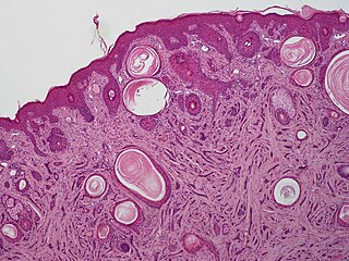
A lipoma is a benign tumor made of fat tissue. They are generally soft to the touch, movable, and painless. They usually occur just under the skin, but occasionally may be deeper. Most are less than 5 cm (2.0 in) in size. Common locations include upper back, shoulders, and abdomen. It is possible to have a number of lipomas.

<span class="mw-page-title-main">Cutaneous squamous-cell carcinoma</span> Medical condition

Cutaneous squamous-cell carcinoma (cSCC), or squamous-cell carcinoma of the skin, also known as squamous-cell skin cancer, is, with basal-cell carcinoma and melanoma, one of the three principal types of skin cancer. cSCC typically presents as a hard lump with a scaly top layer, but it may instead form an ulcer. Onset often occurs over a period of months. Cutaneous squamous-cell carcinoma is more likely to spread to distant areas than basal cell cancer. When confined to the outermost layer of the skin, a pre-invasive, or in situ, form of cSCC is known as Bowen's disease.

<span class="mw-page-title-main">Acral lentiginous melanoma</span> Medical condition

Acral lentiginous melanoma is an aggressive type of skin cancer. Melanoma is a group of serious skin cancers that arise from pigment cells (melanocytes); acral lentiginous melanoma is a kind of lentiginous skin melanoma. Acral lentiginous melanoma is the most common subtype in people with darker skins and is rare in people with lighter skin types. It is not caused by exposure to sunlight or UV radiation, and wearing sunscreen does not protect against it. Acral lentiginous melanoma is commonly found on the palms, soles, under the nails, and in the oral mucosa. It occurs on non-hair-bearing surfaces of the body, which have not necessarily been exposed to sunlight. It is also found on mucous membranes.

<span class="mw-page-title-main">Dermatofibrosarcoma protuberans</span> Medical condition

Dermatofibrosarcoma protuberans (DFSP) is a rare locally aggressive malignant cutaneous soft-tissue sarcoma. DFSP develops in the connective tissue cells in the middle layer of the skin (dermis). Estimates of the overall occurrence of DFSP in the United States are 0.8 to 4.5 cases per million persons per year. In the United States, DFSP accounts for between 1 and 6 percent of all soft-tissue sarcomas and 18 percent of all cutaneous soft-tissue sarcomas. In the Surveillance, Epidemiology and End Results (SEER) tumor registry from 1992 through 2004, DFSP was second only to Kaposi sarcoma.

<span class="mw-page-title-main">Invasive carcinoma of no special type</span> Medical condition

Invasive carcinoma of no special type, invasive breast carcinoma of no special type (IBC-NST), invasive ductal carcinoma (IDC), infiltrating ductal carcinoma (IDC) or invasive ductal carcinoma, not otherwise specified (NOS) is a disease. For international audiences this article will use "invasive carcinoma NST" because it is the preferred term of the World Health Organization (WHO).

<span class="mw-page-title-main">Hemangioendothelioma</span> Medical condition

Hemangioendotheliomas are a family of vascular neoplasms of intermediate malignancy.

<span class="mw-page-title-main">Lentigo maligna</span> Medical condition

Lentigo maligna is where melanocyte cells have become malignant and grow continuously along the stratum basale of the skin, but have not invaded below the epidermis. Lentigo maligna is not the same as lentigo maligna melanoma, as detailed below. It typically progresses very slowly and can remain in a non-invasive form for years.

<span class="mw-page-title-main">Hidradenoma</span> Medical condition

Hidradenoma refers to a benign adnexal tumor of the apical sweat gland. These are 1–3 cm translucent blue cystic nodules. It usually presents as a single, small skin-colored lesion, and may be considered closely related to or a variant of poromas. Hidradenomas are often sub-classified based on subtle histologic differences, for example:

<span class="mw-page-title-main">Extramammary Paget's disease</span> Medical condition

Extramammary Paget's disease (EMPD) is a rare and slow-growing malignancy which occurs within the epithelium and accounts for 6.5% of all Paget's disease. The clinical presentation of this disease is similar to the characteristics of mammary Paget's disease (MPD). However, unlike MPD, which occurs in large lactiferous ducts and then extends into the epidermis, EMPD originates in glandular regions rich in apocrine secretions outside the mammary glands. EMPD incidence is increasing by 3.2% every year, affecting hormonally-targeted tissues such as the vulva and scrotum. In women, 81.3% of EMPD cases are related to the vulva, while for men, 43.2% of the manifestations present at the scrotum.

Infantile digital fibromatosis (IDF), also termed inclusion body fibromatosis, Reye tumor, or Reye's tumor, usually occurs as a single, small, asymptomatic, nodule in the dermis on a finger or toe of infants and young children. IMF is a rare disorder with approximately 200 cases reported in the medical literature as of 2021. The World Health Organization in 2020 classified these nodules as a specific benign tumor type in the category of fibroblastic and myofibroblastic tumors. IDF was first described by the Australian pathologist, Douglas Reye, in 1965.

Atypical fibroxanthoma of the skin is a low-grade malignancy related to malignant fibrous histiocytoma, which it resembles histologically.

<span class="mw-page-title-main">Sebaceous carcinoma</span> Medical condition

Sebaceous carcinoma, also known as sebaceous gland carcinoma (SGc), sebaceous cell carcinoma, and meibomian gland carcinoma is an uncommon malignant cutaneous tumor. Most are typically about 1.4 cm at presentation. SGc originates from sebaceous glands in the skin and, therefore, may originate anywhere in the body where these glands are found. SGc can be divided into 2 types: periocular and extraocular. The periocular region is rich in sebaceous glands making it a common site of origin. The cause of these lesions in the vast majority of cases is unknown. Occasional cases may be associated with Muir-Torre syndrome. SGc accounts for approximately 0.7% of all skin cancers, and the incidence of SGc is highest in Caucasian, Asian, and Indian populations. Due to the rarity of this tumor and variability in clinical and histological presentation, SGc is often misdiagnosed as an inflammatory condition or a more common neoplasm. SGc is commonly treated with wide local excision or Mohs micrographic surgery, and the relative survival rates at 5 and 10 years are 92.72 and 86.98%, respectively.

<span class="mw-page-title-main">Poroma</span>

Poromas are rare, benign, cutaneous adnexal tumors. Cutaneous adnexal tumors are a group of skin tumors consisting of tissues that have differentiated towards one or more of the four primary adnexal structures found in normal skin: hair follicles, sebaceous sweat glands, apocrine sweat glands, and eccrine sweat glands. Poromas are eccrine or apocrine sweat gland tumors derived from the cells in the terminal portion of these glands' ducts. This part of the sweat gland duct is termed the acrosyringium and had led to grouping poromas in the acrospiroma class of skin tumors. Here, poromas are regarded as distinct sweat gland tumors that differ from other sweat gland tumors by their characteristic clinical presentations, microscopic histopathology, and the genetic mutations that their neoplastic cells have recently been found to carry.

Porocarcinoma (PCA) is a rare form of skin cancer that develops in eccrine sweat glands, i.e. the body's widely distributed major type of sweat glands, as opposed to the apocrine sweat glands which are located primarily in the armpits and perineal area. This cancer typically develops in individuals as a single cutaneous tumor in the intraepidermal spiral part of these sweat glands' ducts at or near to where they open on the skin's surface. PCA tumors are classified as one form of the cutaneous adnexal tumors; in a study of 2,205 cases, PCA was the most common (11.8%) form of these tumors.

<span class="mw-page-title-main">Spiradenoma</span> Medical condition

Spiradenomas (SA) are rare, benign cutaneous adnexal tumors that may progress to become their malignant counterparts, i.e. spiradenocarcinomas (SAC). Cutaneous adnexal tumors are a group of skin tumors consisting of tissues that have differentiated towards one of the four primary adnexal structures found in normal skin: hair follicles, sebaceous sweat glands, apocrine sweat glands, and eccrine sweat glands. SA and SAC tumors were regarded as eccrine gland tumors and termed eccrine spiradenomas and eccrine spiradenocarcinomas, respectively. However, more recent studies have found them to be hair follicle tumors and commonly term them spiradenomas and spiradenocarcinomas, respectively. Further confusing the situation, SA-like and SAC-like tumors are also 1) manifestations of the inherited disorder, CYLD cutaneous syndrome (CCS), and 2) have repeatedly been confused with an entirely different tumor, adenoid cystic carcinomas of the salivary gland. Here, SA and SAC are strictly defined as sporadic hair follicle tumors that do not include the hereditary CCS spiradenomas and heridtary spiradenocarcinoms of CCS or the adenoid cystic carcinomas.

<span class="mw-page-title-main">Microcystic adnexal carcinoma</span> Medical condition

Microcystic adnexal carcinoma (MAC) is a rare sweat gland cancer, which often appears as a yellow spot or bump in the skin. It usually occurs in the neck or head, although cases have been documented in other areas of the body. Most diagnosis occur past the age of 50. Although considered an invasive cancer, metastasis rarely occurs. If the tumor spreads, it can grow and invade fat, muscles, and other types of tissue. Main treatments are wide local excision or Mohs micrographic surgery, which ensures that most, if not all, cancer cells are removed surgically.

Skin cancer, or neoplasia, is the most common type of cancer diagnosed in horses, accounting for 45 to 80% of all cancers diagnosed. Sarcoids are the most common type of skin neoplasm and are the most common type of cancer overall in horses. Squamous-cell carcinoma is the second-most prevalent skin cancer, followed by melanoma. Squamous-cell carcinoma and melanoma usually occur in horses greater than 9-years-old, while sarcoids commonly affect horses 3 to 6 years old. Surgical biopsy is the method of choice for diagnosis of most equine skin cancers, but is contraindicated for cases of sarcoids. Prognosis and treatment effectiveness varies based on type of cancer, degree of local tissue destruction, evidence of spread to other organs (metastasis) and location of the tumor. Not all cancers metastasize and some can be cured or mitigated by surgical removal of the cancerous tissue or through use of chemotherapeutic drugs.

<span class="mw-page-title-main">Myoepithelioma of the head and neck</span> Medical condition

Myoepithelioma of the head and neck, also myoepithelioma, is a salivary gland tumour of the head and neck that is usually benign. When malignant, which is exceedingly rare, they are known as malignant myoepithelioma or Myoepithelial carcinoma, and they account for 1% of the salivary tumors with poor prognosis.

Ectomesenchymal chondromyxoid tumor (ECT) is a benign intraoral tumor with presumed origin from undifferentiated (ecto)mesenchymal cells. There are some who think it is a myoepithelial tumor type.

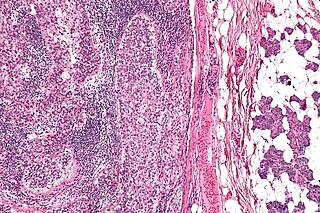
Ovarian germ cell tumors (OGCTs) are heterogeneous tumors that are derived from the primitive germ cells of the embryonic gonad, which accounts for about 2.6% of all ovarian malignancies. There are four main types of OGCTs, namely dysgerminomas, yolk sac tumor, teratoma, and choriocarcinoma.

References

  1. 1 2 James, William D.; Berger, Timothy G.; et al. (2006). Andrews' Diseases of the Skin: clinical Dermatology. Saunders Elsevier. ISBN   0-7216-2921-0.
  2. 1 2 3 4 5 6 7 8 9 10 Malik R, Saxena A, Kamath N (July 2013). "A rare case of malignant chondroid syringoma of scalp". Indian Dermatology Online Journal. 4 (3): 236–8. doi: 10.4103/2229-5178.115533 . PMC   3752488 . PMID   23984246.
  3. 1 2 3 4 5 6 7 8 9 Garcia AM, Atun JM, Fernando G (2016). "Multifocal, recurrent malignant chondroid syringoma with visceral metastases: A case report and literature review". International Journal of Cancer Therapy and Oncology. 4: 4119. doi: 10.14319/ijcto.41.19 via Research Gate.
  4. 1 2 3 4 5 6 7 Mathiasen RA, Rasgon BM, Rumore G (August 2005). "Malignant chondroid syringoma of the face: a first reported case". Otolaryngology–Head and Neck Surgery. 133 (2): 305–7. doi:10.1016/j.otohns.2004.09.139. PMID   16087034.
  5. 1 2 3 4 Sivamani R, Wadhera A, Craig E (September 2006). "Chondroid syringoma: case report and review of the literature". Dermatology Online Journal. 12 (5): 8. PMID   16962023.
  6. Obaidat NA, Alsaad KO, Ghazarian D (February 2007). "Skin adnexal neoplasms--part 2: an approach to tumours of cutaneous sweat glands". Journal of Clinical Pathology. 60 (2): 145–59. doi:10.1136/jcp.2006.041608. PMC   1860616 . PMID   16882695.
  7. Steinmetz JC, Russo BA, Ginsburg RE (May 1990). "Malignant chondroid syringoma with widespread metastasis". Journal of the American Academy of Dermatology. 22 (5 Pt 1): 845–7. doi:10.1016/s0190-9622(08)81178-4. PMID   2161437.
  8. 1 2 3 4 5 6 7 8 9 10 11 12 Tural D, Selçukbiricik F, Günver F, Karışmaz A, Serdengecti S (2013). "Facial localization of malignant chondroid syringoma: a rare case report". Case Reports in Oncological Medicine. 2013: 907980. doi: 10.1155/2013/907980 . PMC   3586475 . PMID   23476847.
  9. 1 2 3 4 5 6 Ka S, Gnangnon F, Diouf D, Dieng MM, Thiam J, Gaye M, Dem A (2016-01-01). "Malignant chondroid syringoma in a West African cancer institute: A case report". International Journal of Surgery Case Reports. 25: 137–8. doi:10.1016/j.ijscr.2016.06.029. PMC   4929341 . PMID   27372026.
  10. 1 2 3 4 5 6 7 8 9 10 11 12 13 14 15 Watarai A, Amoh Y, Aki R, Takasu H, Katsuoka K (September 2011). "Malignant chondroid syringoma: report of a case with lymph node metastasis 12 years after local excision". Dermatology Online Journal. 17 (9): 5. PMID   21971270.
  11. 1 2 3 4 5 6 7 8 9 10 Shashikala P, Chandrashekhar HR, Sharma S, Suresh KK (2004-05-01). "Malignant chondroid syringoma". Indian Journal of Dermatology, Venereology and Leprology. 70 (3): 175–6. PMID   17642601.
  12. 1 2 3 Metzler G, Schaumburg-Lever G, Hornstein O, Rassner G (February 1996). "Malignant chondroid syringoma: immunohistopathology". The American Journal of Dermatopathology. 18 (1): 83–9. doi:10.1097/00000372-199602000-00014. PMID   8721597.
  13. Lu H, Chen LF, Chen Q, Shen H, Liu Z (February 2018). "A rare large cutaneous chondroid syringoma involving a toe: A case report". Medicine. 97 (5): e9825. doi:10.1097/MD.0000000000009825. PMC   5805459 . PMID   29384887.
  14. Chauvel-Picard J, Pierrefeu A, Harou O, Breton P, Sigaux N (June 2018). "Unusual cystic lesion of the eyebrow: A case report of malignant chondroid syringoma". Journal of Stomatology, Oral and Maxillofacial Surgery. 119 (3): 232–235. doi:10.1016/j.jormas.2018.02.008. PMID   29477759.