Related Research Articles

The retina is the innermost, light-sensitive layer of tissue of the eye of most vertebrates and some molluscs. The optics of the eye create a focused two-dimensional image of the visual world on the retina, which then processes that image within the retina and sends nerve impulses along the optic nerve to the visual cortex to create visual perception. The retina serves a function which is in many ways analogous to that of the film or image sensor in a camera.
Macropsia is a neurological condition affecting human visual perception, in which objects within an affected section of the visual field appear larger than normal, causing the person to feel smaller than they actually are. Macropsia, along with its opposite condition, micropsia, can be categorized under dysmetropsia. Macropsia is related to other conditions dealing with visual perception, such as aniseikonia and Alice in Wonderland Syndrome. Macropsia has a wide range of causes, from prescription and illicit drugs, to migraines and (rarely) complex partial epilepsy, and to different retinal conditions, such as epiretinal membrane. Physiologically, retinal macropsia results from the compression of cones in the eye. It is the compression of receptor distribution that results in greater stimulation and thus a larger perceived image of an object.

Peripheral vision, or indirect vision, is vision as it occurs outside the point of fixation, i.e. away from the center of gaze or, when viewed at large angles, in the "corner of one's eye". The vast majority of the area in the visual field is included in the notion of peripheral vision. "Far peripheral" vision refers to the area at the edges of the visual field, "mid-peripheral" vision refers to medium eccentricities, and "near-peripheral", sometimes referred to as "para-central" vision, exists adjacent to the center of gaze.

The field of view (FOV) is the angular extent of the observable world that is seen at any given moment. In the case of optical instruments or sensors, it is a solid angle through which a detector is sensitive to electromagnetic radiation. It is further relevant in photography.

The occipital lobe is one of the four major lobes of the cerebral cortex in the brain of mammals. The name derives from its position at the back of the head, from the Latin ob, 'behind', and caput, 'head'.

Visual acuity (VA) commonly refers to the clarity of vision, but technically rates an animal's ability to recognize small details with precision. Visual acuity depends on optical and neural factors. Optical factors of the eye influence the sharpness of an image on its retina. Neural factors include the health and functioning of the retina, of the neural pathways to the brain, and of the interpretative faculty of the brain.

The fovea centralis is a small, central pit composed of closely packed cones in the eye. It is located in the center of the macula lutea of the retina.
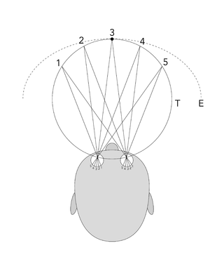
The visual field is "that portion of space in which objects are visible at the same moment during steady fixation of the gaze in one direction"; in ophthalmology and neurology the emphasis is mostly on the structure inside the visual field and it is then considered “the field of functional capacity obtained and recorded by means of perimetry”.
The horopter was originally defined in geometric terms as the locus of points in space that make the same angle at each eye with the fixation point, although more recently in studies of binocular vision it is taken to be the locus of points in space that have the same disparity as fixation. This can be defined theoretically as the points in space that project on corresponding points in the two retinas, that is, on anatomically identical points. The horopter can be measured empirically in which it is defined using some criterion.

Eye movement includes the voluntary or involuntary movement of the eyes. Eye movements are used by a number of organisms to fixate, inspect and track visual objects of interests. A special type of eye movement, rapid eye movement, occurs during REM sleep.

Retinotopy is the mapping of visual input from the retina to neurons, particularly those neurons within the visual stream. For clarity, 'retinotopy' can be replaced with 'retinal mapping', and 'retinotopic' with 'retinally mapped'.

Troxler's fading, also called Troxler fading or the Troxler effect, is an optical illusion affecting visual perception. When one fixates on a particular point for even a short period of time, an unchanging stimulus away from the fixation point will fade away and disappear. Research suggests that at least some portion of the perceptual phenomena associated with Troxler's fading occurs in the brain.
In vision, filling-in phenomena are those responsible for the completion of missing information across the physiological blind spot, and across natural and artificial scotomata. There is also evidence for similar mechanisms of completion in normal visual analysis. Classical demonstrations of perceptual filling-in involve filling in at the blind spot in monocular vision, and images stabilized on the retina either by means of special lenses, or under certain conditions of steady fixation. For example, naturally in monocular vision at the physiological blind spot, the percept is not a hole in the visual field, but the content is “filled-in” based on information from the surrounding visual field. When a textured stimulus is presented centered on but extending beyond the region of the blind spot, a continuous texture is perceived. This partially inferred percept is paradoxically considered more reliable than a percept based on external input..

Fixation or visual fixation is the maintaining of the gaze on a single location. An animal can exhibit visual fixation if it possess a fovea in the anatomy of their eye. The fovea is typically located at the center of the retina and is the point of clearest vision. The species in which fixational eye movement has been verified thus far include humans, primates, cats, rabbits, turtles, salamanders, and owls. Regular eye movement alternates between saccades and visual fixations, the notable exception being in smooth pursuit, controlled by a different neural substrate that appears to have developed for hunting prey. The term "fixation" can either be used to refer to the point in time and space of focus or the act of fixating. Fixation, in the act of fixating, is the point between any two saccades, during which the eyes are relatively stationary and virtually all visual input occurs. In the absence of retinal jitter, a laboratory condition known as retinal stabilization, perceptions tend to rapidly fade away. To maintain visibility, the nervous system carries out a procedure called fixational eye movement, which continuously stimulates neurons in the early visual areas of the brain responding to transient stimuli. There are three categories of fixational eye movement: microsaccades, ocular drifts, and ocular microtremor. At small amplitudes the boundaries between categories become unclear, particularly between drift and tremor.
Binocular disparity refers to the difference in image location of an object seen by the left and right eyes, resulting from the eyes’ horizontal separation (parallax). The mind uses binocular disparity to extract depth information from the two-dimensional retinal images in stereopsis. In computer vision, binocular disparity refers to the difference in coordinates of similar features within two stereo images.
Preferential hyperacuity perimetry (PHP) is a psychophysical test used to identify and quantify visual abnormalities such as metamorphopsia and scotoma. It is a type of perimetry.

The Divided Visual Field Paradigm is an experimental technique that involves measuring task performance when visual stimuli are presented on the left or right visual hemifields. If a visual stimulus appears in the left visual field (LVF), the visual information is initially projected to the right cerebral hemisphere (RH), and conversely, if a visual stimulus appears in the right visual field (RVF), the visual information is initially received by the left cerebral hemisphere (LH). In this way, if a cerebral hemisphere has functional advantages with some aspect of a particular task, an experimenter might observe improvements in task performance when the visual information is presented on the contralateral visual field.

Parafovea or the parafoveal belt is a region in the retina that circumscribes the fovea and is part of the macula lutea. It is circumscribed by the perifovea.
Microperimetry, sometimes called fundus-controlled perimetry, is a type of visual field test which uses one of several technologies to create a "retinal sensitivity map" of the quantity of light perceived in specific parts of the retina in people who have lost the ability to fixate on an object or light source. The main difference with traditional perimetry instruments is that, microperimetry includes a system to image the retina and an eye tracker to compensate eye movements during visual field testing.

Humphrey field analyser (HFA) is a tool for measuring the human visual field that is commonly used by optometrists, orthoptists and ophthalmologists, particularly for detecting monocular visual field.
References
- ↑ Rajimehr, R.; Tootell, R. B. H. (2009). "Does retinotopy influence cortical folding in primate visual cortex?". J. Neurosci. 29 (36): 11149–52. doi:10.1523/JNEUROSCI.1835-09.2009. PMC 2785715 . PMID 19741121.
IPS Perimetry Standards 1978. (2002). Author. Department of Ophthalmology & Visual Sciences, The University of Iowa. Retrieved March 4, 2005, from http://webeye.ophth.uiowa.edu/ips/GEN-INFO/standards/STANDARD.HTM