Related Research Articles

Positron emission tomography (PET) is a functional imaging technique that uses radioactive substances known as radiotracers to visualize and measure changes in metabolic processes, and in other physiological activities including blood flow, regional chemical composition, and absorption. Different tracers are used for various imaging purposes, depending on the target process within the body. For example, 18
F
-FDG is commonly used to detect cancer, NaF18
F
is widely used for detecting bone formation, and oxygen-15 is sometimes used to measure blood flow.

Medical imaging is the technique and process of imaging the interior of a body for clinical analysis and medical intervention, as well as visual representation of the function of some organs or tissues (physiology). Medical imaging seeks to reveal internal structures hidden by the skin and bones, as well as to diagnose and treat disease. Medical imaging also establishes a database of normal anatomy and physiology to make it possible to identify abnormalities. Although imaging of removed organs and tissues can be performed for medical reasons, such procedures are usually considered part of pathology instead of medical imaging.

Nuclear medicine or nucleology is a medical specialty involving the application of radioactive substances in the diagnosis and treatment of disease. Nuclear imaging, in a sense, is "radiology done inside out" because it records radiation emitting from within the body rather than radiation that is generated by external sources like X-rays. In addition, nuclear medicine scans differ from radiology, as the emphasis is not on imaging anatomy, but on the function. For such reason, it is called a physiological imaging modality. Single photon emission computed tomography (SPECT) and positron emission tomography (PET) scans are the two most common imaging modalities in nuclear medicine.
Edward Joseph Hoffman helped invent the first human PET scanner, a commonly used whole-body scanning procedure for detecting diseases like cancer. Hoffman, with Michel Ter-Pogossian and Michael E. Phelps, developed the Positron Emission Tomography scanner in 1973.
The first neuroimaging technique ever is the so-called 'human circulation balance' invented by Angelo Mosso in the 1880s and able to non-invasively measure the redistribution of blood during emotional and intellectual activity. Then, in the early 1900s, a technique called pneumoencephalography was set. This process involved draining the cerebrospinal fluid from around the brain and replacing it with air, altering the relative density of the brain and its surroundings, to cause it to show up better on an x-ray, and it was considered to be incredibly unsafe for patients. A form of magnetic resonance imaging (MRI) and computed tomography (CT) were developed in the 1970s and 1980s. The new MRI and CT technologies were considerably less harmful and are explained in greater detail below. Next came SPECT and PET scans, which allowed scientists to map brain function because, unlike MRI and CT, these scans could create more than just static images of the brain's structure. Learning from MRI, PET and SPECT scanning, scientists were able to develop functional MRI (fMRI) with abilities that opened the door to direct observation of cognitive activities.

Molecular imaging is a field of medical imaging that focuses on imaging molecules of medical interest within living patients. This is in contrast to conventional methods for obtaining molecular information from preserved tissue samples, such as histology. Molecules of interest may be either ones produced naturally by the body, or synthetic molecules produced in a laboratory and injected into a patient by a doctor. The most common example of molecular imaging used clinically today is to inject a contrast agent into a patient's bloodstream and to use an imaging modality to track its movement in the body. Molecular imaging originated from the field of radiology from a need to better understand fundamental molecular processes inside organisms in a noninvasive manner.
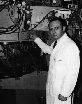
Michel Matthew Ter-Pogossian was an American medical physicist. He was professor of radiology at the Washington University School of Medicine for over 30 years. A pioneer in nuclear medicine, he is best known for his research on the positron emission tomography (PET). He is considered one of its creators and often referred to as the "father of PET."
Joanna Sigfred Fowler is a scientist emeritus at the U.S. Department of Energy's Brookhaven National Laboratory in New York. She served as professor of psychiatry at Mount Sinai School of Medicine and director of Brookhaven's Radiotracer Chemistry, Instrumentation and Biological Imaging Program. Fowler studied the effect of disease, drugs, and aging on the human brain and radiotracers in brain chemistry. She has received many awards for her pioneering work, including the National Medal of Science.
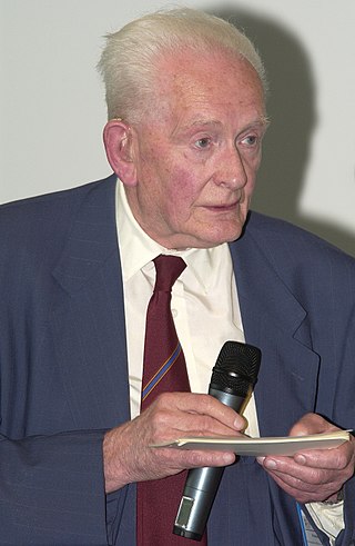
John Rowland Mallard OBE FRSE FREng was an English physicist and professor of Medical Physics at the University of Aberdeen from 1965 until his retirement in 1992. He was known for setting up and leading the team that developed the first magnetic resonance imaging (MRI) full body scanner and, in particular, positron emission tomography (PET). He was born in Kingsthorpe, Northampton, England.
Abass Alavi is an Iranian-American physician-scientist specializing in the field of molecular imaging, most notably in the imaging modality of positron emission tomography (PET). In August 1976, he was part of the team that performed the first human PET studies of the brain and whole body using the radiotracer [18F]Fluorodeoxyglucose (FDG). Alavi holds the position of Professor of Radiology and Neurology, as well as Director of Research Education in the Department of Radiology at the University of Pennsylvania. Over a career spanning five decades, he has amassed over 2,300 publications and 60,000 citations, earning an h-index of 125 and placing his publication record in the top percentile of scientists.
Nuclear medicine physicians, also called nuclear radiologists or simply nucleologists, are medical specialists that use tracers, usually radiopharmaceuticals, for diagnosis and therapy. Nuclear medicine procedures are the major clinical applications of molecular imaging and molecular therapy. In the United States, nuclear medicine physicians are certified by the American Board of Nuclear Medicine and the American Osteopathic Board of Nuclear Medicine.
Preclinical imaging is the visualization of living animals for research purposes, such as drug development. Imaging modalities have long been crucial to the researcher in observing changes, either at the organ, tissue, cell, or molecular level, in animals responding to physiological or environmental changes. Imaging modalities that are non-invasive and in vivo have become especially important to study animal models longitudinally. Broadly speaking, these imaging systems can be categorized into primarily morphological/anatomical and primarily molecular imaging techniques. Techniques such as high-frequency micro-ultrasound, magnetic resonance imaging (MRI) and computed tomography (CT) are usually used for anatomical imaging, while optical imaging, positron emission tomography (PET), and single photon emission computed tomography (SPECT) are usually used for molecular visualizations.

Brain positron emission tomography is a form of positron emission tomography (PET) that is used to measure brain metabolism and the distribution of exogenous radiolabeled chemical agents throughout the brain. PET measures emissions from radioactively labeled metabolically active chemicals that have been injected into the bloodstream. The emission data from brain PET are computer-processed to produce multi-dimensional images of the distribution of the chemicals throughout the brain.

Zang-Hee Cho is a Korean neuroscientist who developed the first Ring-PET scanner and the scintillation detector BGO. More recently, Cho developed the first PET-MRI fusion molecular imaging device for neuro-molecular imaging.

Sanjiv Sam Gambhir was an American physician–scientist. He was the Virginia and D.K. Ludwig Professor in Cancer Research, Chairman of the Department of Radiology at Stanford University School of Medicine, and a professor by courtesy in the departments of Bioengineering and Materials Science and Engineering at Stanford University. Additionally, he served as the Director of the Molecular Imaging Program at Stanford (MIPS), Canary Center at Stanford for Cancer Early Detection and the Precision Health and Integrated Diagnostics Center (PHIND). He authored 680 publications and had over 40 patents pending or granted. His work was featured on the cover of over 25 journals including the Nature Series, Science, and Science Translational Medicine. He was on the editorial board of several journals including Nano Letters, Nature Clinical Practice Oncology, and Science Translational Medicine. He was founder/co-founder of several biotechnology companies and also served on the scientific advisory board of multiple companies. He mentored over 150 post-doctoral fellows and graduate students from over a dozen disciplines. He was known for his work in molecular imaging of living subjects and early cancer detection.
Sandip Basu is an Indian physician of Nuclear Medicine and the Head, Nuclear Medicine Academic Program at the Radiation Medicine Centre. He is also the Dean-Academic (Health-Sciences), BARC at Homi Bhabha National Institute and is known for his services and research in Nuclear Medicine, particularly on Positron emission tomography diagnostics and Targeted Radionuclide Therapy in Cancer. The Council of Scientific and Industrial Research, the apex agency of the Government of India for scientific research, awarded him the Shanti Swarup Bhatnagar Prize for Science and Technology, one of the highest Indian science awards for his contributions to Nuclear Medicine in 2012.
Philip F. Cohen is a Canadian clinical director of Nuclear Medicine working out of the Lions Gate Hospital in North Vancouver, British Columbia. As a nuclear medicine physician, he is a pioneer in the usage of 3-D imaging techniques to improve diagnosis of bone disease and injury in collaboration with the Medical Imaging Research Group at University of British Columbia. Furthermore, Cohen has been involved in clinical research trials of new radiopharmaceuticals. To that effect, Cohen was the first recipient of a research grant from the Lions Gate Hospital Foundation, one of several peer-reviewed awards that would follow.

Carbon-11 choline is the basis of medical imaging technologies. Because of its involvement in biologic processes, choline is related to diseases, leading to the development of medical imaging techniques to monitor its concentration. When radiolabeled with 11CH3, choline is a useful a tracer in PET imaging. Carbon-11 is radioactive with a half-life of 20.38 minutes. By monitoring the gamma radiation resulting from the decay of carbon-11, the uptake, distribution, and retention of carbon-11 choline can be monitored.
Jason S. Lewis is a British radiochemist whose work relates to oncologic therapy and diagnosis. His research focus is a molecular imaging-based program focused on radiopharmaceutical development as well as the study of multimodality small- and biomolecule-based agents and their clinical translation. He has worked on the development of small molecules as well as radiolabeled peptides and antibodies probing the overexpression of receptors and antigens on tumors.

Jean-Claude Baron is an Emeritus Professor of Stroke Medicine at the University of Cambridge. He is also a Fellow of the Academy of Medical Sciences. He has authored around 450 peer-reviewed articles.
References
- ↑ American Men & Women of Science: M-P. R.R. Bowker. 2003. ISBN 9780787665289.
- ↑ Phelps, M.E.; E.J. Hoffman; N.A. Mullani; M.M. Ter-Pogossian (1 March 1975). "Application of annihilation coincidence detection to transaxial reconstruction tomography". Journal of Nuclear Medicine. 16 (3): 210–24. PMID 1113170.
- ↑ Brice, James (July 2001). "PET pioneer meets life head-on". Diagnostic Medicine.com. Archived from the original on 2017-07-26. Retrieved 2009-12-08.
- ↑ Brice, James (July 2001). "PET pioneer meets life head-on". Diagnostic Medicine.com. Archived from the original on 2017-07-26. Retrieved 2009-12-08.
- ↑ "FERMI Michael e. Phelps, 1998 | U.S. DOE Office of Science (SC)". 28 December 2010.
- ↑ "FERMI Michael e. Phelps, 1998 | U.S. DOE Office of Science (SC)". 28 December 2010.
- ↑ "FERMI Michael e. Phelps, 1998 | U.S. DOE Office of Science (SC)". 28 December 2010.
- ↑ "FERMI Michael e. Phelps, 1998 | U.S. DOE Office of Science (SC)". 28 December 2010.