Related Research Articles

Asperger syndrome (AS), also known as Asperger's syndrome or Asperger's, is a term formerly used to describe a neurodevelopmental condition characterized by significant difficulties in social interaction and nonverbal communication, along with restricted, repetitive patterns of behavior and interests. Asperger syndrome has been merged with other conditions into autism spectrum disorder (ASD) and is no longer considered a diagnosis. It was considered milder than other diagnoses which were merged into ASD due to relatively unimpaired spoken language and intelligence.

Facial perception is an individual's understanding and interpretation of the face. Here, perception implies the presence of consciousness and hence excludes automated facial recognition systems. Although facial recognition is found in other species, this article focuses on facial perception in humans.

The superior temporal gyrus (STG) is one of three gyri in the temporal lobe of the human brain, which is located laterally to the head, situated somewhat above the external ear.
Neuroplasticity, also known as neural plasticity or brain plasticity, is the ability of neural networks in the brain to change through growth and reorganization. It is when the brain is rewired to function in some way that differs from how it previously functioned. These changes range from individual neuron pathways making new connections, to systematic adjustments like cortical remapping or neural oscillation. Other forms of neuroplasticity include homologous area adaptation, cross modal reassignment, map expansion, and compensatory masquerade. Examples of neuroplasticity include circuit and network changes that result from learning a new ability, information acquisition, environmental influences, pregnancy, caloric intake, practice/training, and psychological stress.
Mind-blindness, mindblindness or mind blindness is a theory initially proposed in 1990 that claims that all autistic people have a lack or developmental delay of theory of mind (ToM), meaning they are unable to attribute mental states to others. According to the theory, a lack of ToM is considered equivalent to a lack of both cognitive and affective empathy. In the context of the theory, mind-blindness implies being unable to predict behavior and attribute mental states including beliefs, desires, emotions, or intentions of other people. The mind-blindness theory asserts that children who delay in this development will often develop autism.
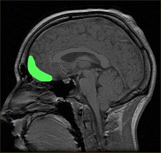
The orbitofrontal cortex (OFC) is a prefrontal cortex region in the frontal lobes of the brain which is involved in the cognitive process of decision-making. In non-human primates it consists of the association cortex areas Brodmann area 11, 12 and 13; in humans it consists of Brodmann area 10, 11 and 47.

The posterior cingulate cortex (PCC) is the caudal part of the cingulate cortex, located posterior to the anterior cingulate cortex. This is the upper part of the "limbic lobe". The cingulate cortex is made up of an area around the midline of the brain. Surrounding areas include the retrosplenial cortex and the precuneus.

The sensorimotor mu rhythm, also known as mu wave, comb or wicket rhythms or arciform rhythms, are synchronized patterns of electrical activity involving large numbers of neurons, probably of the pyramidal type, in the part of the brain that controls voluntary movement. These patterns as measured by electroencephalography (EEG), magnetoencephalography (MEG), or electrocorticography (ECoG), repeat at a frequency of 7.5–12.5 Hz, and are most prominent when the body is physically at rest. Unlike the alpha wave, which occurs at a similar frequency over the resting visual cortex at the back of the scalp, the mu rhythm is found over the motor cortex, in a band approximately from ear to ear. People suppress mu rhythms when they perform motor actions or, with practice, when they visualize performing motor actions. This suppression is called desynchronization of the wave because EEG wave forms are caused by large numbers of neurons firing in synchrony. The mu rhythm is even suppressed when one observes another person performing a motor action or an abstract motion with biological characteristics. Researchers such as V. S. Ramachandran and colleagues have suggested that this is a sign that the mirror neuron system is involved in mu rhythm suppression, although others disagree.

The fusiform face area is a part of the human visual system that is specialized for facial recognition. It is located in the inferior temporal cortex (IT), in the fusiform gyrus.
Frontostriatal circuits are neural pathways that connect frontal lobe regions with the striatum and mediate motor, cognitive, and behavioural functions within the brain. They receive inputs from dopaminergic, serotonergic, noradrenergic, and cholinergic cell groups that modulate information processing. Frontostriatal circuits are part of the executive functions. Executive functions include the following: selection and perception of important information, manipulation of information in working memory, planning and organization, behavioral control, adaptation to changes, and decision making. These circuits are involved in neurodegenerative disorders such as Alzheimer's disease and Parkinson's disease as well as neuropsychiatric disorders including schizophrenia, depression, obsessive compulsive disorder (OCD), and in neurodevelopmental disorder such as attention-deficit hyperactivity disorder (ADHD).

In human neuroanatomy, brain asymmetry can refer to at least two quite distinct findings:
Marcel Just is D. O. Hebb Professor of Psychology at Carnegie Mellon University. His research uses brain imaging (fMRI) in high-level cognitive tasks to study the neuroarchitecture of cognition. Just's areas of expertise include psycholinguistics, object recognition, and autism, with particular attention to cognitive and neural substrates. Just directs the Center for Cognitive Brain Imaging and is a member of the Center for the Neural Basis of Cognition at CMU.
In psychology and neuroscience, executive dysfunction, or executive function deficit, is a disruption to the efficacy of the executive functions, which is a group of cognitive processes that regulate, control, and manage other cognitive processes. Executive dysfunction can refer to both neurocognitive deficits and behavioural symptoms. It is implicated in numerous psychopathologies and mental disorders, as well as short-term and long-term changes in non-clinical executive control. Executive dysfunction is the mechanism underlying ADHD paralysis, and in a broader context, it can encompass other cognitive difficulties like planning, organizing, initiating tasks and regulating emotions. It is a core characteristic of ADHD and can elucidate numerous other recognized symptoms.
The causes of schizophrenia that underlie the development of schizophrenia, a psychiatric disorder, are complex and not clearly understood. A number of hypotheses including the dopamine hypothesis, and the glutamate hypothesis have been put forward in an attempt to explain the link between altered brain function and the symptoms and development of schizophrenia.
Autism spectrum disorder (ASD) refers to a variety of conditions typically identified by challenges with social skills, communication, speech, and repetitive sensory-motor behaviors. The 11th International Classification of Diseases (ICD-11), released in January 2021, characterizes ASD by the associated deficits in the ability to initiate and sustain two-way social communication and restricted or repetitive behavior unusual for the individual's age or situation. Although linked with early childhood, the symptoms can appear later as well. Symptoms can be detected before the age of two and experienced practitioners can give a reliable diagnosis by that age. However, official diagnosis may not occur until much older, even well into adulthood. There is a large degree of variation in how much support a person with ASD needs in day-to-day life. This can be classified by a further diagnosis of ASD level 1, level 2, or level 3. Of these, ASD level 3 describes people requiring very substantial support and who experience more severe symptoms. ASD-related deficits in nonverbal and verbal social skills can result in impediments in personal, family, social, educational, and occupational situations. This disorder tends to have a strong correlation with genetics along with other factors. More research is identifying ways in which epigenetics is linked to autism. Epigenetics generally refers to the ways in which chromatin structure is altered to affect gene expression. Mechanisms such as cytosine regulation and post-translational modifications of histones. Of the 215 genes contributing, to some extent in ASD, 42 have been found to be involved in epigenetic modification of gene expression. Some examples of ASD signs are specific or repeated behaviors, enhanced sensitivity to materials, being upset by changes in routine, appearing to show reduced interest in others, avoiding eye contact and limitations in social situations, as well as verbal communication. When social interaction becomes more important, some whose condition might have been overlooked suffer social and other exclusion and are more likely to have coexisting mental and physical conditions. Long-term problems include difficulties in daily living such as managing schedules, hypersensitivities, initiating and sustaining relationships, and maintaining jobs.
The relationship between autism and memory, specifically memory functions in relation to Autism Spectrum Disorder (ASD), is an ongoing topic of research. ASD is a neurodevelopmental disorder characterised by social communication and interaction impairments, along with restricted and repetitive patterns of behavior. In this article, the word autism is used to refer to the whole range of conditions on the autism spectrum, which are not uncommon.
The mechanisms of autism are the molecular and cellular processes believed to cause or contribute to the symptoms of autism. Multiple processes are hypothesized to explain different autism spectrum features. These hypotheses include defects in synapse structure and function, reduced synaptic plasticity, disrupted neural circuit function, gut–brain axis dyshomeostasis, neuroinflammation, and altered brain structure or connectivity.
Network neuroscience is an approach to understanding the structure and function of the human brain through an approach of network science, through the paradigm of graph theory. A network is a connection of many brain regions that interact with each other to give rise to a particular function. Network Neuroscience is a broad field that studies the brain in an integrative way by recording, analyzing, and mapping the brain in various ways. The field studies the brain at multiple scales of analysis to ultimately explain brain systems, behavior, and dysfunction of behavior in psychiatric and neurological diseases. Network neuroscience provides an important theoretical base for understanding neurobiological systems at multiple scales of analysis.
The pathophysiology of autism is the study of the physiological processes that cause or are otherwise associated with autism spectrum disorders.
Bradley S. Peterson is an American psychiatrist, developmental neuroscientist, academic and author. He is the Inaugural Director of the Institute for the Developing Mind at Children's Hospital Los Angeles (CHLA), and holds the positions of Vice Chair for Research and Chief of the Division of Child & Adolescent Psychiatry in the Department of Psychiatry at the Keck School of Medicine at the University of Southern California.
References
- ↑ Just MA, Cherkassky VL, Keller TA, Kana RK, Minshew NJ (2007). "Functional and anatomical cortical underconnectivity in autism: evidence from an FMRI study of an executive function task and corpus callosum morphometry". Cereb Cortex. 17 (4): 951–61. doi:10.1093/cercor/bhl006. PMC 4500121 . PMID 16772313.
- ↑ Williams DL, Goldstein G, Minshew NJ (2006). "Neuropsychologic functioning in children with autism: further evidence for disordered complex information-processing". Child Neuropsychol. 12 (4–5): 279–98. doi:10.1080/09297040600681190. PMC 1803025 . PMID 16911973.
- ↑ Murias M, Webb SJ, Greenson J, Dawson G (2007). "Resting state cortical connectivity reflected in EEG coherence in individuals with autism". Biol Psychiatry. 62 (3): 270–3. doi:10.1016/j.biopsych.2006.11.012. PMC 2001237 . PMID 17336944.