Nanoshell

Last updated

Figure 1. s-polarization and p-polarization Polarization.gif
Figure 1. s-polarization and p-polarization

A nanoshell, or rather a nanoshell plasmon, is a type of spherical nanoparticle consisting of a dielectric core which is covered by a thin metallic shell (usually gold). [1] These nanoshells involve a quasiparticle called a plasmon which is a collective excitation or quantum plasma oscillation where the electrons simultaneously oscillate with respect to all the ions.

Contents

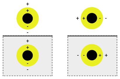
The simultaneous oscillation can be called plasmon hybridization where the tunability of the oscillation is associated with mixture of the inner and outer shell where they hybridize to give a lower energy or higher energy. This lower energy couples strongly to incident light, whereas the higher energy is an anti-bonding and weakly combines to incident light. The hybridization interaction is stronger for thinner shell layers, hence, the thickness of the shell and overall particle radius determines which wavelength of light it couples with. [2] Nanoshells can be varied across a broad range of the light spectrum that spans the visible and near infrared regions. The interaction of light and nanoparticles affects the placement of charges which affects the coupling strength. Incident light polarized parallel to the substrate gives a s-polarization (Figure 1b), hence the charges are further from the substrate surface which gives a stronger interaction between the shell and core. Otherwise, a p-polarization is formed which gives a more strongly shifted plasmon energy causing a weaker interaction and coupling.

Discovery

The discovery of the nanoshell was made by Professor Naomi J. Halas and her team at Rice University in 2003. When she and her team discovered nanoshells, they weren't initially sure what potential such nanoshells held. "We said, 'Gee, what could it be good for?'" Halas told CNN. After many suggestions, cancer therapy came out of ongoing collaborations with bioengineers looking for different types of biomedical applications. [3] "One of our visions", Halas stated, "no less than single visit diagnosis and treatment of cancer". [4] In 2003 Halas was awarded for Best Discovery of 2003 by Nanotechnology Now. [4]

Production

A state of the art method for synthesizing gold nanoshells is the use of the Microfluidic Composite Foams. This method has the potential to replace the standard lithographic method of synthesizing plasmonic nanoshells. The production process described below was an experiment performed by Suhanya Duraiswamy and Saif A. Khan of the Department of Chemical and Biomolecular Engineering in Singapore. Although this method was an experiment, it represents the future of nanoshells synthesis.

The materials required for the production of the nanoshells are the following; Tetraethyl orthosilicate, ammonium hydroxide, hydroxylamine hydrochloride, 3-aminopropyl tris, hydrogentetrachloroaurate(III) trihydrate, tetrakis(hydroxymethyl) phosphonium chloride, sodium hydroxide, potassium carbonate, ethanol, Ultrapure water and glassware washed in aqua regia and rinsed thoroughly in water. [5] )

The first step in synthesizing nanoshells in this method is by creating the device for the reaction to take place within. Microfluidic device patterns were fabricated onto silicon wafers by standard photolithography using negative photoresist SU-8 2050. Devices were subsequently molded in poly(dimethyl siloxane) (PDMS) using the soft lithography technique.(40) Briefly, PDMS was molded onto the SU-8 masters at 70 °C for 4 h, peeled, cut, and cleaned. Inlet and outlet holes (1/16-in. o.d.) were punched into the device. The microchannels were irreversibly bonded to a glass slide precoated with a thin layer of PDMS after a brief 35 s air plasma treatment. The microchannels have rectangular cross-section and are 300 μm wide, 155 μm deep, and 0.45 m long. [5]

The actual production of the nanoparticles involves pumping "silicone oil, a mixture of gold-seeded silica particles and gold-plating solution and reducing agent solution to the microfluidic device while nitrogen gas was delivered from a cylinder." [5] The plating solution was then left to age, in a controlled environment, for longer than 24 hours. After the aging process, the fluid is collected from the Microfluidic Device and placed in a centrifuge. The resulting liquid has a layer of oil on the surface with a solution below that contains the nanoshells.

The reason this method is revolutionary is that the size and relative thickness of the gold nanoshell can be controlled by changing the amount of time the reaction is allowed to take place as well as the concentration of the plating solution. Thereby allowing researchers to tailor the particles to suit their given needs. Albeit for optics or cancer treatment.

Cancer treatment

Gold-shelled nanoparticles, which are spherical nanoparticles with silica and/or liposome cores [6] and gold shells, are used in cancer therapy and bio-imaging enhancement. Theranostic probes – capable of detection and treatment of cancer in a single treatment – are nanoparticles that have binding sites on their shell that allow them to attach to a desired location (typically cancerous cells) then can be imaged through dual modality imagery (an imaging strategy that uses x-rays and radionuclide imaging) and through near-infrared fluorescence. [7] The reason gold nanoparticles are used is due to their vivid optical properties which are controlled by their size, geometry, and their surface plasmons. Gold nanoparticles (such as AuNPs) have the benefit of being biocompatible and the flexibility to have multiple different molecules, and fundamental materials, attached to their shell (almost anything that can normally be attached to gold can be attached to the gold nano-shell, which can be used in helping identifying and treating cancer). The treatment of cancer is possible only because of the scattering and absorption that occurs for plasmonics. Under scattering, the gold-plated nano-particles become visible to imaging processes that are tuned to the correct wavelength which is dependent upon the size and geometry of the particles. Under absorption, photothermal ablation occurs, which heats the nanoparticles and their immediate surroundings to temperatures capable of killing the cancer cells. This is accomplished with minimal damage to cells in the body due to the utilization of the "water window" (the spectral range between 800 and 1300 nm). [1] As the human body is mostly water, this optimizes the light used versus the effects rendered.

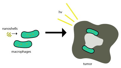
These gold nanoshells are shuttled into tumors by the use of phagocytosis, where phagocytes engulf the nanoshells through the cell membrane to form an internal phagosome, or macrophage. After this it is shuttled into a cell and enzymes are usually used to metabolize it and shuttle it back out of the cell. These nanoshells are not metabolized so for them to be effective they just need to be within the tumor cells and photo-induced cell death (as described above) is used to terminate the tumor cells. This scheme is shown in Figure 2.

Figure 2. Nanoshells taken into tumors. Attack-cancer.png
Figure 2. Nanoshells taken into tumors.

Nanoparticle-based therapeutics have been successfully delivered into tumors by exploiting the enhanced permeability and retention effect, a property that permits nanoscale structures to be taken up passively into tumors without the assistance of antibodies.[4] Delivery of nanoshells into the important regions of tumors can be very difficult. This is where most nanoshells try to exploit the tumor's natural recruitment of monocytes for delivery as seen in the above figure. This delivery system is called a "Trojan Horse". [8]

This process works so well since tumors are about ¾ macrophages and once monocytes are brought into the tumor, it differentiates into macrophages which would also be need to maintain the cargo nanoparticles. Once the nanoshells are at the necrotic center, near-infrared illumination is used to destroy the tumor associated macrophages.

Additionally, these nanoparticles can be made to release antisense DNA oligonucleotides when under photo-activation. These oligonucleotides are used in conjunction with the photo-thermal ablation treatments to perform gene-therapy. This is accomplished because nanoparticle complexes are delivered inside of cells then undergo light induced release of DNA from their surface. This will allow for the internal manipulation of a cell and provide a means for monitoring a group cells return to equilibrium. [9]

Another example of nanoshell plasmonics in cancer treatment involves placing drugs inside of the nanoparticle and using it as a vehicle to deliver toxic drugs to cancerous sites only. [10] This is accomplished by coating the outside of a nanoparticle with iron oxide (allowing for easy tracking with an MRI machine), then once the area of the tumor is coated with the drug-filled nanoparticles, the nanoparticles can be activated using resonant light waves to release the drug.

See also

Related Research Articles

Nanomedicine is the medical application of nanotechnology. Nanomedicine ranges from the medical applications of nanomaterials and biological devices, to nanoelectronic biosensors, and even possible future applications of molecular nanotechnology such as biological machines. Current problems for nanomedicine involve understanding the issues related to toxicity and environmental impact of nanoscale materials.

<span class="mw-page-title-main">Plasmon</span> Quasiparticle of charge oscillations in condensed matter

In physics, a plasmon is a quantum of plasma oscillation. Just as light consists of photons, the plasma oscillation consists of plasmons. The plasmon can be considered as a quasiparticle since it arises from the quantization of plasma oscillations, just like phonons are quantizations of mechanical vibrations. Thus, plasmons are collective oscillations of the free electron gas density. For example, at optical frequencies, plasmons can couple with a photon to create another quasiparticle called a plasmon polariton.

Naomi J. Halas is the Stanley C. Moore Professor in Electrical and Computer Engineering, and professor of biomedical engineering, chemistry and physics at Rice University. She is also the founding director of Rice University Laboratory for Nanophotonics, and the Smalley-Curl Institute. She invented the first nanoparticle with tunable plasmonic resonances, which are controlled by their shape and structure, and has won numerous awards for her pioneering work in the field of nanophotonics and plasmonics. She was also part of a team that developed the first dark pulse soliton in 1987 while working for IBM.

<span class="mw-page-title-main">Colloidal gold</span> Suspension of gold nanoparticles in a liquid

Colloidal gold is a sol or colloidal suspension of nanoparticles of gold in a fluid, usually water. The colloid is coloured usually either wine red or blue-purple . Due to their optical, electronic, and molecular-recognition properties, gold nanoparticles are the subject of substantial research, with many potential or promised applications in a wide variety of areas, including electron microscopy, electronics, nanotechnology, materials science, and biomedicine.

<span class="mw-page-title-main">Surface plasmon resonance</span> Physical phenomenon of electron resonance

Surface plasmon resonance (SPR) is a phenomenon that occurs where electrons in a thin metal sheet become excited by light that is directed to the sheet with a particular angle of incidence, and then travel parallel to the sheet. Assuming a constant light source wavelength and that the metal sheet is thin, the angle of incidence that triggers SPR is related to the refractive index of the material and even a small change in the refractive index will cause SPR to not be observed. This makes SPR a possible technique for detecting particular substances (analytes) and SPR biosensors have been developed to detect various important biomarkers.

<span class="mw-page-title-main">Surface-enhanced Raman spectroscopy</span>

Surface-enhanced Raman spectroscopy or surface-enhanced Raman scattering (SERS) is a surface-sensitive technique that enhances Raman scattering by molecules adsorbed on rough metal surfaces or by nanostructures such as plasmonic-magnetic silica nanotubes. The enhancement factor can be as much as 1010 to 1011, which means the technique may detect single molecules.

Photothermal therapy (PTT) refers to efforts to use electromagnetic radiation for the treatment of various medical conditions, including cancer. This approach is an extension of photodynamic therapy, in which a photosensitizer is excited with specific band light. This activation brings the sensitizer to an excited state where it then releases vibrational energy (heat), which is what kills the targeted cells.

<span class="mw-page-title-main">Surface plasmon</span>

Surface plasmons (SPs) are coherent delocalized electron oscillations that exist at the interface between any two materials where the real part of the dielectric function changes sign across the interface. SPs have lower energy than bulk plasmons which quantise the longitudinal electron oscillations about positive ion cores within the bulk of an electron gas.

<span class="mw-page-title-main">Janus particles</span> Type of nanoparticle or microparticle

Janus particles are special types of nanoparticles or microparticles whose surfaces have two or more distinct physical properties. This unique surface of Janus particles allows two different types of chemistry to occur on the same particle. The simplest case of a Janus particle is achieved by dividing the particle into two distinct parts, each of them either made of a different material, or bearing different functional groups. For example, a Janus particle may have one half of its surface composed of hydrophilic groups and the other half hydrophobic groups, the particles might have two surfaces of different color, fluorescence, or magnetic properties. This gives these particles unique properties related to their asymmetric structure and/or functionalization.

<span class="mw-page-title-main">Silver nanoparticle</span> Ultrafine particles of silver between 1 nm and 100 nm in size

Silver nanoparticles are nanoparticles of silver of between 1 nm and 100 nm in size. While frequently described as being 'silver' some are composed of a large percentage of silver oxide due to their large ratio of surface to bulk silver atoms. Numerous shapes of nanoparticles can be constructed depending on the application at hand. Commonly used silver nanoparticles are spherical, but diamond, octagonal, and thin sheets are also common.

A plasmonic-enhanced solar cell, commonly referred to simply as plasmonic solar cell, is a type of solar cell that converts light into electricity with the assistance of plasmons, but where the photovoltaic effect occurs in another material.

A plasmonic metamaterial is a metamaterial that uses surface plasmons to achieve optical properties not seen in nature. Plasmons are produced from the interaction of light with metal-dielectric materials. Under specific conditions, the incident light couples with the surface plasmons to create self-sustaining, propagating electromagnetic waves known as surface plasmon polaritons (SPPs). Once launched, the SPPs ripple along the metal-dielectric interface. Compared with the incident light, the SPPs can be much shorter in wavelength.

Multiple layered plasmonics use electronically responsive media to change and manipulate the plasmonic properties of plasmons. The properties typically being manipulated can include the directed scattering of light and light absorption. The use of these to use “changeable” plasmonics is currently undergoing development in the academic community by allowing them to have multiple sets of functions that are dependent on how they are being manipulated or excited. Under these new manipulations, such as multiple layers that respond to different resonant frequencies, their new functions were designed to accomplish multiple objectives in a single application.

<span class="mw-page-title-main">Plasmonic nanoparticles</span>

Plasmonic nanoparticles are particles whose electron density can couple with electromagnetic radiation of wavelengths that are far larger than the particle due to the nature of the dielectric-metal interface between the medium and the particles: unlike in a pure metal where there is a maximum limit on what size wavelength can be effectively coupled based on the material size.

<span class="mw-page-title-main">Localized surface plasmon</span>

A localized surface plasmon (LSP) is the result of the confinement of a surface plasmon in a nanoparticle of size comparable to or smaller than the wavelength of light used to excite the plasmon. When a small spherical metallic nanoparticle is irradiated by light, the oscillating electric field causes the conduction electrons to oscillate coherently. When the electron cloud is displaced relative to its original position, a restoring force arises from Coulombic attraction between electrons and nuclei. This force causes the electron cloud to oscillate. The oscillation frequency is determined by the density of electrons, the effective electron mass, and the size and shape of the charge distribution. The LSP has two important effects: electric fields near the particle's surface are greatly enhanced and the particle's optical absorption has a maximum at the plasmon resonant frequency. Surface plasmon resonance can also be tuned based on the shape of the nanoparticle. The plasmon frequency can be related to the metal dielectric constant. The enhancement falls off quickly with distance from the surface and, for noble metal nanoparticles, the resonance occurs at visible wavelengths. Localized surface plasmon resonance creates brilliant colors in metal colloidal solutions.

<span class="mw-page-title-main">Gold nanoparticles in chemotherapy</span> Drug delivery technique using gold nanoparticles as vectors

Gold nanoparticles in chemotherapy and radiotherapy is the use of colloidal gold in therapeutic treatments, often for cancer or arthritis. Gold nanoparticle technology shows promise in the advancement of cancer treatments. Some of the properties that gold nanoparticles possess, such as small size, non-toxicity and non-immunogenicity make these molecules useful candidates for targeted drug delivery systems. With tumor-targeting delivery vectors becoming smaller, the ability to by-pass the natural barriers and obstacles of the body becomes more probable. To increase specificity and likelihood of drug delivery, tumor specific ligands may be grafted onto the particles along with the chemotherapeutic drug molecules, to allow these molecules to circulate throughout the tumor without being redistributed into the body.

Plasmon coupling is a phenomenon that occurs when two or more plasmonic particles approach each other to a distance below approximately one diameter's length. Upon the occurrence of plasmon coupling, the resonance of individual particles start to hybridize, and their resonance spectrum peak wavelength will shift, depending on how surface charge density distributes over the coupled particles. At a single particle's resonance wavelength, the surface charge densities of close particles can either be out of phase or in phase, causing repulsion or attraction and thus leading to increase (blueshift) or decrease (redshift) of hybridized mode energy. The magnitude of the shift, which can be the measure of plasmon coupling, is dependent on the interparticle gap as well as particles geometry and plasmonic resonances supported by individual particles. A larger redshift is usually associated with smaller interparticle gap and larger cluster size.

Prashant Jain is an Indian-born American scientist and a professor of chemistry at the University of Illinois Urbana–Champaign where his research laboratory studies the interaction of light with matter, designs nanoparticle catalysts, and develops methods for mimicking plant photosynthesis. He is a Fellow of the American Association for the Advancement of Science and the Royal Society of Chemistry, a TR35 inventor, a Sloan Fellow, a PECASE recipient, a Royal Society of Chemistry Beilby medalist, and a top-cited researcher in chemical sciences.

Rizia Bardhan is an Indian origin American biomolecular engineer who is an Associate Professor of Chemical & Biological Engineering at Iowa State University. She is Associate Editor of ACS Applied Materials & Interfaces.

In chemistry, plasmonic catalysis is a type of catalysis that uses plasmons to increase the rate of a chemical reaction. A plasmonic catalyst is made up of a metal nanoparticle surface which generates localized surface plasmon resonances (LSPRs) when excited by light. These plasmon oscillations create an electron-rich region near the surface of the nanoparticle, which can be used to excite the electrons of nearby molecules.

References

  1. 1 2 Loo, C; Lin, A; Hirsch, L; Lee, Mh; Barton, J; Halas, N; West, J; Drezek, R (February 2004). "Nanoshell-enabled photonics-based imaging and therapy of cancer". Technology in Cancer Research & Treatment. 3 (1): 33–40. doi:10.1177/153303460400300104. PMID   14750891. S2CID   17523671. Archived from the original (Free full text) on 23 October 2007. Retrieved 6 August 2009.
  2. Brinson, Be; Lassiter, Jb; Levin, Cs; Bardhan, R; Mirin, N; Halas, Nj (November 2008). "Nanoshells Made Easy: Improving Au Layer Growth on Nanoparticle Surfaces". Langmuir. 24 (24): 14166–14171. doi:10.1021/la802049p. PMC   5922771 . PMID   19360963.
  3. CNN. "Biography: Naomi Halas." CNN. Cable News Network, 11 Mar. 2008. Web. 7 May 2012. <http://edition.cnn.com/2007/TECH/science/06/11/halas.biog/>.
  4. 1 2 "Best Discoveries." – Best of Nanotechnology. Nanotechnology Now, 29 Mar. 2008. Web. 7 May 2012. <http://www.nanotech-now.com/2003-Awards/Best-Discoveries-2003.htm>.
  5. 1 2 3 Duraiswamy, Suhanya; Khan, Saif (23 August 2010). "Plasmonic Nanoshell Synthesis in Microfluidic Composite Foams". Nano Letters. 9. 10 (9): 3757–3763. Bibcode:2010NanoL..10.3757D. doi:10.1021/nl102478q. PMID   20731386.
  6. Abbasi, Akram; Park, Keunhan; Bose, Arijit; Bothun, Geoffrey D. (30 May 2017). "Near-Infrared Responsive Gold–Layersome Nanoshells". Langmuir. 33 (21): 5321–5327. doi:10.1021/acs.langmuir.7b01273. ISSN   0743-7463. PMID   28486807.
  7. Bardhan, R; Grady, Nk; Halas, Nj (September 2008). "Nanoscale Control of Near-Infrared Fluorescence Enhancement Using Au Nanoshells". Nano Micro Small. 4 (10): 1716–1722. doi: 10.1002/smll.200800405 . PMID   18819167.
  8. Choi, Mr; Stanton-Maxey, Kj; Stanley, Jk; Levin, Cs; Bardhan, R; Akin, D; Badve, S; Sturgis, J; Robinson, Jp; Bashir, R; Halas, Nj; Clare, Se (December 2007). "A cellular Trojan Horse for delivery of therapeutic nanoparticles into tumors". Nano Letters. 7 (12): 3759–65. Bibcode:2007NanoL...7.3759C. doi:10.1021/nl072209h. PMID   17979310.
  9. Bardan, R; Lal, S; Joshi, A; Halas, Nj (May 2011). "Theranostic Nanoshells: From Probe Design to Imaging and Treatment of Cancer". Accounts of Chemical Research. 44 (10): 936–946. doi:10.1021/ar200023x. PMC   3888233 . PMID   21612199.
  10. "Nanoparticles Used To Target Brain Cancer -- ScienceDaily".