Neuroendocrine adenoma middle ear

Last updated

Neuroendocrine adenoma middle ear (NAME) is a tumor which arises from a specific anatomic site, the middle ear. [1] NAME is a benign glandular neoplasm of middle ear showing histologic and immunohistochemical neuroendocrine and mucin-secreting differentiation (biphasic or dual differentiation). [2] [3]

Contents

Classification

Neuroendocrine adenoma of the middle ear has gone by several different names, including middle ear adenoma, carcinoid tumor, [4] amphicrine adenoma, [5] adenocarcinoid, and adenomatoid tumor of middle ear. [6] The various names have created some confusion about this uncommon middle ear tumor. Regardless of the name applied, the middle ear anatomic site must be known or confirmed.

Signs and symptoms

This uncommon tumor accounts for less than 2% of all ear tumors. While patients present with symptoms related to the middle ear cavity location of the tumor, the tumor may expand into the adjacent structures (external auditory canal, mastoid bone, and eustachian tube). [7] [8] Patients come to clinical attention with unilateral (one sided) hearing loss, usually associated with decreased auditory acuity, and particularly conductive hearing loss if the ossicular bone chain (middle ear bones) is involved. [2] Tinnitus (ringing), otitis media, pressure or occasionally ear discharge are seen. At the time of otoscopic exam, the tympanic membrane is usually intact, with a fluid level or mass noted behind the ear drum. Even though this is a "neuroendocrine" type tumor, there is almost never evidence of neuroendocrine function clinically or by laboratory examination.

Imaging findings

In general, an axial and coronal bone computed tomography study without contrast will yield the most information for this tumor. The tumor appears as a soft tissue mass usually within a well-aerated mastoid bone. The features of chronic otitis media are not usually seen. Bone invasion and destruction are usually not seen in this tumor which expands within the mesotympanum (middle ear cavity). Encasement of the ossicles is usually present. There may be an irregular margin at the periphery, especially if the tumor has been present for a long time, with associated bone remodeling. [2] [7]

Pathology findings

A hematoxylin and eosin stained intermediate power showing an infiltrating tumor with glandular and neuroendocrine features in a neuroendocrine adenoma of the middle ear. Ear Neuroendocrine Adenoma Middle Ear H&E LDRT.tif
A hematoxylin and eosin stained intermediate power showing an infiltrating tumor with glandular and neuroendocrine features in a neuroendocrine adenoma of the middle ear.

At the time of surgery, the tumor tends to peel away from the adjacent bones, although not the ossicles. It is usually fragmented, soft, rubbery and white to gray-tan. Due to the anatomic confines of the region, tumors are usually <1 cm. [8] The tumors arise below the surface, are unencapsulated, and have an infiltrative pattern of growth, composed of many different patterns (glandular, trabecular, cords, festoons, single cells). The tumor shows duct-like structures with inner luminal, flattened cells and outer, basal, cuboidal cells. The cells may have an eccentrically placed nucleus. The nuclear chromatin distribution is "salt-and-pepper", giving a delicate, fine appearance. Nucleoli are small with inconspicuous mitoses. There may be secretions in the gland tumor. It is possible to see a concurrent cholesteatoma. [2]

Histochemistry

It is possible to see intracytoplasmic as well as luminal mucinous material highlighted by a Periodic acid-Schiff (PAS) or Alcian blue stain. [9] [10]

Immunohistochemistry

The tumor cells differential stain with markers for epithelial and neuroendocrine immunohistochemistry markers.

Electron microscopy

Scanning electron microscopy shows two distinct cell types:

In a few areas, transitional forms with features of both cell types may be present. [12]

Diagnosis

From a pathology perspective, several tumors need to be considered in the differential diagnosis, including paraganglioma, ceruminous adenoma, metastatic adenocarcinoma, and meningioma. [2]

Management

The tumor must be removed with as complete a surgical excision as possible. In nearly all cases, the ossicular chain must be included if recurrences are to be avoided. Due to the anatomic site of involvement, facial nerve paralysis and/or paresthesias may be seen or develop; this is probably due to mass effect rather than nerve invasion. In a few cases, reconstructive surgery may be required. Since this is a benign tumor, no radiation is required. Patients experience an excellent long term outcome, although recurrences can be seen (up to 15%), [2] especially if the ossicular chain is not removed. Although controversial, [13] metastases are not seen in this tumor. There are reports of disease in the neck lymph nodes, but these patients have also had other diseases or multiple surgeries, such that it may represent iatrogenic disease. [2]

Epidemiology

Most individuals come to clinical attention during the 5th decade, although the age range is broad (20 to 80 years). There is an equal gender distribution. [7]

Related Research Articles

<span class="mw-page-title-main">Adenoma</span> Type of benign tumor

An adenoma is a benign tumor of epithelial tissue with glandular origin, glandular characteristics, or both. Adenomas can grow from many glandular organs, including the adrenal glands, pituitary gland, thyroid, prostate, and others. Some adenomas grow from epithelial tissue in nonglandular areas but express glandular tissue structure. Although adenomas are benign, they should be treated as pre-cancerous. Over time adenomas may transform to become malignant, at which point they are called adenocarcinomas. Most adenomas do not transform. However, even though benign, they have the potential to cause serious health complications by compressing other structures and by producing large amounts of hormones in an unregulated, non-feedback-dependent manner. Some adenomas are too small to be seen macroscopically but can still cause clinical symptoms.

<span class="mw-page-title-main">Benign tumor</span> Mass of cells which cannot spread throughout the body

A benign tumor is a mass of cells (tumor) that does not invade neighboring tissue or metastasize. Compared to malignant (cancerous) tumors, benign tumors generally have a slower growth rate. Benign tumors have relatively well differentiated cells. They are often surrounded by an outer surface or stay contained within the epithelium. Common examples of benign tumors include moles and uterine fibroids.

<span class="mw-page-title-main">Pseudomyxoma peritonei</span> Medical condition

Pseudomyxoma peritonei (PMP) is a clinical condition caused by cancerous cells that produce abundant mucin or gelatinous ascites. The tumors cause fibrosis of tissues and impede digestion or organ function, and if left untreated, the tumors and mucin they produce will fill the abdominal cavity. This will result in compression of organs and will destroy the function of the colon, small intestine, stomach, or other organs. Prognosis with treatment in many cases is optimistic, but the disease is lethal if untreated, with death occurring via cachexia, bowel obstruction, or other types of complications.

<span class="mw-page-title-main">Carcinoid</span> Slow-growing type of neuroendocrine tumor

A carcinoid is a slow-growing type of neuroendocrine tumor originating in the cells of the neuroendocrine system. In some cases, metastasis may occur. Carcinoid tumors of the midgut are associated with carcinoid syndrome.

The International Classification of Diseases for Oncology (ICD-O) is a domain-specific extension of the International Statistical Classification of Diseases and Related Health Problems for tumor diseases. This classification is widely used by cancer registries.

Carcinoid syndrome is a paraneoplastic syndrome comprising the signs and symptoms that occur secondary to neuroendocrine tumors. The syndrome is caused by neuroendocrine tumors most often found in the gut releasing biologically active substances into the blood causing symptoms such as flushing and diarrhea, and less frequently, heart failure, vomiting and bronchoconstriction.

<span class="mw-page-title-main">Neuroendocrine tumor</span> Medical condition

Neuroendocrine tumors (NETs) are neoplasms that arise from cells of the endocrine (hormonal) and nervous systems. They most commonly occur in the intestine, where they are often called carcinoid tumors, but they are also found in the pancreas, lung, and the rest of the body.

<span class="mw-page-title-main">Appendix cancer</span> Medical condition

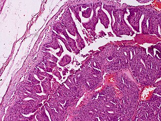
Appendix cancer, also known as appendiceal cancer, is a very rare malignant tumor that forms in the vermiform appendix.

<span class="mw-page-title-main">Neprilysin</span> Mammalian protein found in Homo sapiens

Neprilysin is an enzyme that in humans is encoded by the MME gene. Neprilysin is a zinc-dependent metalloprotease that cleaves peptides at the amino side of hydrophobic residues and inactivates several peptide hormones including glucagon, enkephalins, substance P, neurotensin, oxytocin, and bradykinin. It also degrades the amyloid beta peptide whose abnormal folding and aggregation in neural tissue has been implicated as a cause of Alzheimer's disease. Synthesized as a membrane-bound protein, the neprilysin ectodomain is released into the extracellular domain after it has been transported from the Golgi apparatus to the cell surface.

<span class="mw-page-title-main">Adenocarcinoma in situ of the lung</span> Medical condition

Adenocarcinoma in situ (AIS) of the lung —previously included in the category of "bronchioloalveolar carcinoma" (BAC)—is a subtype of lung adenocarcinoma. It tends to arise in the distal bronchioles or alveoli and is defined by a non-invasive growth pattern. This small solitary tumor exhibits pure alveolar distribution and lacks any invasion of the surrounding normal lung. If completely removed by surgery, the prognosis is excellent with up to 100% 5-year survival.

<span class="mw-page-title-main">Mucinous cystadenoma</span> Medical condition

Mucinous cystadenoma is a benign cystic tumor lined by a mucinous epithelium. It is a type of cystic adenoma (cystadenoma).

<span class="mw-page-title-main">Aggressive digital papillary adenocarcinoma</span> Medical condition

Aggressive digital papillary adenocarcinoma, also known as digital papillary adenocarcinoma and papillary adenoma is a cutaneous condition characterized by an aggressive malignancy involving the digit between the nailbed and the distal interphalangeal joint spaces. Genetic studies reveal that human papilloma virus HPV42 is the likely driving virus in this cancer.

<span class="mw-page-title-main">Metanephric adenoma</span> Medical condition

Metanephric adenoma (MA) is a rare, benign tumour of the kidney, that can have a microscopic appearance similar to a nephroblastoma, or a papillary renal cell carcinoma.

A sialoblastoma is a low-grade salivary gland neoplasm that recapitulates primitive salivary gland anlage. It has previously been referred to as congenital basal cell adenoma, embryoma, or basaloid adenocarcinoma. It is an extremely rare tumor, with less than 100 cases reported worldwide.

A ceruminous adenoma is a benign glandular neoplasm which arises from the ceruminous glands located within the external auditory canal. These glands are found within the outer one third to one half of the external auditory canal, more common along the posterior surface; therefore, the tumor develops within a very specific location.

<span class="mw-page-title-main">Endolymphatic sac tumor</span>

An endolymphatic sac tumor (ELST) is a very uncommon papillary epithelial neoplasm arising within the endolymphatic sac or endolymphatic duct. This tumor shows a very high association with Von Hippel–Lindau syndrome (VHL).

<span class="mw-page-title-main">Ceruminous adenocarcinoma</span> Medical condition

Ceruminous adenocarcinoma is a malignant neoplasm derived from ceruminous glands of the external auditory canal. This tumor is rare, with several names used in the past. Synonyms have included cylindroma, ceruminoma, ceruminous adenocarcinoma, not otherwise specified (NOS), ceruminous adenoid cystic carcinoma (ACC), and ceruminous mucoepidermoid carcinoma.

Neuroendocrine differentiation is a term primarily used in relation to prostate cancers that display a significant neuroendocrine cell population on histopathological examination. These types of prostate cancer comprise true neuroendocrine cancers, such as small cell carcinoma, carcinoid and carcinoid-like tumors, as well as prostatic adenocarcinoma exhibiting focal neuroendocrine phenotype.

Diffuse idiopathic pulmonary neuroendocrine cell hyperplasia (DIPNECH) is a diffuse parenchymal lung disease which often presents with symptoms of cough and shortness of breath. The pathological definition published by the World Health Organization is “a generalized proliferation of scattered single cells, small nodules, or linear proliferations of pulmonary neuroendocrine (PNE) cells that may be confined to the bronchial and bronchiolar epithelium.” The true prevalence of this disease is not known. To date, just under 200 cases have been reported in the literature. However, with an increase in recognition of this disease by radiologists and pulmonologists, the number of cases has been increasing. DIPNECH predominantly affects middle-aged women with slowly progressive lung obstruction. DIPNECH is usually discovered in one of two ways: 1) as an unexpected finding following a lung surgery; or 2) by evaluation of a patient in a pulmonary clinic with longstanding, unexplained symptoms.

<span class="mw-page-title-main">Histopathology of colorectal adenocarcinoma</span> Analysis of tissue from a biopsy or surgery to identify colorectal cancer characteristics

The histopathology of colorectal cancer of the adenocarcinoma type involves analysis of tissue taken from a biopsy or surgery. A pathology report contains a description of the microscopical characteristics of the tumor tissue, including both tumor cells and how the tumor invades into healthy tissues and finally if the tumor appears to be completely removed. The most common form of colon cancer is adenocarcinoma, constituting between 95% and 98% of all cases of colorectal cancer. Other, rarer types include lymphoma, adenosquamous and squamous cell carcinoma. Some subtypes have been found to be more aggressive.

References

  1. 1 2 Devaney, Kenneth O.; Ferlito, Alfio; Rinaldo, Alessandra (2003). "Epithelial Tumors of the Middle Ear--Are Middle Ear Carcinoids Really Distinct from Middle Ear Adenomas?". Acta Oto-Laryngologica. 123 (6): 678–682. doi:10.1080/00016480310001862. ISSN   0001-6489. PMID   12953765. S2CID   24772063.
  2. 1 2 3 4 5 6 7 8 Torske KR, Thompson LD (May 2002). "Adenoma versus carcinoid tumor of the middle ear: a study of 48 cases and review of the literature". Mod. Pathol. 15 (5): 543–55. doi: 10.1038/modpathol.3880561 . PMID   12011260.
  3. Hale, R J; McMahon, R F; Whittaker, J S (1991). "Middle ear adenoma: tumour of mixed mucinous and neuroendocrine differentiation". Journal of Clinical Pathology. 44 (8): 652–654. doi:10.1136/jcp.44.8.652. ISSN   0021-9746. PMC   496756 . PMID   1890199.
  4. Stanley MW, Horwitz CA, Levinson RM, Sibley RK (1987). "Carcinoid tumors of the middle ear". American Journal of Clinical Pathology . 87 (5): 592–600. doi:10.1093/ajcp/87.5.592. PMID   3578133.
  5. Ketabchi S, Massi D, Franchi A, Vannucchi P, Santucci M (2001). "Middle ear adenoma is an amphicrine tumor: why call it adenoma?". Ultrastruct. Pathol. 25 (1): 73–8. doi:10.1080/019131201300004717. PMID   11297323. S2CID   218868445.
  6. Amble FR, Harner SG, Weiland LH, McDonald TJ, Facer GW (1993). "Middle ear adenoma and adenocarcinoma". Otolaryngol Head Neck Surg. 109 (5): 871–6. doi:10.1177/019459989310900516. PMID   8247568. S2CID   23006159.
  7. 1 2 3 Leong K, Haber MM, Divi V, Sataloff RT (2009). "Neuroendocrine adenoma of the middle ear (NAME)". Ear Nose Throat J. 88 (4): 874–9. doi: 10.1177/014556130908800412 . PMID   19358129. S2CID   35987188.
  8. 1 2 Thompson L.D. (Sep 2005). "Neuroendocrine adenoma of the middle ear". Ear Nose Throat J. 84 (9): 560–1. doi:10.1177/014556130508400908. PMID   16261754. S2CID   13251769.
  9. Wassef M, et al. (Oct 1989). "Middle ear adenoma. A tumor displaying mucinous and neuroendocrine differentiation". Am. J. Surg. Pathol. 13 (10): 838–47. doi:10.1097/00000478-198910000-00003. PMID   2782545. S2CID   6515115.
  10. Davies JE, Semeraro D, Knight LC, Griffiths GJ (1989). "Middle ear neoplasms showing adenomatous and neuroendocrine components". J Laryngol Otol. 103 (4): 404–7. doi:10.1017/S0022215100109065. PMID   2715695.
  11. Bold EL, Wanamaker JR, Hughes GB, Rhee CK, Sebek BA, Kinney SE (1995). "Adenomatous lesions of the temporal bone immunohistochemical analysis and theories of histogenesis". Am J Otol. 16 (2): 146–52. PMID   8572112.
  12. McNutt MA, Bolen JW (1985). "Adenomatous tumor of the middle ear: An ultrastructural and immunocytochemical study". American Journal of Clinical Pathology. 84 (4): 541–7. doi:10.1093/ajcp/84.4.541. PMID   4036884.
  13. Ramsey MJ, Nadol JB, Pilch BZ, McKenna MJ (2005). "Carcinoid tumor of the middle ear: clinical features, recurrences, and metastases". Laryngoscope. 115 (9): 1660–6. doi:10.1097/01.mlg.0000175069.13685.37. PMID   16148713. S2CID   22056068.

Further reading