Related Research Articles

Cranial nerves are the nerves that emerge directly from the brain, of which there are conventionally considered twelve pairs. Cranial nerves relay information between the brain and parts of the body, primarily to and from regions of the head and neck, including the special senses of vision, taste, smell, and hearing.

The abducens nerve or abducent nerve, also known as the sixth cranial nerve, cranial nerve VI, or simply CN VI, is a cranial nerve in humans and various other animals that controls the movement of the lateral rectus muscle, one of the extraocular muscles responsible for outward gaze. It is a somatic efferent nerve.

The oculomotor nerve, also known as the third cranial nerve, cranial nerve III, or simply CN III, is a cranial nerve that enters the orbit through the superior orbital fissure and innervates extraocular muscles that enable most movements of the eye and that raise the eyelid. The nerve also contains fibers that innervate the intrinsic eye muscles that enable pupillary constriction and accommodation. The oculomotor nerve is derived from the basal plate of the embryonic midbrain. Cranial nerves IV and VI also participate in control of eye movement.

The midbrain or mesencephalon is the rostral-most portion of the brainstem connecting the diencephalon and cerebrum with the pons. It consists of the cerebral peduncles, tegmentum, and tectum.

The vestibulo-ocular reflex (VOR) is a reflex that acts to stabilize gaze during head movement, with eye movement due to activation of the vestibular system, it is also known as the Cervico-ocular reflex. The reflex acts to stabilize images on the retinas of the eye during head movement. Gaze is held steadily on a location by producing eye movements in the direction opposite that of head movement. For example, when the head moves to the right, the eyes move to the left, meaning the image a person sees stays the same even though the head has turned. Since slight head movement is present all the time, VOR is necessary for stabilizing vision: people with an impaired reflex find it difficult to read using print, because the eyes do not stabilise during small head tremors, and also because damage to reflex can cause nystagmus.

The medial longitudinal fasciculus (MLF) is a prominent bundle of nerve fibres which pass within the ventral/anterior portion of periaqueductal gray of the mesencephalon (midbrain). It contains the interstitial nucleus of Cajal, responsible for oculomotor control, head posture, and vertical eye movement.

The accommodation reflex is a reflex action of the eye, in response to focusing on a near object, then looking at a distant object, comprising coordinated changes in vergence, lens shape (accommodation) and pupil size. It is dependent on cranial nerve II, superior centers (interneuron) and cranial nerve III. The change in the shape of the lens is controlled by ciliary muscles inside the eye. Changes in contraction of the ciliary muscles alter the focal distance of the eye, causing nearer or farther images to come into focus on the retina; this process is known as accommodation. The reflex, controlled by the parasympathetic nervous system, involves three responses: pupil constriction, lens accommodation, and convergence.

In neuroanatomy, the pretectal area, or pretectum, is a midbrain structure composed of seven nuclei and comprises part of the subcortical visual system. Through reciprocal bilateral projections from the retina, it is involved primarily in mediating behavioral responses to acute changes in ambient light such as the pupillary light reflex, the optokinetic reflex, and temporary changes to the circadian rhythm. In addition to the pretectum's role in the visual system, the anterior pretectal nucleus has been found to mediate somatosensory and nociceptive information.

The abducens nucleus is the originating nucleus from which the abducens nerve (VI) emerges—a cranial nerve nucleus. This nucleus is located beneath the fourth ventricle in the caudal portion of the pons near the midline, medial to the sulcus limitans.

The fastigial nucleus is located in each hemisphere of the cerebellum. It is one of the four deep cerebellar nuclei.

Parinaud's syndrome is a constellation of neurological signs indicating injury to the dorsal midbrain. More specifically, compression of the vertical gaze center at the rostral interstitial nucleus of medial longitudinal fasciculus (riMLF).
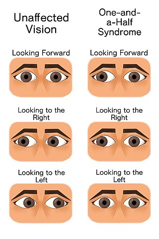
The one and a half syndrome is a rare weakness in eye movement affecting both eyes, in which one cannot move laterally at all, and the other can move only in outward direction. More formally, it is characterized by "a conjugate horizontal gaze palsy in one direction and an internuclear ophthalmoplegia in the other". Nystagmus is also present when the eye on the opposite side of the lesion is abducted. Convergence is classically spared as cranial nerve III and its nucleus is spared bilaterally.

The paramedian pontine reticular formation (PPRF) is a subset of neurons of the oral and caudal pontine reticular nuclei mediating horizontal gaze. It is situated in the pons adjacent to the abducens nucleus. It projects to the ipsilateral abducens nucleus, and contralateral oculomotor nucleus to mediate conjugate horizontal eye movements and saccades.
The rostral interstitial nucleus of medial longitudinal fasciculus (riMLF) is a collection of neurons in the mesencephalon (midbrain) responsible for mediating vertical conjugate eye movements and vertical saccades. It mostly projects efferents to the ipsilateral oculomotor and trochlear nuclei.
The term gaze is frequently used in physiology to describe coordinated motion of the eyes and neck. The lateral gaze is controlled by the paramedian pontine reticular formation (PPRF). The vertical gaze is controlled by the rostral interstitial nucleus of medial longitudinal fasciculus and the interstitial nucleus of Cajal.

The nucleus prepositus or nucleus prepositus hypoglossi is one of the largest of the three perihypoglossal nuclei. It is situated in the caudal pons and rostral medulla oblongata. It contributes to several aspects of gaze control including the horizontal gaze holding system.
Perihypoglossal nuclei are three prominent groups of neurons in the caudal medulla oblongata near the hypoglossal nucleus: the nucleus prepositus hypoglossi, intercalated nucleus, and sublingual nucleus. They are involved in controling eye movements: they send their principal projections to the three cranial nerve nuclei controling extrinsic eye muscles via the medial longitudinal fasciculus.
The interstitial nucleus of Cajal is a collection of neurons in the mesencephalon (midbrain) which are involved in integrating eye position-velocity information in order to coordinate head-eye movements - especially those related to vertical and torsional conjugate eye movements (gaze). It also mediates vertical gaze holding.
The accessory oculomotor nuclei are a group of nuclei situated in the rostral mesencephalon (midbrain) near its junction with the diencephalon, and consist of:
The nucleus of Darkschewitsch is an accessory oculomotor nucleus situated in the ventrolateral portion of the periaqueductal gray of the mesencephalon (midbrain) near its junction with the diencephalon. It is involved in mediating vertical eye movements. It projects to the trochlear nucleus, receives afferents from the visual cortex, and forms a reciprocal (looping) connection with the cerebellum by way of the inferior olive.
References
- 1 2 Kiernan, John A.; Rajakumar, Nagalingam (2013). Barr's The Human Nervous System: An Anatomical Viewpoint (10th ed.). Philadelphia: Wolters Kluwer Lippincott Williams & Wilkins. p. 156. ISBN 978-1-4511-7327-7.
- 1 2 3 Patestas, Maria A.; Gartner, Leslie P. (2016). A Textbook of Neuroanatomy (2nd ed.). Hoboken, New Jersey: Wiley-Blackwell. pp. 321–322. ISBN 978-1-118-67746-9.
- ↑ Standring, Susan (2020). Gray's Anatomy: The Anatomical Basis of Clinical Practice (42th ed.). New York: Elsevier. p. 455. ISBN 978-0-7020-7707-4. OCLC 1201341621.