
Otorhinolaryngology is a surgical subspecialty within medicine that deals with the surgical and medical management of conditions of the head and neck. Doctors who specialize in this area are called otorhinolaryngologists, otolaryngologists, head and neck surgeons, or ENT surgeons or physicians. Patients seek treatment from an otorhinolaryngologist for diseases of the ear, nose, throat, base of the skull, head, and neck. These commonly include functional diseases that affect the senses and activities of eating, drinking, speaking, breathing, swallowing, and hearing. In addition, ENT surgery encompasses the surgical management and reconstruction of cancers and benign tumors of the head and neck as well as plastic surgery of the face and neck.
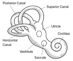
Ménière's disease (MD) is a disorder of the inner ear that is characterized by episodes of feeling like the world is spinning (vertigo), ringing in the ears (tinnitus), hearing loss, and a fullness in the ear. Typically, only one ear is affected initially; however, over time both ears may become involved. Episodes generally last from 20 minutes to a few hours. The time between episodes varies. The hearing loss and ringing in the ears can become constant over time.

Cholesteatoma is a destructive and expanding growth consisting of keratinizing squamous epithelium in the middle ear and/or mastoid process. Cholesteatomas are not cancerous as the name may suggest, but can cause significant problems because of their erosive and expansile properties. This can result in the destruction of the bones of the middle ear (ossicles), as well as growth through the base of the skull into the brain. They often become infected and can result in chronically draining ears. Treatment almost always consists of surgical removal.
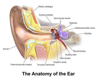
In the anatomy of humans and various other tetrapods, the eardrum, also called the tympanic membrane or myringa, is a thin, cone-shaped membrane that separates the external ear from the middle ear. Its function is to transmit sound from the air to the ossicles inside the middle ear, and then to the oval window in the fluid-filled cochlea. Hence, it ultimately converts and amplifies vibration in air to vibration in cochlear fluid. The malleus bone bridges the gap between the eardrum and the other ossicles.
Vocal cord nodules are bilaterally symmetrical benign white masses that form at the midpoint of the vocal folds. Although diagnosis involves a physical examination of the head and neck, as well as perceptual voice measures, visualization of the vocal nodules via laryngeal endoscopy remains the primary diagnostic method. Vocal fold nodules interfere with the vibratory characteristics of the vocal folds by increasing the mass of the vocal folds and changing the configuration of the vocal fold closure pattern. Due to these changes, the quality of the voice may be affected. As such, the major perceptual signs of vocal fold nodules include vocal hoarseness and breathiness. Other common symptoms include vocal fatigue, soreness or pain lateral to the larynx, and reduced frequency and intensity range. Airflow levels during speech may also be increased. Vocal fold nodules are thought to be the result of vocal fold tissue trauma caused by excessive mechanical stress, including repeated or chronic vocal overuse, abuse, or misuse. Predisposing factors include profession, gender, dehydration, respiratory infection, and other inflammatory factors.
Vocal fold cysts are benign masses of the membranous vocal folds. These cysts are enclosed, sac-like structures that are typically of a yellow or white colour. They occur unilaterally on the midpoint of the medial edge of the vocal folds. They can also form on the upper/superior, surface of the vocal folds. There are two types of vocal fold cysts:
- Sub-epithelial vocal fold cysts- located in the superficial lamina propria of the vocal folds.
- Ligament vocal fold cysts- located within the deeper layers of the lamina propria or on the vocal ligament.

Reinke's edema is the swelling of the vocal cords due to fluid (edema) collected within the Reinke's space. First identified by the German anatomist Friedrich B. Reinke in 1895, the Reinke's space is a gelatinous layer of the vocal cord located underneath the outer cells of the vocal cord. When a person speaks, the Reinke's space vibrates to allow for sound to be produced (phonation). The Reinke's space is sometimes referred to as the superficial lamina propria.
Otitis media is a group of inflammatory diseases of the middle ear. One of the two main types is acute otitis media (AOM), an infection of rapid onset that usually presents with ear pain. In young children this may result in pulling at the ear, increased crying, and poor sleep. Decreased eating and a fever may also be present. The other main type is otitis media with effusion (OME), typically not associated with symptoms, although occasionally a feeling of fullness is described; it is defined as the presence of non-infectious fluid in the middle ear for more than three months. Chronic suppurative otitis media (CSOM) is middle ear inflammation that results in discharge from the ear for more than three months. It may be a complication of acute otitis media. Pain is rarely present. All three types of otitis media may be associated with hearing loss. The hearing loss in OME, due to its chronic nature, may affect a child's ability to learn.

Earwax, also known by the medical term cerumen, is a brown, orange, red, yellowish or gray waxy substance secreted in the ear canal of humans and other mammals. It protects the skin of the human ear canal, assists in cleaning and lubrication, and provides protection against bacteria, fungi, and water.

An adenoma is a benign tumor of epithelial tissue with glandular origin, glandular characteristics, or both. Adenomas can grow from many glandular organs, including the adrenal glands, pituitary gland, thyroid, prostate, and others. Some adenomas grow from epithelial tissue in nonglandular areas but express glandular tissue structure. Although adenomas are benign, they should be treated as pre-cancerous. Over time adenomas may transform to become malignant, at which point they are called adenocarcinomas. Most adenomas do not transform. However, even though benign, they have the potential to cause serious health complications by compressing other structures and by producing large amounts of hormones in an unregulated, non-feedback-dependent manner. Some adenomas are too small to be seen macroscopically but can still cause clinical symptoms.

A hoarse voice, also known as dysphonia or hoarseness, is when the voice involuntarily sounds breathy, raspy, or strained, or is softer in volume or lower in pitch. A hoarse voice, can be associated with a feeling of unease or scratchiness in the throat. Hoarseness is often a symptom of problems in the vocal folds of the larynx. It may be caused by laryngitis, which in turn may be caused by an upper respiratory infection, a cold, or allergies. Cheering at sporting events, speaking loudly in noisy situations, talking for too long without resting one's voice, singing loudly, or speaking with a voice that's too high or too low can also cause temporary hoarseness. A number of other causes for losing one's voice exist, and treatment is generally by resting the voice and treating the underlying cause. If the cause is misuse or overuse of the voice, drinking plenty of water may alleviate the problems.

A bone-anchored hearing aid (BAHA) is a type of hearing aid based on bone conduction. It is primarily suited for people who have conductive hearing losses, unilateral hearing loss, single-sided deafness and people with mixed hearing losses who cannot otherwise wear 'in the ear' or 'behind the ear' hearing aids. They are more expensive than conventional hearing aids, and their placement involves invasive surgery which carries a risk of complications, although when complications do occur, they are usually minor.

Contact granuloma is a condition that develops due to persistent tissue irritation in the posterior larynx. Benign granulomas, not to be confused with other types of granulomas, occur on the vocal process of the vocal folds, where the vocal ligament attaches. Signs and symptoms may include hoarseness of the voice, or a sensation of having a lump in the throat, but contact granulomas may also be without symptoms. There are two common causes associated with contact granulomas; the first common cause is sustained periods of increased pressure on the vocal folds, and is commonly seen in people who use their voice excessively, such as singers. Treatment typically includes voice therapy and changes to lifestyle factors. The second common cause of granulomas is gastroesophageal reflux and is controlled primarily through the use of anti-reflux medication. Other associated causes are discussed below.

The tonsils are a set of lymphoid organs facing into the aerodigestive tract, which is known as Waldeyer's tonsillar ring and consists of the adenoid tonsil, two tubal tonsils, two palatine tonsils, and the lingual tonsils. These organs play an important role in the immune system.
A mastoidectomy is a procedure performed to remove the mastoid air cells, air bubbles in the skull, near the inner ears. This can be done as part of treatment for mastoiditis, chronic suppurative otitis media or cholesteatoma. In addition, it is sometimes performed as part of other procedures or for access to the middle ear. There are classically 5 different types of mastoidectomy:

Kimura's disease is a benign rare chronic inflammatory disorder. Its primary symptoms are subdermal lesions in the head or neck or painless unilateral inflammation of cervical lymph nodes.

Marginal zone B-cell lymphomas, also known as marginal zone lymphomas (MZLs), are a heterogeneous group of lymphomas that derive from the malignant transformation of marginal zone B-cells. Marginal zone B cells are innate lymphoid cells that normally function by rapidly mounting IgM antibody immune responses to antigens such as those presented by infectious agents and damaged tissues. They are lymphocytes of the B-cell line that originate and mature in secondary lymphoid follicles and then move to the marginal zones of mucosa-associated lymphoid tissue, the spleen, or lymph nodes. Mucosa-associated lymphoid tissue is a diffuse system of small concentrations of lymphoid tissue found in various submucosal membrane sites of the body such as the gastrointestinal tract, mouth, nasal cavity, pharynx, thyroid gland, breast, lung, salivary glands, eye, skin and the human spleen.

| name = | synonyms = Myringosclerosis, intratympanic tympanosclerosis | field = ENT surgery | symptoms = | complications = | onset = | duration = | types = | causes = | risks = | diagnosis = | differential = | prevention = | treatment = | medication = | prognosis = | frequency = | deaths = }} Tympanosclerosis is a condition caused by hyalinization and subsequent calcification of subepithelial connective tissue of TM and middle ear, sometimes resulting in a detrimental effect to hearing.

Ceruminous adenocarcinoma is a malignant neoplasm derived from ceruminous glands of the external auditory canal. This tumor is rare, with several names used in the past. Synonyms have included cylindroma, ceruminoma, ceruminous adenocarcinoma, not otherwise specified (NOS), ceruminous adenoid cystic carcinoma (ACC), and ceruminous mucoepidermoid carcinoma.
Granular myringitis is a long term condition in which there is inflammation of the tympanic membrane in the ear and formation of granulation tissue within the tympanic membrane. It is a type of otitis externa.
