
The peritoneum is the serous membrane forming the lining of the abdominal cavity or coelom in amniotes and some invertebrates, such as annelids. It covers most of the intra-abdominal organs, and is composed of a layer of mesothelium supported by a thin layer of connective tissue. This peritoneal lining of the cavity supports many of the abdominal organs and serves as a conduit for their blood vessels, lymphatic vessels, and nerves.

The abdominal cavity is a large body cavity in humans and many other animals that contain organs. It is a part of the abdominopelvic cavity. It is located below the thoracic cavity, and above the pelvic cavity. Its dome-shaped roof is the thoracic diaphragm, a thin sheet of muscle under the lungs, and its floor is the pelvic inlet, opening into the pelvis.

In medicine, a shunt is a hole or a small passage that moves, or allows movement of, fluid from one part of the body to another. The term may describe either congenital or acquired shunts; acquired shunts may be either biological or mechanical.

The mesothelium is a membrane composed of simple squamous epithelial cells of mesodermal origin, which forms the lining of several body cavities: the pleura, peritoneum and pericardium.

The dura mater, is the outermost of the three meningeal membranes. The dura mater has two layers, an outer periosteal layer closely adhered to the neurocranium, and an inner meningeal layer known as the dural border cell layer. The two dural layers are for the most part fused together forming a thick fibrous tissue membrane that covers the brain and the vertebrae of the spinal column. But the layers are separated at the dural venous sinuses to allow blood to drain from the brain. The dura covers the arachnoid mater and the pia mater the other two meninges in protecting the central nervous system.
The peritoneal cavity is a potential space located between the two layers of the peritoneum—the parietal peritoneum, the serous membrane that lines the abdominal wall, and visceral peritoneum, which surrounds the internal organs. While situated within the abdominal cavity, the term peritoneal cavity specifically refers to the potential space enclosed by these peritoneal membranes. The cavity contains a thin layer of lubricating serous fluid that enables the organs to move smoothly against each other, facilitating the movement and expansion of internal organs during digestion.

Cardiac catheterization is the insertion of a catheter into a chamber or vessel of the heart. This is done both for diagnostic and interventional purposes.

The jugular veins are veins that take blood from the head back to the heart via the superior vena cava. The internal jugular vein descends next to the internal carotid artery and continues posteriorly to the sternocleidomastoid muscle.
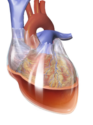
Beck's triad is a collection of three medical signs associated with acute cardiac tamponade, a medical emergency when excessive fluid accumulates in the pericardial sac around the heart and impairs its ability to pump blood. The signs are low arterial blood pressure, distended neck veins, and distant, muffled heart sounds.

The internal jugular vein is a paired jugular vein that collects blood from the brain and the superficial parts of the face and neck. This vein runs in the carotid sheath with the common carotid artery and vagus nerve.

Superficial veins are veins that are close to the surface of the body, as opposed to deep veins, which are far from the surface.

Transjugular intrahepatic portosystemic shunt is an artificial channel within the liver that establishes communication between the inflow portal vein and the outflow hepatic vein. It is used to treat portal hypertension which frequently leads to intestinal bleeding, life-threatening esophageal bleeding and the buildup of fluid within the abdomen (ascites).

The sigmoid sinuses, also known as the pars sigmoid, are paired dural venous sinuses within the skull that receive blood from posterior transverse sinuses.
The anterior jugular vein is a vein in the neck.

The posterior auricular vein is a vein of the head. It begins from a plexus with the occipital vein and the superficial temporal vein, descends behind the auricle, and drains into the external jugular vein.
The occipital vein is a vein of the scalp. It originates from a plexus around the external occipital protuberance and superior nuchal line to the back part of the vertex of the skull. It usually drains into the internal jugular vein, but may also drain into the posterior auricular vein. It drains part of the scalp.

The right lymphatic duct is an important lymphatic vessel that drains the right upper quadrant of the human body. It forms various combinations with the right subclavian vein and right internal jugular vein.

The bronchomediastinal lymph trunks are essential components of the human lymphatic system, tasked with draining lymph from the tracheobronchial, internal mammary, and anterior mediastinal lymph nodes.
Peritoneal recesses are the spaces formed by peritoneum draping over viscera.
Lymph sacs are a part of the development of the lymphatic system, known as lymphangiogenesis. The lymph sacs are precursors of the lymph vessels. These sacs develop through the processes of vasculogenesis and angiogenesis. However, there is evidence of both of these processes in different organisms. In mice, it is thought that the lymphatic components form through an angiogenic process. But, there is evidence from bird embryos that gives rise to the idea that lymphatic vessels arise in the embryos through a vasculogenesis-like process from the lymphangioblastic endothelial precursor cells.