Pigment dispersion syndrome

Last updated
Pigment dispersion syndrome
Other namesPigmentary glaucoma
Schematic diagram of the human eye en.svg
The iris(cells) plays a role in this condition
Specialty Ophthalmology
Medication Latanaprost (eye drops)

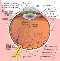
Pigment dispersion syndrome (PDS) is an eye disorder that can lead to a form of glaucoma known as pigmentary glaucoma. It takes place when pigment cells slough off from the back of the iris and float around in the aqueous humor. Over time, these pigment cells can accumulate in the anterior chamber in such a way that they begin to clog the trabecular meshwork (the major site of aqueous humour drainage), which can in turn prevent the aqueous humour from draining and therefore increases the pressure inside the eye. [1] A common finding in PDS are central, vertical corneal endothelial pigment deposits, known as Krukenberg spindle. [2] With PDS, the intraocular pressure tends to spike at times and then can return to normal. Exercise has been shown to contribute to spikes in pressure as well. When the pressure is great enough to cause damage to the optic nerve, this is called pigmentary glaucoma. [1] As with all types of glaucoma, when damage happens to the optic nerve fibers, the vision loss that occurs is irreversible and painless.

Contents

Risk factors

This condition is rare, but occurs most often in Caucasians, particularly men, and the age of onset is relatively low: mid 20s to 40s. As the crystalline lens hardens with age, the lens zonules pull away from the iris and the syndrome lessens and stops. The main risk factor is nearsightedness. [1] Genetic factors may also play a part in the transition from syndrome to the glaucoma condition. [3]

Diagnosis

Diagnosis for pigment dispersion syndrome is made with characteristic slit lamp and gonioscopy findings. [4]

Management

There is no cure, but pigmentary glaucoma can be managed with eye drops or treated with simple surgeries. If caught early and monitored, chances of glaucoma are greatly reduced.

A 2016 Cochrane Review sought to determine the effectiveness of YAG laser iridotomy versus no laser iridotomy for pigment dispersion syndrome and pigmentary glaucoma, in 195 participants, across five studies. [5] No clear benefits in preventing loss of visual field were found for eyes treated with peripheral laser iridotomy. [5] There was weak evidence suggesting that laser iridotomy could be more effective in lowering intraocular pressure in eyes versus no treatment. [5]

Related Research Articles

<span class="mw-page-title-main">Glaucoma</span> Group of eye diseases

Glaucoma is a group of eye diseases that lead to damage of the optic nerve, which is important for transmitting visual information from the eye to the brain. This damage is often caused by increased pressure within the eye, known as intraocular pressure (IOP) and may cause vision loss if left untreated. The word glaucoma originated from the Greek word ΓλαύV̇ξ (glaukos), which means "to glow". Glaucoma has been called the "silent thief of sight" because the loss of vision usually occurs slowly over a long period of time. It is associated with old age, a family history of glaucoma, and certain medical conditions or medications.

<span class="mw-page-title-main">Optic nerve</span> Second cranial nerve, which connects the eyes to the brain

In neuroanatomy, the optic nerve, also known as the second cranial nerve, cranial nerve II, or simply CN II, is a paired cranial nerve that transmits visual information from the retina to the brain. In humans, the optic nerve is derived from optic stalks during the seventh week of development and is composed of retinal ganglion cell axons and glial cells; it extends from the optic disc to the optic chiasma and continues as the optic tract to the lateral geniculate nucleus, pretectal nuclei, and superior colliculus.

<span class="mw-page-title-main">Aqueous humour</span> Fluid in the anterior segment of the eye

The aqueous humour is a transparent water-like fluid similar to blood plasma, but containing low protein concentrations. It is secreted from the ciliary body, a structure supporting the lens of the eyeball. It fills both the anterior and the posterior chambers of the eye, and is not to be confused with the vitreous humour, which is located in the space between the lens and the retina, also known as the posterior cavity or vitreous chamber. Blood cannot normally enter the eyeball.

<span class="mw-page-title-main">Phacoemulsification</span> Method of cataract surgery

Phacoemulsification is a cataract surgery method in which the internal lens of the eye which has developed a cataract is emulsified with the tip of an ultrasonic handpiece and aspirated from the eye. Aspirated fluids are replaced with irrigation of balanced salt solution to maintain the volume of the anterior chamber during the procedure. This procedure minimises the incision size and reduces the recovery time and risk of surgery induced astigmatism.

<span class="mw-page-title-main">Phakic intraocular lens</span> Lens implanted in eye in addition to the natural lens

A phakic intraocular lens (PIOL) is a special kind of intraocular lens that is implanted surgically into the eye to correct myopia (nearsightedness). It is called "phakic" because the eye's natural lens is left untouched. Intraocular lenses that are implanted into eyes after the eye's natural lens has been removed during cataract surgery are known as pseudophakic.

<span class="mw-page-title-main">Ciliary body</span> Part of the eye

The ciliary body is a part of the eye that includes the ciliary muscle, which controls the shape of the lens, and the ciliary epithelium, which produces the aqueous humor. The aqueous humor is produced in the non-pigmented portion of the ciliary body. The ciliary body is part of the uvea, the layer of tissue that delivers oxygen and nutrients to the eye tissues. The ciliary body joins the ora serrata of the choroid to the root of the iris.

<span class="mw-page-title-main">Optic disc</span> Optic nerve head, the point of exit for ganglion cell axons leaving the eye

The optic disc or optic nerve head is the point of exit for ganglion cell axons leaving the eye. Because there are no rods or cones overlying the optic disc, it corresponds to a small blind spot in each eye.

<span class="mw-page-title-main">Latanoprost</span> Chemical compound

Latanoprost, sold under the brand name Xalatan among others, is a medication used to treat increased pressure inside the eye. This includes ocular hypertension and open angle glaucoma. It is applied as eye drops to the eyes. Onset of effects is usually within four hours, and they last for up to a day.

<span class="mw-page-title-main">Hyphema</span> Hemorrhage in the front chamber of the eye

Hyphema is the medical condition of bleeding in the anterior chamber of the eye between the iris and the cornea. People usually first notice a loss or decrease in vision. The eye may also appear to have a reddish tinge, or it may appear as a small pool of blood at the bottom of the iris in the cornea. A traumatic hyphema is caused by a blow to the eye. A hyphema can also occur spontaneously.

<span class="mw-page-title-main">Schlemm's canal</span> Lymphatic-like vessel in the eye

Schlemm's canal is a circular lymphatic-like vessel in the eye. It collects aqueous humor from the anterior chamber and delivers it into the episcleral blood vessels. Canaloplasty may be used to widen it.

Ocular hypertension is the presence of elevated fluid pressure inside the eye, usually with no optic nerve damage or visual field loss.

<span class="mw-page-title-main">Iridectomy</span> Surgical removal of part of the iris

An iridectomy, also known as a surgical iridectomy or corectomy, is the surgical removal of part of the iris. These procedures are most frequently performed in the treatment of closed-angle glaucoma and iris melanoma.

<span class="mw-page-title-main">Posterior chamber of eyeball</span> Region of the eyeball between the iris and lens

The posterior chamber is a narrow space behind the peripheral part of the iris, and in front of the suspensory ligament of the lens and the ciliary processes. The posterior chamber consists of small space directly posterior to the iris but anterior to the lens. The posterior chamber is part of the anterior segment and should not be confused with the vitreous chamber.

<span class="mw-page-title-main">Apraclonidine</span> Chemical compound

Apraclonidine (INN), also known under the brand name Iopidine, is a sympathomimetic used in glaucoma therapy. It is an α2 adrenergic receptor agonist and a weak α1 adrenergic receptor agonist.

<span class="mw-page-title-main">Glaucoma surgery</span> Type of eye surgery

Glaucoma is a group of diseases affecting the optic nerve that results in vision loss and is frequently characterized by raised intraocular pressure (IOP). There are many glaucoma surgeries, and variations or combinations of those surgeries, that facilitate the escape of excess aqueous humor from the eye to lower intraocular pressure, and a few that lower IOP by decreasing the production of aqueous humor.

Pseudoexfoliation syndrome, often abbreviated as PEX and sometimes as PES or PXS, is an aging-related systemic disease manifesting itself primarily in the eyes which is characterized by the accumulation of microscopic granular amyloid-like protein fibers. Its cause is unknown, although there is speculation that there may be a genetic basis. It is more prevalent in women than men, and in persons past the age of seventy. Its prevalence in different human populations varies; for example, it is prevalent in Scandinavia. The buildup of protein clumps can block normal drainage of the eye fluid called the aqueous humor and can cause, in turn, a buildup of pressure leading to glaucoma and loss of vision. As worldwide populations become older because of shifts in demography, PEX may become a matter of greater concern.

<span class="mw-page-title-main">Secondary glaucoma</span>

Secondary glaucoma is a collection of progressive optic nerve disorders associated with a rise in intraocular pressure (IOP) which results in the loss of vision. In clinical settings, it is defined as the occurrence of IOP above 21 mmHg requiring the prescription of IOP-managing drugs. It can be broadly divided into two subtypes: secondary open-angle glaucoma and secondary angle-closure glaucoma, depending on the closure of the angle between the cornea and the iris. Principal causes of secondary glaucoma include optic nerve trauma or damage, eye disease, surgery, neovascularization, tumours and use of steroid and sulfa drugs. Risk factors for secondary glaucoma include uveitis, cataract surgery and also intraocular tumours. Common treatments are designed according to the type and the underlying causative condition, in addition to the consequent rise in IOP. These include drug therapy, the use of miotics, surgery or laser therapy.

Uveitis–glaucoma–hyphaema (UGH) syndrome, also known as Ellingson syndrome, is a complication of cataract surgery, caused by intraocular lens subluxation or dislocation. The chafing of mispositioned intraocular lens over iris, ciliary body or iridocorneal angle cause elevated intraocular pressure (IOP) anterior uveitis and hyphema. It is most commonly caused by anterior chamber IOLs and sulcus IOLs but, the condition can be seen with any type of IOL, including posterior chamber lenses and cosmetic iris implants.

<span class="mw-page-title-main">Uveitic glaucoma</span> Glaucoma caused by uveitis or its treatments

Uveitic glaucoma is most commonly a progression stage of noninfectious anterior uveitis or iritis.

<span class="mw-page-title-main">Phacomorphic glaucoma</span> Disorder of the human eye

Phacomorphic glaucoma is an eye disease that can occur due to a neglected advanced cataract. In this, the mature cataractous lens cause secondary angle closure glaucoma. The presence of an asymmetric mature cataractous lens, shallow or closed anterior chamber angle, raised intraocular pressure (IOP) and other typical signs and symptoms of angle-closure glaucoma in the eye may lead to a diagnosis of phacomorphic glaucoma. Cataract surgery after initial IOP control with medication is the only treatment.

References

  1. 1 2 3 "Pigment-dispersion syndrome". National Institutes of Health. Retrieved October 7, 2018.
  2. Friedman, Neil J. (2021). The Massachusetts eye and ear infirmary illustrated manual of ophthalmology (Fifth ed.). St. Louis, Missouri. ISBN   9780323613323.{{cite book}}: CS1 maint: location missing publisher (link)
  3. Lascaratos G, Shah A, Garway-Heath DF (2013). "The genetics of pigment dispersion syndrome and pigmentary glaucoma". Surv Ophthalmol. 58 (2): 164–75. doi:10.1016/j.survophthal.2012.08.002. PMID   23218808.
  4. "Pigment Dispersion Syndrome Diagnosis". American Academy of Ophthalmology. 28 April 2018. Retrieved 18 November 2018.
  5. 1 2 3 Michelessi M, Lindsley K (2016). "Peripheral iridotomy for pigmentary glaucoma". Cochrane Database Syst Rev. 2 (9): CD005655. doi:10.1002/14651858.CD005655.PUB2. PMC   5032906 . PMID   26871761.