
The mandibular nerve (V3) is the largest of the three divisions of the trigeminal nerve, the fifth cranial nerve (CN V).

The sphenoid bone is an unpaired bone of the neurocranium. It is situated in the middle of the skull towards the front, in front of the temporal bone and the basilar part of the occipital bone. The sphenoid bone is one of the seven bones that articulate to form the orbit. Its shape somewhat resembles that of a butterfly or bat with its wings extended.
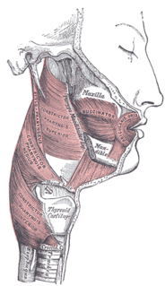
There are four classical muscles of mastication. During mastication, three muscles of mastication are responsible for adduction of the jaw, and one helps to abduct it. All four move the jaw laterally. Other muscles, usually associated with the hyoid, such as the mylohyoid muscle, are responsible for opening the jaw in addition to the lateral pterygoid.

In human anatomy, the masseter is one of the muscles of mastication. Found only in mammals, it is particularly powerful in herbivores to facilitate chewing of plant matter. The most obvious muscle of mastication is the masseter muscle, since it is the most superficial and one of the strongest.

The medial pterygoid, is a thick, quadrilateral muscle of mastication.

The temporal fossa is a shallow depression on the side of the skull bounded by the temporal lines and terminating below the level of the zygomatic arch.
The tensor veli palatini muscle is a broad, thin, ribbon-like muscle in the head that tenses the soft palate.

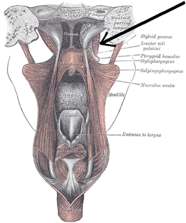
In human anatomy, the pterygopalatine fossa is a fossa in the skull. A human skull contains two pterygopalatine fossae—one on the left side, and another on the right side. Each fossa is a cone-shaped paired depression deep to the infratemporal fossa and posterior to the maxilla on each side of the skull, located between the pterygoid process and the maxillary tuberosity close to the apex of the orbit. It is the indented area medial to the pterygomaxillary fissure leading into the sphenopalatine foramen. It communicates with the nasal and oral cavities, infratemporal fossa, orbit, pharynx, and middle cranial fossa through eight foramina.

The greater wing of the sphenoid bone, or alisphenoid, is a bony process of the sphenoid bone; there is one on each side, extending from the side of the body of the sphenoid and curving upward, laterally, and backward.

The infratemporal fossa is an irregularly shaped cavity, situated below and medial to the zygomatic arch. It is not fully enclosed by bone in all directions, and it contains superficial muscles that are visible during dissection after removing skin and fascia: namely, the lower part of the temporalis muscle, the lateral pterygoid, and the medial pterygoid.
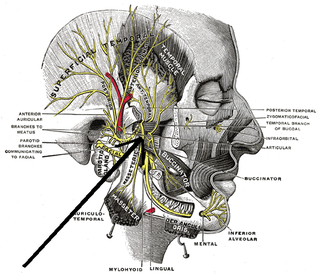
The medial pterygoid nerve is a branch of the mandibular nerve that innervates the medial pterygoid muscle, tensor veli palatini and tensor tympani.

The sphenoidal process of the palatine bone is a thin, compressed plate, much smaller than the orbital, and directed upward and medialward.
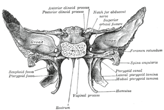
The pterygoid fossa is an anatomical term for the fossa formed by the divergence of the lateral pterygoid plate and the medial pterygoid plate of the sphenoid bone.
The pterygomandibular raphe is a ligamentous band of the buccopharyngeal fascia, attached superiorly to the pterygoid hamulus of the medial pterygoid plate, and inferiorly to the posterior end of the mylohyoid line of the mandible.

At the lower part of the infratemporal surface of the maxilla is a rounded eminence, the maxillary tuberosity, especially prominent after the growth of the wisdom tooth; it is rough on its lateral side for articulation with the pyramidal process of the palatine bone and in some cases articulates with the lateral pterygoid plate of the sphenoid.
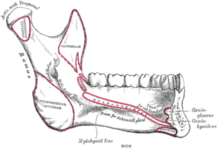
The pterygoid fovea is a concave surface on the uppermost medial side of the ramus of the mandible.
Pterygoid notch is a notch on the inferior portion of the pterygoid processes of the sphenoid bone, between the medial and lateral plates into which the pyramidal process of the palatine bone is fitted.
The Infratemporal space is a fascial space of the head and neck. It is a potential space in the side of the head, and is paired on either side. It is located posterior to the maxilla, between the lateral pterygoid plate of the sphenoid bone medially and by the base of skull superiorly. The term is derived from infra- meaning below and temporal which refers to the temporalis muscle.
