
An axon, or nerve fiber, is a long, slender projection of a nerve cell, or neuron, in vertebrates, that typically conducts electrical impulses known as action potentials away from the nerve cell body. The function of the axon is to transmit information to different neurons, muscles, and glands. In certain sensory neurons, such as those for touch and warmth, the axons are called afferent nerve fibers and the electrical impulse travels along these from the periphery to the cell body, and from the cell body to the spinal cord along another branch of the same axon. Axon dysfunction has caused many inherited and acquired neurological disorders which can affect both the peripheral and central neurons. Nerve fibers are classed into three types – group A nerve fibers, group B nerve fibers, and group C nerve fibers. Groups A and B are myelinated, and group C are unmyelinated. These groups include both sensory fibers and motor fibers. Another classification groups only the sensory fibers as Type I, Type II, Type III, and Type IV.

Wallerian degeneration is an active process of degeneration that results when a nerve fiber is cut or crushed and the part of the axon distal to the injury degenerates. A related process of dying back or retrograde degeneration known as 'Wallerian-like degeneration' occurs in many neurodegenerative diseases, especially those where axonal transport is impaired. Primary culture studies suggest that a failure to deliver sufficient quantities of the essential axonal protein NMNAT2 is a key initiating event.
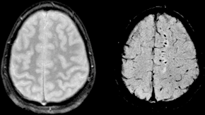
Diffuse axonal injury (DAI) is a brain injury in which scattered lesions in white matter tracts as well as gray matter occur over a widespread area. DAI is one of the most common and devastating types of traumatic brain injury and is a major cause of unconsciousness and persistent vegetative state after severe head trauma. It occurs in about half of all cases of severe head trauma and may be the primary damage that occurs in concussion. The outcome is frequently coma, with over 90% of patients with severe DAI never regaining consciousness. Those who do wake up often remain significantly impaired.

Astrogliosis is an abnormal increase in the number of astrocytes due to the destruction of nearby neurons from CNS trauma, infection, ischemia, stroke, autoimmune responses, and neurodegenerative disease. In healthy neural tissue, astrocytes play critical roles in energy provision, regulation of blood flow, homeostasis of extracellular fluid, homeostasis of ions and transmitters, regulation of synapse function, and synaptic remodeling. Astrogliosis changes the molecular expression and morphology of astrocytes, causing scar formation and, in severe cases, inhibition of axon regeneration.
Axon guidance is a subfield of neural development concerning the process by which neurons send out axons to reach the correct targets. Axons often follow very precise paths in the nervous system, and how they manage to find their way so accurately is being researched.

Netrins are a class of proteins involved in axon guidance. They are named after the Sanskrit word "netr", which means "one who guides." Netrins are genetically conserved across nematode worms, fruit flies, frogs, mice, and humans. Structurally, netrin resembles the extracellular matrix protein laminin.

A growth cone is a large actin-supported extension of a developing or regenerating neurite seeking its synaptic target. Their existence was originally proposed by Spanish histologist Santiago Ramón y Cajal based upon stationary images he observed under the microscope. He first described the growth cone based on fixed cells as "a concentration of protoplasm of conical form, endowed with amoeboid movements". Growth cones are situated on the tips of neurites, either dendrites or axons, of the nerve cell. The sensory, motor, integrative, and adaptive functions of growing axons and dendrites are all contained within this specialized structure.
A neurite or neuronal process refers to any projection from the cell body of a neuron. This projection can be either an axon or a dendrite. The term is frequently used when speaking of immature or developing neurons, especially of cells in culture, because it can be difficult to tell axons from dendrites before differentiation is complete.
Gliosis is a nonspecific reactive change of glial cells in response to damage to the central nervous system (CNS). In most cases, gliosis involves the proliferation or hypertrophy of several different types of glial cells, including astrocytes, microglia, and oligodendrocytes. In its most extreme form, the proliferation associated with gliosis leads to the formation of a glial scar.
Neuroregeneration refers to the regrowth or repair of nervous tissues, cells or cell products. Such mechanisms may include generation of new neurons, glia, axons, myelin, or synapses. Neuroregeneration differs between the peripheral nervous system (PNS) and the central nervous system (CNS) by the functional mechanisms and especially the extent and speed. When an axon is damaged, the distal segment undergoes Wallerian degeneration, losing its myelin sheath. The proximal segment can either die by apoptosis or undergo the chromatolytic reaction, which is an attempt at repair. In the CNS, synaptic stripping occurs as glial foot processes invade the dead synapse.
Neural tissue engineering is a specific sub-field of tissue engineering. Neural tissue engineering is primarily a search for strategies to eliminate inflammation and fibrosis upon implantation of foreign substances. Often foreign substances in the form of grafts and scaffolds are implanted to promote nerve regeneration and to repair damage caused to nerves of both the central nervous system (CNS) and peripheral nervous system (PNS) by an injury.
Glial scar formation (gliosis) is a reactive cellular process involving astrogliosis that occurs after injury to the central nervous system. As with scarring in other organs and tissues, the glial scar is the body's mechanism to protect and begin the healing process in the nervous system.

Repulsive guidance molecule A (RGMa) is a bone morphogenetic protein (BMP) co-receptor of the repulsive guidance molecule family. Together with BMPR1A and BMPR1B, as well as ACVR2A and BMPR2, it binds BMPs thereby activating the intracellular SMAD1/5/8 signalling pathway. In humans this protein is encoded by the RGMA gene.
Slit is a family of secreted extracellular matrix proteins which play an important signalling role in the neural development of most bilaterians. While lower animal species, including insects and nematode worms, possess a single Slit gene, humans, mice and other vertebrates possess three Slit homologs: Slit1, Slit2 and Slit3. Human Slits have been shown to be involved certain pathological conditions, such as cancer and inflammation.
Collapsin response mediator protein family or CRMP family consists of five intracellular phosphoproteins of similar molecular size and high (50–70%) amino acid sequence identity. CRMPs are predominantly expressed in the nervous system during development and play important roles in axon formation from neurites and in growth cone guidance and collapse through their interactions with microtubules. Cleaved forms of CRMPs have also been linked to neuron degeneration after trauma induced injury.

Repulsive guidance molecule B (RGMb), also known as DRAGON, is a bone morphogenetic protein (BMP) co-receptor of the repulsive guidance molecule family. In humans this protein is encoded by the RGMB gene.
In molecular biology, the FEZ-like protein family is a family of eukaryotic proteins thought to be involved in axonal outgrowth and fasciculation. The N-terminal regions of these sequences are less conserved than the C-terminal regions, and are highly acidic. The Caenorhabditis elegans homologue, UNC-76, may play structural and signalling roles in the control of axonal extension and adhesion and these roles have also been postulated for other FEZ family proteins. Certain homologues have been definitively found to interact with the N-terminal variable region (V1) of PKC-zeta, and this interaction causes cytoplasmic translocation of the FEZ family protein in mammalian neuronal cells. The C-terminal region probably participates in the association with the regulatory domain of PKC-zeta. The members of this family are predicted to form coiled-coil structures which may interact with members of the RhoA family of signalling proteins, but are not thought to contain other characteristic protein motifs. Certain members of this family are expressed almost exclusively in the brain, whereas others are expressed in other tissues, and are thought to perform similar but unknown functions in these tissues.

The growth cone is a highly dynamic structure of the developing neuron, changing directionality in response to different secreted and contact-dependent guidance cues; it navigates through the developing nervous system in search of its target. The migration of the growth cone is mediated through the interaction of numerous trophic and tropic factors; netrins, slits, ephrins and semaphorins are four well-studied tropic cues (Fig.1). The growth cone is capable of modifying its sensitivity to these guidance molecules as it migrates to its target; this sensitivity regulation is an important theme seen throughout development.
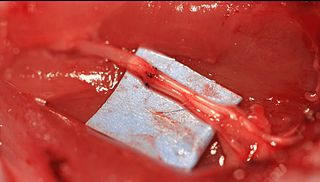
Epineurial repair is a common surgical procedure to repair a nerve laceration via the epineurium, the connective tissue surrounding nerve fibers originating from the spinal cord. It is intended to allow the restoration of sensory function. When a nerve is lacerated or cut, repair is done by sewing the cut ends together through the epineurium to increase the potential of the proximal part growing correctly along the route the degrading distal part leaves behind. Usual sensation and mobility will not be an immediate result because nerves grow at a rate of approximately 1 millimeter per day, so it will take a few months to notice the final outcome. Research in use of nerve grafts and nerve growth factors is being done to speed recovery time.

