Hemojuvelin

Last updated
HJV
Available structures
PDB Ortholog search: PDBe RCSB
Identifiers
Aliases HJV , HFE2, hemojuvelin BMP co-receptor, HFE2A, hemochromatosis type 2 (juvenile), JH, RGMC
External IDs OMIM: 608374 MGI: 1916835 HomoloGene: 17060 GeneCards: HJV
Orthologs
SpeciesHumanMouse
Entrez
Ensembl
UniProt
RefSeq (mRNA)

NM_027126

RefSeq (protein)

NP_081402

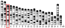
Location (UCSC) Chr 1: 146.02 – 146.04 Mb Chr 3: 96.43 – 96.44 Mb
PubMed search [3] [4]
Wikidata
View/Edit Human View/Edit Mouse

Hemojuvelin (HJV), also known as repulsive guidance molecule C (RGMc) or hemochromatosis type 2 protein (HFE2), is a membrane-bound and soluble protein in mammals that is responsible for the iron overload condition known as juvenile hemochromatosis in humans, a severe form of hemochromatosis. In humans, the hemojuvelin protein is encoded by the HFE2 gene. [5] [6] Hemojuvelin is a member of the repulsive guidance molecule family of proteins. [7] [8] Both RGMa and RGMb are found in the nervous system, [9] [10] while hemojuvelin is found in skeletal muscle and the liver. [10] [11]

Contents

Function

For many years the signal transduction pathways that regulate systemic iron homeostasis have been unknown. However it has been demonstrated that hemojuvelin interacts with bone morphogenetic protein (BMP), possibly as a co-receptor, and may signal via the SMAD pathway to regulate hepcidin expression. [12] Associations with BMP2 and BMP4 have been described. [13]

Mouse HJV knock-out models confirmed that HJV is the gene responsible for juvenile hemochromatosis. Hepcidin levels in the liver are dramatically depressed in these knockout animals. [14] [15]

A soluble form of HJV may be a molecule that suppresses hepcidin expression. [16]

RGMs may play inhibitory roles in prostate cancer by suppressing cell growth, adhesion, migration and invasion. RGMs can coordinate Smad-dependent and Smad-independent signalling of BMPs in prostate cancer and breast cancer cells. [17] [18] Furthermore, aberrant expression of RGMs was indicated in breast cancer. The perturbed expression was associated with disease progression and poor prognosis. [19]

Gene structure and transcription

RGMc/HJV is a 4-exon gene in mammals that undergoes alternative RNA splicing to yield 3 mRNAs with different 5’ untranslated regions (5’UTRs). [11] Gene transcription is induced during myoblast differentiation, producing all 3 mRNAs. There are three critical promoter elements responsible for transcriptional activation in skeletal muscle (the tissue that has the highest level of RGMc expressesion per weight), comprising paired E-boxes, a putative Stat and/or Ets element, and a MEF2 site, and muscle transcription factors myogenin and MEF2C stimulate RGMc promoter function in non-muscle cells. As these elements are conserved in RGMc genes from multiple species, these results suggest that RGMc has been a muscle-enriched gene throughout its evolutionary history. [11]

RGMc/HJV, is transcriptionally regulated during muscle differentiation. [11]

Isoforms

Two classes of GPI-anchored and glycosylated HJV molecules are targeted to the membrane and undergo distinct fates. [20]

RGMc appears to undergo a complex processing that generates 2 soluble, single-chain forms, and two membrane-bound forms found as a (i) single-chain, and (ii) two-chain species which appears to be cleaved at a site within a partial von Willebrand factor domain. [20]

Using a combination of biochemical and cell-based approaches, it has demonstrated that BMP-2 could interact in biochemical assays with the single-chain HJV species, and also could bind to cell-associated HJV. Two mouse HJV amino acid substitution mutants, D165E and G313V (corresponding to human D172E and G320V), also could bind BMP-2, but less effectively than wild-type HJV, while G92V (human G99V) could not. In contrast, the membrane-spanning protein, neogenin, a receptor for the related molecule, RGMa, preferentially bound membrane-associated heterodimeric RGMc and was able to interact on cells only with wild-type RGMc and G92V. These results show that different isoforms of RGMc/HJV may play unique physiological roles through defined interactions with distinct signaling proteins and demonstrate that, in some disease-linked HJV mutants, these interactions are defective. [21]

Structure

In 2009, the Rosetta ab initio protein structure prediction software has been used to create a three-dimensional model of the RGM family of proteins., [8] In 2011, a crystal structure of a fragment of hemojuvelin binding to neogenin was completed [22] showing similar structures to the ab initio model and further informing the view of the RGM family of proteins.

Mechanism of action

Furin-like proprotein convertases (PPC) are responsible for conversion of 50 kDa HJV to a 40 kDa protein with a truncated COOH-terminus, at a conserved polybasic RNRR site. This suggests a potential mechanism to generate the soluble forms of HJV/hemojuvelin (s-hemojuvelin) found in the blood of rodents and humans. [23] [24]

Clinical significance

Mutations in HJV are responsible for the vast majority of juvenile hemochromatosis patients. A small number of patients have mutations in the hepcidin (HAMP) gene. The gene was positionally cloned. [6] Hemojuvelin is highly expressed in skeletal muscle and heart, and to a lesser extent in the liver. One insight into the pathogenesis of juvenile hemochromatosis is that patients have low to undetectable urinary hepcidin levels, suggesting that hemojuvelin is a positive regulator of hepcidin, the central iron regulatory hormone. As a result, low hepcidin levels would result in increased intestinal iron absorption. Thus, HJV/RGMc appears to play a critical role in iron metabolism.[ citation needed ]

Related Research Articles

<span class="mw-page-title-main">Iron overload</span> Human disease

Iron overload or haemochromatosis indicates increased total accumulation of iron in the body from any cause and resulting organ damage. The most important causes are hereditary haemochromatosis, a genetic disorder, and transfusional iron overload, which can result from repeated blood transfusions.

<span class="mw-page-title-main">Human iron metabolism</span> Iron metabolism in the body

Human iron metabolism is the set of chemical reactions that maintain human homeostasis of iron at the systemic and cellular level. Iron is both necessary to the body and potentially toxic. Controlling iron levels in the body is a critically important part of many aspects of human health and disease. Hematologists have been especially interested in systemic iron metabolism, because iron is essential for red blood cells, where most of the human body's iron is contained. Understanding iron metabolism is also important for understanding diseases of iron overload, such as hereditary hemochromatosis, and iron deficiency, such as iron-deficiency anemia.

<span class="mw-page-title-main">Hepcidin</span> Protein-coding gene in the species Homo sapiens

Hepcidin is a protein that in humans is encoded by the HAMP gene. Hepcidin is a key regulator of the entry of iron into the circulation in mammals.

<span class="mw-page-title-main">Ferroportin</span> Protein

Ferroportin-1, also known as solute carrier family 40 member 1 (SLC40A1) or iron-regulated transporter 1 (IREG1), is a protein that in humans is encoded by the SLC40A1 gene. Ferroportin is a transmembrane protein that transports iron from the inside of a cell to the outside of the cell. Ferroportin is the only known iron exporter.

<span class="mw-page-title-main">HFE (gene)</span> Mammalian protein found in Homo sapiens

Human homeostatic iron regulator protein, also known as the HFE protein, is a transmembrane protein that in humans is encoded by the HFE gene. The HFE gene is located on short arm of chromosome 6 at location 6p22.2

<span class="mw-page-title-main">Furin</span> Enzyme found in humans

Furin is a protease, a proteolytic enzyme that in humans and other animals is encoded by the FURIN gene. Some proteins are inactive when they are first synthesized, and must have sections removed in order to become active. Furin cleaves these sections and activates the proteins. It was named furin because it was in the upstream region of an oncogene known as FES. The gene was known as FUR and therefore the protein was named furin. Furin is also known as PACE. A member of family S8, furin is a subtilisin-like peptidase.

Proprotein convertases (PPCs) are a family of proteins that activate other proteins. Many proteins are inactive when they are first synthesized, because they contain chains of amino acids that block their activity. Proprotein convertases remove those chains and activate the protein. The prototypical proprotein convertase is furin. Proprotein convertases have medical significance, because they are involved in many important biological processes, such as cholesterol synthesis. Compounds called proprotein convertase inhibitors can block their action, and block the target proteins from becoming active. Many proprotein convertases, especially furin and PACE4, are involved in pathological processes such as viral infection, inflammation, hypercholesterolemia, and cancer, and have been postulated as therapeutic targets for some of these diseases.

<span class="mw-page-title-main">Bone morphogenetic protein 6</span> Protein-coding gene in the species Homo sapiens

Bone morphogenetic protein 6 is a protein that in humans is encoded by the BMP6 gene.

<span class="mw-page-title-main">Mothers against decapentaplegic homolog 4</span> Mammalian protein found in Homo sapiens

SMAD4, also called SMAD family member 4, Mothers against decapentaplegic homolog 4, or DPC4 is a highly conserved protein present in all metazoans. It belongs to the SMAD family of transcription factor proteins, which act as mediators of TGF-β signal transduction. The TGFβ family of cytokines regulates critical processes during the lifecycle of metazoans, with important roles during embryo development, tissue homeostasis, regeneration, and immune regulation.

<span class="mw-page-title-main">Mothers against decapentaplegic homolog 7</span> Protein-coding gene in the species Homo sapiens

Mothers against decapentaplegic homolog 7 or SMAD7 is a protein that in humans is encoded by the SMAD7 gene.

Smads comprise a family of structurally similar proteins that are the main signal transducers for receptors of the transforming growth factor beta (TGF-B) superfamily, which are critically important for regulating cell development and growth. The abbreviation refers to the homologies to the Caenorhabditis elegans SMA and MAD family of genes in Drosophila.

<span class="mw-page-title-main">Carnitine palmitoyltransferase I</span> Protein-coding gene in the species Homo sapiens

Carnitine palmitoyltransferase I (CPT1) also known as carnitine acyltransferase I, CPTI, CAT1, CoA:carnitine acyl transferase (CCAT), or palmitoylCoA transferase I, is a mitochondrial enzyme responsible for the formation of acyl carnitines by catalyzing the transfer of the acyl group of a long-chain fatty acyl-CoA from coenzyme A to l-carnitine. The product is often Palmitoylcarnitine, but other fatty acids may also be substrates. It is part of a family of enzymes called carnitine acyltransferases. This "preparation" allows for subsequent movement of the acyl carnitine from the cytosol into the intermembrane space of mitochondria.

<span class="mw-page-title-main">ADAM9</span> Protein-coding gene in the species Homo sapiens

Disintegrin and metalloproteinase domain-containing protein 9 is an enzyme that in humans is encoded by the ADAM9 gene.

<span class="mw-page-title-main">Repulsive guidance molecule A</span> Protein-coding gene in the species Homo sapiens

Repulsive guidance molecule A (RGMa) is a bone morphogenetic protein (BMP) co-receptor of the repulsive guidance molecule family. Together with BMPR1A and BMPR1B, as well as ACVR2A and BMPR2, it binds BMPs thereby activating the intracellular SMAD1/5/8 signalling pathway. In humans this protein is encoded by the RGMA gene.

<span class="mw-page-title-main">Iron metabolism disorder</span> Medical condition

Iron metabolism disorders may involve a number of genes including HFE and TFR2.

Juvenile hemochromatosis, also known as hemochromatosis type 2, is a rare form of hereditary hemochromatosis, which emerges in young individuals, typically between 15 and 30 years of age, but occasionally later. It is characterized by an inability to control how much iron is absorbed by the body, in turn leading to iron overload, where excess iron accumulates in many areas of the body and causes damage to the places it accumulates.

Repulsive guidance molecules (RGMs) are members of a three gene family composed of RGMa, RGMb, and RGMc . RGMa has been implicated to play an important role in the developing brain and in the scar tissue that forms after a brain injury. For example, RGMa helps guide retinal ganglion cell (RGC) axons to the tectum in the midbrain. It has also been demonstrated that after induced spinal cord injury RGMa accumulates in the scar tissue around the lesion. Further research has shown that RGMa is an inhibitor of axonal outgrowth. Taken together, these findings highlight the importance of RGMa in axonal guidance and outgrowth.

Haemochromatosis type 3 is a type of iron overload disorder associated with deficiencies in transferrin receptor 2. It exhibits an autosomal recessive inheritance pattern. The first confirmed case was diagnosed in 1865 by French doctor Trousseau. Later in 1889, the German doctor von Recklinghausen indicated that the liver contains iron, and due to bleeding being considered to be the cause, he called the pigment "Haemochromatosis." In 1935, English doctor Sheldon's groundbreaking book titled, Haemochromatosis, reviewed 311 patient case reports and presented the idea that haemochromatosis was a congenital metabolic disorder. Hereditary haemochromatosis is a congenital disorder which affects the regulation of iron metabolism thus causing increased gut absorption of iron and a gradual build-up of pathologic iron deposits in the liver and other internal organs, joint capsules and the skin. The iron overload could potentially cause serious disease from the age of 40–50 years. In the final stages of the disease, the major symptoms include liver cirrhosis, diabetes and bronze-colored skin. There are four types of hereditary hemochromatosis which are classified depending on the age of onset and other factors such as genetic cause and mode of inheritance.

<span class="mw-page-title-main">Repulsive guidance molecule B</span> Protein-coding gene in the species Homo sapiens

Repulsive guidance molecule B (RGMb), also known as DRAGON, is a bone morphogenetic protein (BMP) co-receptor of the repulsive guidance molecule family. In humans this protein is encoded by the RGMB gene.

Hemochromatosis type 4 is a hereditary iron overload disorder that affects ferroportin, an iron transport protein needed to export iron from cells into circulation. Although the disease is rare, it is found throughout the world and affects people from various ethnic groups. While the majority of individuals with type 4 hemochromatosis have a relatively mild form of the disease, some affected individuals have a more severe form. As the disease progresses, iron may accumulate in the tissues of affected individuals over time, potentially resulting in organ damage.

References

  1. 1 2 3 GRCh38: Ensembl release 89: ENSG00000168509 - Ensembl, May 2017
  2. 1 2 3 GRCm38: Ensembl release 89: ENSMUSG00000038403 - Ensembl, May 2017
  3. "Human PubMed Reference:". National Center for Biotechnology Information, U.S. National Library of Medicine.
  4. "Mouse PubMed Reference:". National Center for Biotechnology Information, U.S. National Library of Medicine.
  5. Roetto A, Totaro A, Cazzola M, Cicilano M, Bosio S, D'Ascola G, Carella M, Zelante L, Kelly AL, Cox TM, Gasparini P, Camaschella C (May 1999). "Juvenile hemochromatosis locus maps to chromosome 1q". Am. J. Hum. Genet. 64 (5): 1388–93. doi:10.1086/302379. PMC   1377875 . PMID   10205270.
  6. 1 2 Papanikolaou G, Samuels ME, Ludwig EH, MacDonald ML, Franchini PL, Dubé MP, Andres L, MacFarlane J, Sakellaropoulos N, Politou M, Nemeth E, Thompson J, Risler JK, Zaborowska C, Babakaiff R, Radomski CC, Pape TD, Davidas O, Christakis J, Brissot P, Lockitch G, Ganz T, Hayden MR, Goldberg YP (January 2004). "Mutations in HFE2 cause iron overload in chromosome 1q-linked juvenile hemochromatosis". Nat. Genet. 36 (1): 77–82. doi: 10.1038/ng1274 . PMID   14647275.
  7. Corradini E, Babitt JL, Lin HY (October 2009). "The RGM/DRAGON family of BMP co-receptors". Cytokine & Growth Factor Reviews. 20 (5–6): 389–398. doi:10.1016/j.cytogfr.2009.10.008. PMC   3715994 . PMID   19897400.
  8. 1 2 Severyn CJ, Shinde U, Rotwein P (September 2009). "Molecular biology, genetics and biochemistry of the repulsive guidance molecule family". Biochem. J. 422 (3): 393–403. doi:10.1042/BJ20090978. PMC   4242795 . PMID   19698085.
  9. Samad TA, Srinivasan A, Karchewski LA, Jeong SJ, Campagna JA, Ji RR, Fabrizio DA, Zhang Y, Lin HY, Bell E, Woolf CJ (February 2004). "DRAGON: a member of the repulsive guidance molecule-related family of neuronal- and muscle-expressed membrane proteins is regulated by DRG11 and has neuronal adhesive properties". J. Neurosci. 24 (8): 2027–36. doi:10.1523/JNEUROSCI.4115-03.2004. PMC   6730385 . PMID   14985445.
  10. 1 2 Schmidtmer J, Engelkamp D (January 2004). "Isolation and expression pattern of three mouse homologues of chick Rgm". Gene Expr. Patterns. 4 (1): 105–10. doi:10.1016/S1567-133X(03)00144-3. PMID   14678836.
  11. 1 2 3 4 Severyn CJ, Rotwein P (December 2010). "Conserved proximal promoter elements control repulsive guidance molecule c/hemojuvelin (Hfe2) gene transcription in skeletal muscle". Genomics. 96 (6): 342–51. doi:10.1016/j.ygeno.2010.09.001. PMC   2988867 . PMID   20858542.
  12. Babitt JL, Huang FW, Wrighting DM, Xia Y, Sidis Y, Samad TA, Campagna JA, Chung RT, Schneyer AL, Woolf CJ, Andrews NC, Lin HY (May 2006). "Bone morphogenetic protein signaling by hemojuvelin regulates hepcidin expression". Nat. Genet. 38 (5): 531–9. doi:10.1038/ng1777. PMID   16604073. S2CID   19282860.
  13. Zhang AS, Yang F, Meyer K, Hernandez C, Chapman-Arvedson T, Bjorkman PJ, Enns CA (June 2008). "Neogenin-mediated hemojuvelin shedding occurs after hemojuvelin traffics to the plasma membrane". J. Biol. Chem. 283 (25): 17494–502. doi: 10.1074/jbc.M710527200 . PMC   2427329 . PMID   18445598.
  14. Huang FW, Pinkus JL, Pinkus GS, Fleming MD, Andrews NC (August 2005). "A mouse model of juvenile hemochromatosis". J. Clin. Invest. 115 (8): 2187–91. doi:10.1172/JCI25049. PMC   1180543 . PMID   16075059.
  15. Niederkofler V, Salie R, Arber S (August 2005). "Hemojuvelin is essential for dietary iron sensing, and its mutation leads to severe iron overload". J. Clin. Invest. 115 (8): 2180–6. doi:10.1172/JCI25683. PMC   1180556 . PMID   16075058.
  16. Lin L, Goldberg YP, Ganz T (October 2005). "Competitive regulation of hepcidin mRNA by soluble and cell-associated hemojuvelin". Blood. 106 (8): 2884–9. doi: 10.1182/blood-2005-05-1845 . PMID   15998830.
  17. Li J, Ye L, Sanders AJ, Jiang WG (March 2012). "Repulsive guidance molecule B (RGMB) plays negative roles in breast cancer by coordinating BMP signaling". J Cell Biochem. 113 (7): 2523–31. doi:10.1002/jcb.24128. PMID   22415859. S2CID   35629616.
  18. Li J, Ye L, Kynaston HG, Jiang WG (February 2012). "Repulsive guidance molecules, novel bone morphogenetic protein co-receptors, are key regulators of the growth and aggressiveness of prostate cancer cells". Int. J. Oncol. 40 (2): 544–50. doi: 10.3892/ijo.2011.1251 . PMID   22076499.
  19. Li J, Ye L, Mansel RE, Jiang WG (May 2011). "Potential prognostic value of repulsive guidance molecules in breast cancer". Anticancer Res. 31 (5): 1703–11. PMID   21617229.
  20. 1 2 3 4 Kuninger D, Kuns-Hashimoto R, Kuzmickas R, Rotwein P (August 2006). "Complex biosynthesis of the muscle-enriched iron regulator RGMc". J. Cell Sci. 119 (Pt 16): 3273–83. doi:10.1242/jcs.03074. PMID   16868025. S2CID   15574534.
  21. Kuns-Hashimoto R, Kuninger D, Nili M, Rotwein P (April 2008). "Selective binding of RGMc/hemojuvelin, a key protein in systemic iron metabolism, to BMP-2 and neogenin". Am. J. Physiol., Cell Physiol. 294 (4): C994–C1003. doi:10.1152/ajpcell.00563.2007. PMID   18287331. S2CID   32158124.
  22. Yang F, West AP, Bjorkman PJ (2011). "Crystal structure of a hemojuvelin-binding fragment of neogenin at 1.8Å". Journal of Structural Biology. 174 (1): 239–244. doi:10.1016/j.jsb.2010.10.005. ISSN   1047-8477. PMC   3074981 . PMID   20971194.
  23. Lin L, Nemeth E, Goodnough JB, Thapa DR, Gabayan V, Ganz T (2008). "Soluble hemojuvelin is released by proprotein convertase-mediated cleavage at a conserved polybasic RNRR site". Blood Cells Mol. Dis. 40 (1): 122–31. doi:10.1016/j.bcmd.2007.06.023. PMC   2211380 . PMID   17869549.
  24. Kuninger D, Kuns-Hashimoto R, Nili M, Rotwein P (2008). "Pro-protein convertases control the maturation and processing of the iron-regulatory protein, RGMc/hemojuvelin". BMC Biochem. 9: 9. doi: 10.1186/1471-2091-9-9 . PMC   2323002 . PMID   18384687.

Further reading