
Sinusitis, also known as rhinosinusitis, is inflammation of the mucous membranes that line the sinuses resulting in symptoms. Common symptoms include thick nasal mucus, a plugged nose, and facial pain. Other signs and symptoms may include fever, headaches, a poor sense of smell, sore throat, and a cough. The cough is often worse at night. Serious complications are rare. It is defined as acute sinusitis if it lasts fewer than 4 weeks, and as chronic sinusitis if it lasts for more than 12 weeks.

The maxilla in vertebrates is the upper fixed bone of the jaw formed from the fusion of two maxillary bones. The upper jaw includes the hard palate in the front of the mouth. The two maxillary bones are fused at the intermaxillary suture, forming the anterior nasal spine. This is similar to the mandible, which is also a fusion of two mandibular bones at the mandibular symphysis. The mandible is the movable part of the jaw.

The glossopharyngeal nerve, known as the ninth cranial nerve, is a mixed nerve that carries afferent sensory and efferent motor information. It exits the brainstem out from the sides of the upper medulla, just anterior to the vagus nerve. The motor division of the glossopharyngeal nerve is derived from the basal plate of the embryonic medulla oblongata, while the sensory division originates from the cranial neural crest.

The sphenoid bone is an unpaired bone of the neurocranium. It is situated in the middle of the skull towards the front, in front of the basilar part of the occipital bone. The sphenoid bone is one of the seven bones that articulate to form the orbit. Its shape somewhat resembles that of a butterfly or bat with its wings extended.

In anatomy, the orbit is the cavity or socket of the skull in which the eye and its appendages are situated. Anatomical term created by Gerard of Cremona. "Orbit" can refer to the bony socket, or it can also be used to imply the contents. In the adult human, the volume of the orbit is 30 millilitres, of which the eye occupies 6.5 ml. The orbital contents comprise the eye, the orbital and retrobulbar fascia, extraocular muscles, cranial nerves II, III, IV, V, and VI, blood vessels, fat, the lacrimal gland with its sac and nasolacrimal duct, the eyelids, medial and lateral palpebral ligaments, check ligaments, the suspensory ligament, septum, ciliary ganglion and short ciliary nerves.

Dura mater is a thick membrane made of dense irregular connective tissue that surrounds the brain and spinal cord. It is the outermost of the three layers of membrane called the meninges that protect the central nervous system. The other two meningeal layers are the arachnoid mater and the pia mater. The dura surrounds the brain and the spinal cord. It envelops the arachnoid mater, which is responsible for keeping in the cerebrospinal fluid. It is derived primarily from the neural crest cell population, with postnatal contributions of the paraxial mesoderm.

The pyramid-shaped maxillary sinus is the largest of the paranasal sinuses, and drains into the middle meatus of the nose through the osteomeatal complex.

The cavernous sinus within the human head is one of the dural venous sinuses creating a cavity called the lateral sellar compartment bordered by the temporal bone of the skull and the sphenoid bone, lateral to the sella turcica.

The ophthalmic nerve is the first branch of the trigeminal nerve. The ophthalmic nerve is a sensory nerve mostly carrying general somatic afferent fibers that transmit sensory information to the CNS from structures of the eyeball, the skin of the upper face and anterior scalp, the lining of the upper part of the nasal cavity and air cells, and the meninges of the anterior cranial fossa. Some of ophthalmic nerve branches also convey parasympathetic fibers.
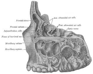
The frontal sinuses are one of the four pairs of paranasal sinuses that are situated behind the brow ridges. Sinuses are mucosa-lined airspaces within the bones of the face and skull. Each opens into the anterior part of the corresponding middle nasal meatus of the nose through the frontonasal duct which traverses the anterior part of the labyrinth of the ethmoid. These structures then open into the semilunar hiatus in the middle meatus.

The ethmoidal labyrinth or lateral mass of the ethmoid bone consists of a number of thin-walled cellular cavities, the ethmoid air cells, arranged in three groups, anterior, middle, and posterior, and interposed between two vertical plates of bone; the lateral plate forms part of the orbit, the medial plate forms part of the nasal cavity. In the disarticulated bone many of these cells are opened into, but when the bones are articulated, they are closed in at every part, except where they open into the nasal cavity.
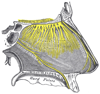
One branch of the pterygopalatine ganglion, longer and larger than the others, is named the nasopalatine nerve.

Below the bulla ethmoidalis, and partly hidden by the inferior end of the uncinate process of ethmoid bone, is the maxillary hiatus ; in a frontal section this opening is seen to be placed near the roof of the sinus. In the articulated skull this aperture is much reduced in size by the following bones: the uncinate process of the ethmoid above, the ethmoidal process of the inferior nasal concha below, the vertical part of the palatine behind, and a small part of the lacrimal above and in front; the sinus communicates with the middle meatus of the nose, generally by two small apertures left between the above-mentioned bones.

Maxillary sinus floor augmentation is a surgical procedure which aims to increase the amount of bone in the posterior maxilla, in the area of the premolar and molar teeth, by lifting the lower Schneiderian membrane and placing a bone graft.

The human nose is the most protruding part of the face. It bears the nostrils and is the first organ of the respiratory system. It is also the principal organ in the olfactory system. The shape of the nose is determined by the nasal bones and the nasal cartilages, including the nasal septum which separates the nostrils and divides the nasal cavity into two. On average the nose of a male is larger than that of a female.
A sinus is a sac or cavity in any organ or tissue, or an abnormal cavity or passage caused by the destruction of tissue. In common usage, "sinus" usually refers to the paranasal sinuses, which are air cavities in the cranial bones, especially those near the nose and connecting to it. Most individuals have four paired cavities located in the cranial bone or skull.

A Le Fort fracture of the skull is a classic transfacial fracture of the midface, involving the maxillary bone and surrounding structures in either a horizontal, pyramidal or transverse direction. The hallmark of Lefort fractures is traumatic pterygomaxillary separation, which signifies fractures between the pterygoid plates, horseshoe shaped bony protuberances which extend from the inferior margin of the maxilla, and the maxillary sinuses. Continuity of this structure is a keystone for stability of the midface, involvement of which impacts surgical management of trauma victims, as it requires fixation to a horizontal bar of the frontal bone. The pterygoid plates lie posterior to the upper dental row, or alveolar ridge, when viewing the face from an anterior view. The fractures are named after French surgeon René Le Fort (1869–1951), who discovered the fracture patterns by examining crush injuries in cadavers.

The respiratory system of the horse is the biological system by which a horse circulates air for the purpose of gaseous exchange.
In anatomy, Underwood's septa are fin-shaped projections of bone that may exist in the maxillary sinus, first described in 1910 by Arthur S. Underwood, an anatomist at King's College in London. The presence of septa at or near the floor of the sinus are of interest to the dental clinician when proposing or performing sinus floor elevation procedures because of an increased likelihood of surgical complications, such as tearing of the Schneiderian membrane.
Endoscopic endonasal surgery is a minimally invasive technique used mainly in neurosurgery and otolaryngology. A neurosurgeon or an otolaryngologist, using an endoscope that is entered through the nose, fixes or removes brain defects or tumors in the anterior skull base. Normally an otolaryngologist performs the initial stage of surgery through the nasal cavity and sphenoid bone; a neurosurgeon performs the rest of the surgery involving drilling into any cavities containing a neural organ such as the pituitary gland.
